Today’s literature sharing features an article titled “Nanosensor-Based Pattern-Generating Probe Accelerates Sepsis Diagnosis” published in ACS Nano by a collaborative team from Nanjing University of Chinese Medicine, China Pharmaceutical University, and Karolinska Institute.
Part 1 Research Background
Sepsis, as a globally high-mortality infectious disease, is caused by pathogen infection and can cause severe damage to human tissues and organs. If appropriate antibiotic treatment is not administered in a timely manner, the mortality rate can increase by 8% per hour, making the rapid and accurate identification of pathogens a core challenge in clinical diagnosis and treatment. Current diagnostic methods struggle to complete the detection of multiple pathogens within minutes, limiting early intervention. Biomimetic optical sensor arrays show great promise in distinguishing subtle differences in complex mixtures and biological samples. However, developing a standalone pattern-generating sensor that can achieve multi-analyte recognition in clinical biological fluids without complex synthesis remains a significant challenge.
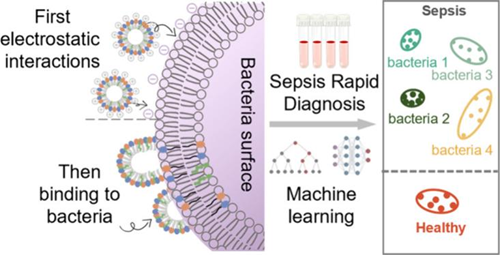
Part 2 Author’s Approach
The authors innovatively constructed a nanosensor-inspired sensor array (NanoSA) composed of nanostructures containing three types of sensors, utilizing intermolecular multiple fluorescence resonance energy transfer (FRET) effects to achieve multi-channel signal output. By validating the array’s ability to recognize bacteria in water, urine, serum, and clinical sepsis samples, and optimizing the model with nine machine learning algorithms, they ultimately achieved rapid identification of 24 sepsis bacteria, capable of distinguishing different concentrations of bacteria, mixed bacteria, and clinical sepsis samples, addressing the time-consuming and limited accuracy issues of traditional diagnostic methods.
Part 3 Visual Guide
Construction of Six-Channel Multi-FRET NanoSA
Three amphiphilic molecules (L1-L3) with different fluorescent groups (ThT, RhB, BODIPY) were designed and synthesized, forming nanostructures through ultrasonic self-assembly, utilizing intermolecular FRET effects to construct a six-channel sensor array. Exciting different fluorescent groups can initiate specific energy transfer, forming unique signal outputs. After adding bacteria, the nanostructures interact with bacterial cell membranes, altering FRET efficiency and causing significant changes in six-channel fluorescence signals (Figure 1).
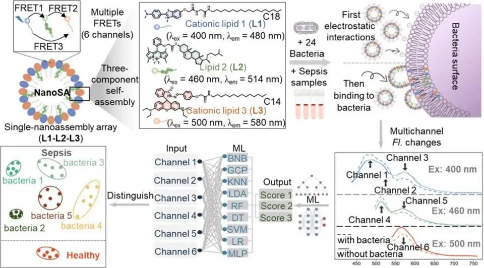
Figure 1. Schematic diagram of the construction of the 6-channel multi-FRET NanoSA for identifying 24 clinical pathogens and sepsis samples.
Simultaneous Recognition of 24 Clinical Pathogens in Water
Testing 24 common pathogenic bacteria in water, the six channels of NanoSA exhibited unique fluorescence responses to different bacteria (Figure 2a): channels 1, 2, and 4 showed increased fluorescence (indicating disassembly of the nanostructures leading to FRET interruption), while channels 3, 5, and 6 showed decreased fluorescence (possibly due to weakened FRET effects and L3 aggregation on the bacterial surface). After optimization with nine machine learning algorithms, the LDA algorithm performed best, achieving an accuracy of 96.9% within 30 seconds, with the same accuracy for blind test samples (Figure 2c), effectively distinguishing 24 bacteria in the score plot.
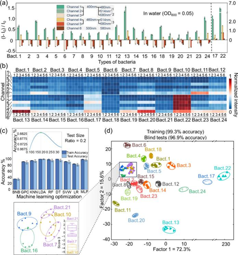
Figure 2. Recognition of 24 bacteria in water.
Recognition of Different Concentrations and Mixed Bacteria
Selecting common clinical bacteria such as Klebsiella pneumoniae and Staphylococcus aureus, the recognition ability of NanoSA for different concentrations of bacteria was verified: as bacterial concentrations changed, the six-channel fluorescence signals exhibited regular variations, and the clustering heatmap visually displayed this correlation (Figure 3a-c). The sensor array can accurately cluster different concentrations of bacteria, with a limit of detection (LOD) as low as OD600=0.011-0.018 (Figure 3d-f). For common clinical mixed bacterial infections, the array can distinguish different proportions of mixed bacterial systems, achieving classification accuracy of 97.0%-100% in typical classification matrices (Figure 3g).
Exploration of Bacterial Recognition Mechanism
Laser confocal microscopy observations showed that L3 had the strongest staining ability for bacteria (Figure 3k). After binding to bacteria, the nanostructures disassemble, releasing L3, leading to FRET interruption, which enhances the fluorescence of L1 and L2 while reducing L3 fluorescence due to aggregation. The differences in affinity of different amphiphilic molecules for bacteria collectively drive the specific signal changes across the six channels, achieving precise bacterial recognition.
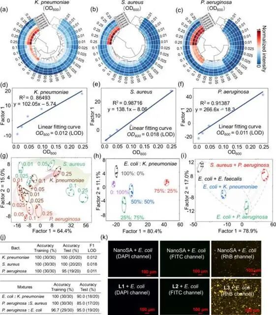
Figure 3. Recognition of different concentrations and mixed proportions of bacteria.
Recognition of Bacteria in Biological Fluids
In urine and serum systems, NanoSA demonstrated excellent resistance to matrix interference and recognition stability: in the detection of 24 pathogenic bacteria in urine (Figure 4a), the six-channel signals exhibited species-specific responses, with an accuracy of 92.0% for LDA algorithm recognition and 86.5% for blind tests, showing clear clustering (Figure 4c); although signal drift occurred in serum due to matrix components, normalization still achieved an accuracy of 93.7% (Figure 4e), with signal variation trends consistent with those in water detection, validating its applicability in complex biological fluids.
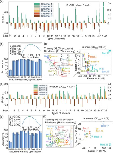
Figure 4. Identification of bacteria in biological fluids.
Recognition of Clinical Sepsis Samples
Selecting 51 sepsis patients (including infections from 5 types of pathogens) and 10 healthy serum samples, NanoSA effectively distinguished patients from healthy individuals, with PCA analysis showing significant inter-group separation (Figure 5b); the MLP algorithm achieved a classification accuracy of 97.6% for the 5 types of infection samples and healthy individuals, with only 1 misclassification (Figure 5e); 3D visualization confirmed significant differences in signal features among different samples (Figure 5k), providing a reliable basis for precise typing of pathogenic bacteria in clinical sepsis.
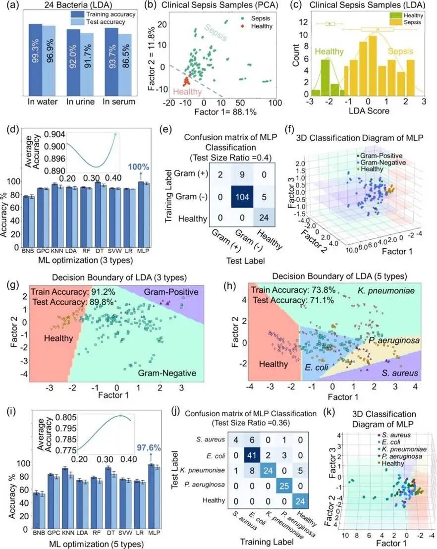
Figure 5. Identification of bacteria in clinical samples.
Part 4 Conclusion
The research team developed a six-channel NanoSA that achieves rapid and precise identification of bacteria in water, biological fluids, and clinical samples through the multiple FRET effects mediated by nanostructures, completing classification of 24 sepsis bacteria and clinical samples within 30 seconds. This system addresses the issues of complex synthesis and weak signals in traditional sensors, while also possessing strong anti-interference capabilities and rapid detection advantages, providing a novel and efficient platform for the rapid clinical diagnosis of infectious diseases such as sepsis, with potential applications in more infectious diseases and cancer detection after optimization.
Original Link
DOI: https://doi.org/10.1021/acsnano.5c14974
Author|Zhai Zhaojie
Proofreader|Tian Yinqi, Ni Fan
Promoter|YC Zhang

Disclaimer:
1. The copyright of this article belongs to the original author. Please contact us for reprints in public accounts and media!
2. Due to limited knowledge, there may be omissions and errors; please feel free to criticize and correct!
3. This article mainly references published literature and is intended for the introduction, commentary, or classroom teaching of related scientific research, and may not be used for commercial purposes. If there are any copyright issues, please feel free to contact us!