Today we share a study published in June 2025 in J Am Soc Nephrol: Molecular Mechanisms of Sepsis-Associated Acute Kidney Injury. This review aims to refine the timeline of sepsis-associated acute kidney injury by analyzing the key molecular events driving disease progression, thereby providing information for rational treatment strategies.
1.Research Background and Core Issues
1.1The Severity and Cognitive Challenges of Sepsis-Associated Acute Kidney Injury
Epidemiological Importance: By 2050, infectious diseases are expected to become the leading cause of death globally, primarily due to the emergence of antibiotic resistance. Sepsis is a major consequence of severe infections, and acute kidney injury is a common and serious complication, serving as an independent risk factor for mortality.
Insufficient Pathobiological Understanding: Due to the complex interactions between the host and microorganisms in the tissue microenvironment, which are difficult to capture, and the rarity of kidney biopsies during the active phase of infection, there is little understanding of the fundamental pathological features and molecular mechanisms of sepsis-associated acute kidney injury. Despite numerous clinical trials targeting potential therapeutic points, none have succeeded, highlighting its complexity.
1.2Core Scientific Issue: The Absence of a Disease Timeline
The rapid progression of sepsis-associated acute kidney injury and the lack of a “molecular clock” to identify disease stages make it extremely challenging to implement precise, stage-specific therapeutic interventions. Many clinical trials targeting upstream inflammatory factors (such as cytokines) have failed, likely because these events have already subsided by the time patients present.
1.3The Core Objective of This Article
This review aims to refine the timeline of sepsis-associated acute kidney injury by analyzing the key molecular events driving disease progression, emphasizing the importance of understanding the temporal order of dynamic interactions between the host and microorganisms.
2. The Disease Timeline and Molecular Mechanisms of Sepsis-Associated Acute Kidney Injury
This article proposes a clear multi-stage model to describe the molecular evolution of sepsis-associated acute kidney injury. The following figure (Figure 1) visually presents the overall timeline of kidney tissue responses.
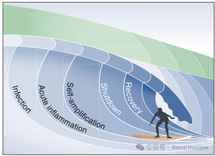
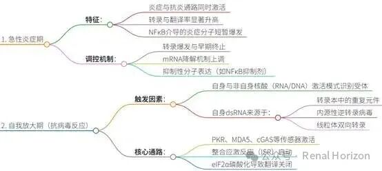
Stage One: Acute Inflammatory Phase
1.Characteristics: Synchronous Outbreak of Inflammatory and Anti-inflammatory Pathways
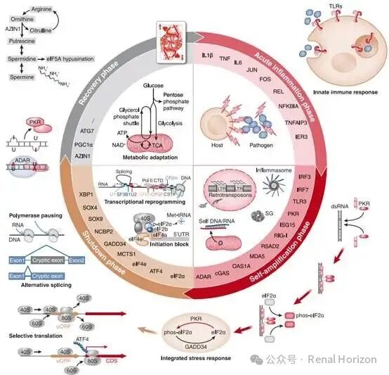
A.This phase is triggered by microorganisms or their mimetics (such as endotoxins), characterized by a significant increase in systemic transcription and translation rates to produce large amounts of defensive molecules (such as pro-inflammatory cytokines) and activate the immune system.
B.The key mechanism is the “left foot brake” technology: the host genome activates pro-inflammatory and anti-inflammatory pathways simultaneously rather than sequentially from the earliest stages of infection (such as NF-κB and its inhibitors), allowing pro-inflammatory genes to be expressed vigorously and rapidly downregulated within hours to prevent excessive tissue damage.
2.Fine-tuning Mechanisms
A.Transcriptional Burst: Gene expression occurs in a pulsed manner, allowing for rapid production of new transcripts.
B.Multi-layer Regulation: Cells simultaneously initiate multiple mechanisms to counteract this pulsed expression, including:
Transcript and Protein Compartmentalization (e.g., forming stress granules to sequester transcripts).
Translational Regulation (e.g., using upstream open reading frames uORF).
Selective Splicing generates isoforms that target nonsense-mediated decay.
C.These mechanisms collectively ensure that cells can mount a dynamic and controlled stress response.
Stage Two: Self-amplifying Phase (Antiviral Response)
1.Core Driver: Confusion Between Self and Non-self Nucleic Acids
A significant portion of the large amounts of host-derived RNA produced during the early inflammatory phase contains repetitive elements (such as retrotransposons), which can form double-stranded RNA through intramolecular or intermolecular base pairing.
Under stress conditions, endogenous retroviruses and mitochondrial-derived transcripts, which were originally epigenetically suppressed, may also be reactivated or released into the cytoplasm, forming dsRNA. This self-derived dsRNA can be misidentified by pattern recognition receptors (such as PKR, MDA5) as viral RNA, triggering a strong antiviral response, regardless of whether the initial infection was bacterial or viral.
2.Key Node: Integration of Stress Response and Translation Shutdown
Once activated, the dsRNA sensor PKR phosphorylates the eukaryotic translation initiation factor2α. Phosphorylated eIF2α obstructs the GTP/GDP exchange reaction necessary for translation initiation, leading to global translation shutdown.
Thus, the antiviral phase initiated by transcription/translation bursts is ultimately terminated by the integrated stress response. In animal models, this phase peaks approximately 8-16 hours post-infection.
The following figure (Figure 2) details the representative molecular pathways of each stage.
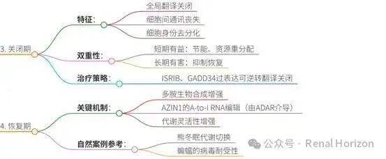
Stage Three: Shutdown Phase
1.Characteristics: Global Translation Shutdown and Organ “Paralysis”
Unlike the asynchronous inflammatory/antiviral responses observed previously, translation shutdown occurs at a distinct time point across all cell types. This phase is characterized by loss of intercellular communication, dedifferentiation of cell identity, and downregulation of physiological functions.
2.Duality: Protective Adaptation or Pathological Damage?
Adaptive Aspect: Brief global metabolic suppression may be beneficial, reducing energy expenditure and enabling resource reallocation. During this time, genome-wide reprogramming is occurring in preparation for recovery (e.g., induction of key renal repair factors such as SOX9).
Pathological Aspect: Prolonged translation shutdown is clearly detrimental. Experiments have shown that using small molecules ISRIB (which promote GTP/GDP exchange on the eIF2 complex) or overexpressing the eIF2α phosphatase GADD34 to reverse translation shutdown can promote renal function recovery. This indicates that an extended shutdown period is part of the pathophysiology of sepsis-associated acute kidney injury.
3.Seeds of Recovery: Shutdown-Resistant Genes
Some genes exhibit high resistance to translation shutdown, and these genes are enriched in mRNA processing and RNA splicing pathways. The isoform diversity brought about by RNA splicing may drive cellular phenotype changes, playing a role in recovery programs.
Stage Four: Recovery Phase
1.Significant Plasticity of the Kidney
Despite severe damage, many clinical and experimental models indicate that the kidney possesses significant plasticity and recovery capacity, capable of restoring normal function.
2.Key Recovery Mechanisms: Polyamine Biosynthesis andA-to-I RNA Editing
Core Role of Polyamines: Polyamines (such as spermidine) are crucial in fundamental biological processes such as transcription, translation, and differentiation. Alterations in polyamine metabolism are a common feature across various kidney injury models.
RNA Editing of AZIN1: The rate-limiting step of polyamine biosynthesis is activated by the AZIN1 protein. Under stress conditions, ADAR enzymes can perform adenosine-to-inosine editing at specific sites of the AZIN1 transcript. The edited AZIN1 exhibits functional gain, promoting polyamine biosynthesis more robustly and synergizing with glycolysis and nicotinamide metabolism to form a metabolically resilient phenotype.
Linking Inflammation and Recovery: Importantly, this A-to-I editing is mediated by IFN signaling and dsRNA stress (i.e., early inflammatory stress). Therefore, the initial inflammatory stress not only leads to translation shutdown but also enhances polyamine biosynthesis through A-to-I editing, laying the foundation for subsequent endogenous recovery programs..
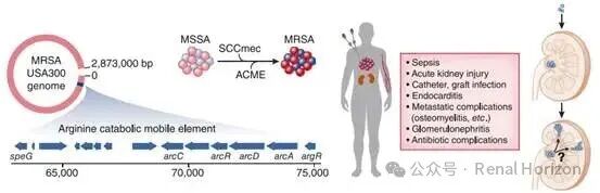
3. The Complexity of Host-Microbe Interactions: A Case Study of Staphylococcus aureus
The article uses methicillin-resistant Staphylococcus aureus infection as an example to illustrate the complex metabolic adaptations and competition between the host and microorganisms.
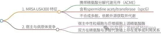
3.1Successful Adaptation of MRSA
MRSA can survive in various host ecological niches through genetic drift, adaptive evolution, and transcriptional reprogramming. One of the key genomic differences between it and methicillin-sensitive Staphylococcus aureus is the presence of arginine catabolism mobile elements.
3.2 ACME Elements and the Polyamine Metabolism Game
ACME Elements contain arginine decarboxylase and spermidine acetyltransferase (speG). Arginine catabolism can provide energy (ATP) and acid resistance for MRSA.
Polyamine Game: Notably, Staphylococcus aureus (including MRSA and MSSA) does not synthesize polyamines and relies entirely on external sources. The speG of MRSA can acetylate and export polyamines (such as spermidine), thereby neutralizing the toxicity of polyamines (Staphylococcus aureus is abnormally sensitive to polyamines).
Host Countermeasures: Host immune cells (neutrophils, macrophages) upregulate arginase and polyamine acetyltransferase (such as SAT1) in response to Staphylococcus aureus infection, competing with MRSA for arginine and polyamine resources. This forms a parallel molecular arms race.
The following figure (Figure 3) illustrates the genomic structure of MRSA USA300 and its situation in kidney infections.
4. Conclusion and Outlook
4.1Core Conclusion: Timing is Everything
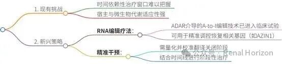
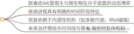
Surviving sepsis requires the kidney to dynamically adapt and evolve during the infection process. The ongoing battle between the host and microorganisms means that any treatment strategy that does not carefully consider the timeline of sepsis may become irrelevant or even harmful.
4.2Therapeutic Insights
Stage-Specific Treatment: Future therapeutic strategies need to be calibrated according to disease stages. For example, after the early inflammatory phase, treatments targeting pro-inflammatory factors may be ineffective; while in the shutdown phase, promoting translation recovery (such as using ISRIB class molecules) may be more beneficial.
Targeting Recovery Mechanisms: Utilizing endogenous recovery pathways, such as ADAR-based RNA editing technologies, may become a new strategy for treating sepsis-associated acute kidney injury at specific time points. This technology has entered clinical trials.
Understanding Host-Microbe Interactions: In-depth studies of the metabolic interactions between the host and microorganisms (such as MRSA) in the unique renal microenvironment will provide key insights for developing new therapies.
4.3Metaphor of the “Red Queen Hypothesis”: The article concludes with a reference to the “Red Queen Hypothesis,” vividly illustrating that to survive and maintain physiological function in sepsis, the kidney must run at full speed like the Red Queen in Alice in Wonderland just to stay in place. To achieve therapeutic breakthroughs, we need to understand the molecular dialogue between the host and pathogens at a faster pace (more deeply and dynamically).
Mind Map
1.Overview

2. Disease Timeline Pathological Stages
3. Host-Microbe Interactions: A Case Study of Staphylococcus aureus
4. Treatment and Outlook
5. Core Conclusions
Note: This article is for academic exchange only and not for any commercial use. Copyright belongs to the original author and the original publication.
Hato T, Dagher PC. Molecular Mechanisms of Sepsis-Associated Acute Kidney Injury. J Am Soc Nephrol. 2025 Nov 1;36(11):2259-2268. doi: 10.1681/ASN.0000000809. Epub 2025 Jul 2. PMID: 40601936; PMCID: PMC12344547.