The exact cause of intrahepatic cholangiocarcinoma (ICC) remains unclear. Well-known risk factors for ICC include congenital choledochal cysts, chronic cholangitis, inflammatory bowel disease, primary sclerosing cholangitis, parasitic infections, chemical carcinogens (such as thorium dioxide and nitrosamines), genetic factors, biliary cirrhosis, cholelithiasis, alcoholic liver disease, and non-specific liver cirrhosis. Recently, studies have found a close association between hepatitis viruses and ICC. It should be noted that some ICC patients do not have a history of exposure to the above factors, thus the pathogenesis of ICC still requires in-depth research.
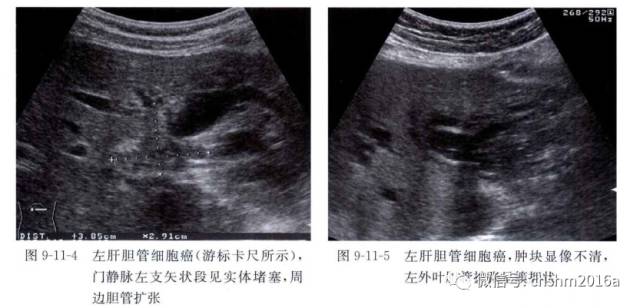
The typical ultrasound imaging of intrahepatic cholangiocarcinoma presents as irregularly shaped, poorly defined low echo heterogeneous masses, with multiple arterial blood flow signals within the lesions.
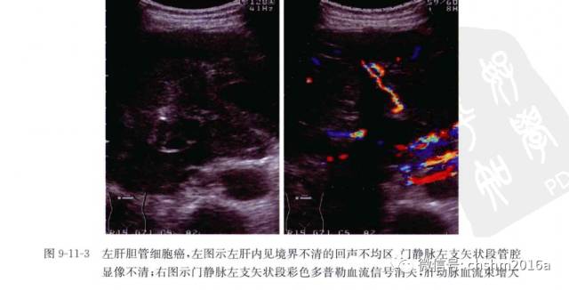
On color Doppler ultrasound, ICC often shows a hypovascular pattern. The ultrasound features of ICC are diverse; although the grayscale ultrasound has certain characteristics, its diagnostic accuracy is relatively low, making preoperative diagnosis still quite challenging. Combining various imaging diagnostic methods aids in the diagnosis of this disease.
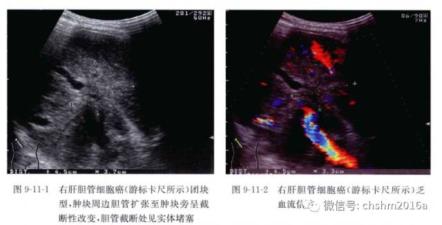
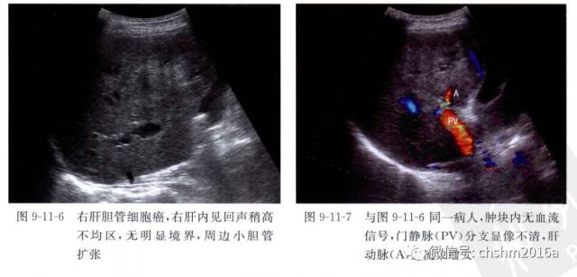
Grossly, ICC can be classified into three types: mass-forming type, periductal infiltrating type, and intraductal growth type.
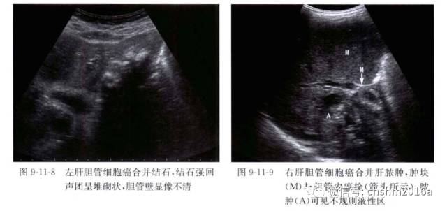
The most common type is the mass-forming type, accounting for 60% to 80% of ICC; the periductal infiltrating type accounts for 15% to 35%, which can infiltrate along the bile duct system and portal vein system, leading to bile duct stenosis and surrounding bile duct dilation; the intraductal growth type accounts for 8% to 29%, often presenting as papillary, polypoid, or granular growths that extend superficially along the bile duct.
Histologically, ICC includes various types such as adenocarcinoma, adenosquamous carcinoma, squamous cell carcinoma, mucinous carcinoma, and signet-ring cell carcinoma. Most ICCs are adenocarcinomas of varying degrees of differentiation, classified as well, moderately, or poorly differentiated. Those occurring in larger bile ducts may form papillary structures. Tumors often exhibit rich stromal responses, and even local calcification may occur. Most tumors show varying amounts of mucus. Cancer cells often invade the portal area, portal area blood vessels, or nerves, potentially leading to intrahepatic metastasis or metastasis to local lymph nodes. Immunohistochemistry for ICC shows positive expression of CK7/CK19, mucin, and partial expression of membrane epithelial antigen, CEA, CK20, whereas CDX-2, TTF-1, ER, PR, BRST-2, PSA, etc. are all negative.
The staging of ICC according to the AJCC Cancer Staging Manual, 7th Edition:
Primary Tumor T:
T1 single tumor without vascular invasion (including major vessels such as the portal vein or hepatic vein or microvessels)
T2a single tumor with vascular invasion T2b multiple tumors with or without vascular invasion
T3 tumor perforating the visceral peritoneum or directly invading extrahepatic tissues
T4 tumor with periductal infiltration
Regional Lymph Nodes N:
N0 no regional lymph node metastasis N1 presence of regional lymph node metastasis
Distance Metastasis M:
M0 no distant metastasis
M1 presence of distant metastasis (presence of lymph node metastasis around the celiac artery, abdominal aorta, and inferior vena cava is considered M1)
TNM Staging:
Stage I: T1N0M0 Stage II: T2N0M0 Stage III: T3N0M0
Stage IVA: T4N0M0 Stage IVB: any TN1M0, any T any NM1
LCSGJ Staging[12]:
Stage I: T1N0M0 Stage II: T2N0M0 Stage III: T3N0M0
Stage IVA: T4N0M0 Stage IVB: any TN1M0 any T any NM1
① tumor diameter ≤2 cm; ② single nodule; ③ no vascular or severe capsule invasion. Tl meets all three criteria; T2 meets two of the three criteria; T3 meets one of the three criteria; T4 meets none. No indicates no lymph node metastasis; N1 indicates lymph node metastasis. M0 indicates no distant metastasis; M indicates distant metastasis.