Technical Terms are often confused with Immunofluorescence, Immunocytochemistry, and Immunohistochemistry. These three terms are often used interchangeably, which can lead to confusion when purchasing antibodies for microscopic experiments.
Immunofluorescence (IF) refers to the detection method used (i.e., using fluorescent dyes to visualize the target of interest), regardless of the type of sample used. IF can be further divided into two categories: direct and indirect methods.
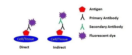
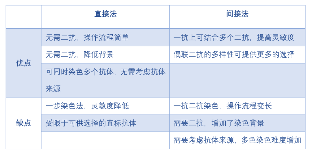
Direct IF uses a single antibody directly conjugated to a fluorescent dye to detect the target of interest. Indirect IF uses two antibodies to detect the target of interest. The primary antibody that binds to the target is unconjugated, and the secondary antibody that reacts with the primary antibody is conjugated to a fluorescent dye.
Immunocytochemistry is most commonly used for cells that are grown in a monolayer in a dish or transferred from suspension to a slide for observation under a microscope. When using adherent cells, the cells are typically grown directly on the coverslip. Depending on the type of cells, they may not adhere to the coverslip on their own, and the coverslip needs to be coated with gelatin or poly-L-lysine or poly-D-lysine to facilitate this process. Suspension cells are typically fixed by directly adding a fixative to the cell culture medium for about 15-20 minutes, washed, resuspended in buffer, and placed directly on gelatin or poly-L/poly-D-lysine coated slides, using the edge of another slide or the side of a pipette tip to spread. Suspension cells can also be fixed onto slides using a centrifugation method called Cytospin™.
Two types of fixatives are commonly used in immunocytochemistry: crosslinking fixatives such as formaldehyde and paraformaldehyde or organic solvents such as methanol or acetone. Crosslinking fixatives are generally preferred because they tend to better preserve cell morphology by crosslinking proteins and forming a cytoskeletal network. However, this protein crosslinking can mask epitopes and reduce the ability of antibodies to bind to their targets, so a permeabilization step is needed to allow antibodies to ultimately approach the target.
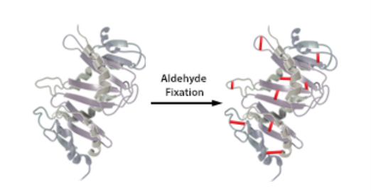
The most common permeabilizing agent used for immunocytochemistry is Triton X-100, at a concentration range of 0.1-0.5% in PBS, but saponin, digitonin, and Tween-20 can also be used. When using organic solvents as fixatives, a permeabilization step is typically not necessary.
HeLa cells were fixed with 1% paraformaldehyde (PFA) for 10 minutes, permeabilized with 0.5% Triton X-100 for 10 minutes, and blocked with 5% FBS for 30 minutes. The cells were then stained intracellularly overnight at 4°C with 2.5μg/ml Ki-67 (clone Ki-67) Alexa Fluor® 594 (red) followed by staining with Alexa Fluor® 488 Phalloidin (green) for 20 minutes. The nuclei were counterstained with DAPI and visualized in blue. Images were taken with a 40x objective.
Organic solvents can preserve cell structure, but they can also dehydrate samples, precipitate proteins, denature proteins you wish to detect, denature GFP and other fluorescent proteins, and remove lipoproteins and other soluble molecules. Organic solvents work well for cytoskeletal marker staining.
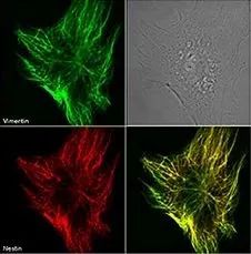
Immunofluorescence of Clone Nestin 20 on methanol-fixed mouse embryonic cells, with Nestin stained red and Vimentin stained green. Photo provided by Dr. R. Goldman of Northwestern University.
Immunohistochemistry (IHC)
Immunohistochemistry is most commonly performed on tissue samples that are either frozen in an optimal cutting temperature (OCT) compound or fixed in neutral formalin and embedded in paraffin. Immunohistochemistry allows you to observe the target of interest within tissue samples while preserving cell structure.
Immunohistochemistry – Frozen Tissue (IHC-F)
Preparing frozen tissue for microscopic experiments typically requires rapid freezing in liquid nitrogen or isopentane. Before slicing the tissue, it is usually placed in a mold and covered with OCT embedding medium. After embedding, you can transfer it to a cryostat and begin slicing. Typically, 5-20μm sections are made and mounted onto gelatin or poly-L/poly-D-lysine coated slides, similar to immunocytochemistry.
You can also place freshly harvested tissue directly into the mold and cover it with OCT, then place the mold containing the embedded tissue directly into liquid nitrogen. Tissue blocks can be stored at -80°C until you are ready to slice. Once ready, the frozen tissue block can be transferred to a cryostat, sliced, and mounted onto coated slides as described above.
Slides can be stored at -80°C for up to a year until staining. When ready for use, the slides are taken out of the freezer and typically fixed with methanol or acetone. Therefore, there is no need for antigen retrieval steps as required for formalin-fixed paraffin-embedded (FFPE) tissues.
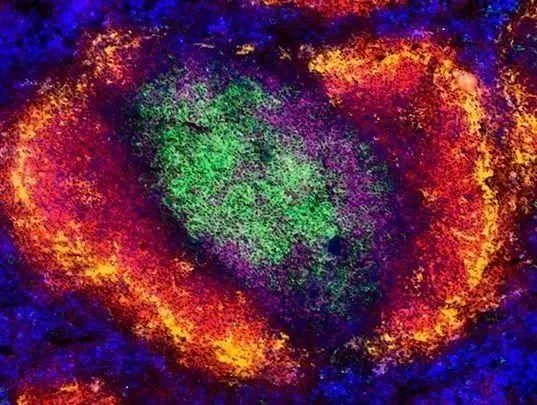
Staining of C57BL/6 mouse spleen tissue for CD4, CD8a, B220, CD169, and F4/80.
Immunohistochemistry – Paraffin Embedded (IHC-P)
Paraffin embedding of tissue is an ideal choice for preserving tissue morphology. If it is critical to maintain tissue morphology for the markers to be stained, it is advisable to perfuse the animal with phosphate-buffered saline, and once most of the blood is expelled, immediately perfuse with 10% formalin or 4% paraformaldehyde. After perfusion, collect the desired tissues and organs, place them in fixative, and store them on ice until ready for embedding.
While perfusion of the animal is recommended due to the high autofluorescence of blood, for standard staining where morphology is less critical, harvested tissues can be placed in 10% formalin or 4% paraformaldehyde solution until ready for embedding.
Before starting the paraffin embedding process, tissues should be placed in fixative for 4-48 hours. However, be careful not to over-fix the tissue, as this can lead to epitope availability (masking) issues and reduce antibody binding.
Embedding tissues in paraffin is a complex process, first requiring dehydration of the tissue with a series of increasing concentrations of alcohol. Paraffin is not soluble in water, which ensures that the water in the tissue is gradually replaced by alcohol. Since paraffin is also not soluble in alcohol, the next step of the process is called clearing, which replaces alcohol with an organic solvent, most commonly xylene, allowing the tissue to accept paraffin. As the name suggests, clearing also makes the tissue clear or transparent. The tissue is then embedded in warm paraffin, which fills the spaces previously occupied by water. Once the paraffin cools, the tissue hardens and is ready to be sliced with a microtome. Sections of about 5-8μm are cut and mounted onto slides. Since most antibody staining solutions are aqueous, you now need to remove the paraffin and replace the water that has just been removed! To do this, you need to reverse the dehydration and clearing steps. The slides are dewaxed with xylene and then the alcohol is reduced.

Antigen Retrieval Methods
Due to the crosslinking fixatives used in paraffin embedding, antibodies may be unable to bind due to epitope masking. To make binding epitopes available or exposed, a process called antigen retrieval is performed. Antigen retrieval reverses the crosslinked proteins, allowing antibodies to approach the epitopes they need to bind to. There are generally two methods of antigen retrieval: heat-induced epitope retrieval (HIER) and proteolytic-induced epitope retrieval (PIER).
Heat-Induced Epitope Retrieval (HIER)
HIER is the most common method of epitope retrieval because it is more convenient than PIER. Several important factors need to be tested and optimized to find the right conditions for the antigen you are using. Incubation time, buffer pH and composition, and temperature are all factors to consider when establishing an experimental protocol. The most commonly used buffers in HIER are low pH (~6.0) citrate or high pH (~9.0) Tris/EDTA, used at ~95-100°C for 10 minutes. Buffer conditions, temperature, and incubation time should be carefully optimized, and may need to be determined for each experiment based on the antigen.
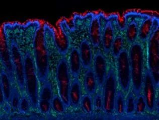
Human paraffin-embedded colon tissue sections were prepared using a standard deparaffinization and rehydration protocol. Antigen retrieval was performed using citrate buffer, pH 6.0 at 95°C for 40 minutes. The tissue was then stained with 10μg/mL Alexa Fluor® 647 anti-Vimentin (clone O91D3) antibody (green) and 10μg/mL Alexa Fluor® 594 anti-CD66d/e (clone 308/3-3) antibody (red) overnight at 4°C. The nuclei were counterstained with DAPI (blue).
Proteolytic-Induced Epitope Retrieval (PIER)
Some antigens may require more stringent methods to expose epitopes and make them available for antibody binding. PIER uses enzymes to digest the protein crosslinks formed during fixation. The most commonly used proteolytic enzymes are trypsin, streptokinase, and proteinase K. As with HIER, conditions need to be tested to find the optimal conditions for antigen retrieval. The incubation time, temperature, and concentration of the proteolytic enzyme used are critical.
Hopefully, after reading this article, you have a better understanding of the differences between IF, ICC, and IHC. In summary:Immunofluorescence is a detection method that is independent of the type of sample you may be using.