Source: YaoDu
Author: Cheng Chuan Yuan Hang Editor: Wan Zi
1
Introduction
Antibody-Drug Conjugates (ADCs) are a novel class of highly effective biopharmaceuticals that link antibodies (Antibody) to biologically active small-molecule cytotoxic payloads (Payload) via linkers (Linker). ADCs aim to enhance the therapeutic index (TI) of chemotherapeutic agents by more selectively delivering cytotoxic drugs to tumor cells while minimizing exposure in normal cells. Despite using a large number of antibodies targeting antigens and/or those expressed only on cancer cells (i.e., target cells), dose-limiting toxicities (DLTs) in normal cells/tissues are frequently reported, even at suboptimal therapeutic doses. The toxicity mechanisms of ADCs in normal cells/tissues remain unclear, but most DLTs are believed to be target-independent.In addition to the instability of ADCs leading to early release of cytotoxic drugs (active payload) in circulation, the uptake/transport of intact ADCs through dependent receptors (FcγRs, FcRn, and C-type lectin receptors) and non-specific endocytosis mechanisms may contribute to off-target toxicity in normal cells.In this article, we summarize the non-targeted uptake of ADCs in normal cells and the potential toxicity mechanisms involved, discussing which components of ADCs influence the occurrence of these mechanisms. This information will help deepen the understanding of the potential mechanisms of off-target toxicity of ADCs and aid in improving the overall therapeutic index (TI) of the next generation of ADCs.
2
Overview
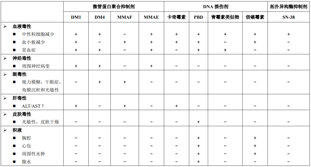
So far, a total of 16 ADC drugs have been approved globally, with one (Trastuzumab duocarmazine) in the application stage for market approval. In recent years, the ADC field continues to expand with a significant increase in IND applications submitted to the FDA. Currently, over 900 different ADCs are in various stages of research and development, primarily targeting hematological malignancies and solid tumors.In the development of ADC drugs, despite using antibodies targeting tumor-specific and/or overexpressed antigens, dose-limiting toxicities (DLTs) at suboptimal therapeutic doses remain a major challenge for the clinical application of ADCs. DLTs lead to relatively narrow therapeutic indices (TIs) and are a primary reason limiting dose escalation of ADCs to achieve maximum efficacy.Table 1 summarizes the preclinical toxicities and clinical DLTs or SAEs of four ADCs approved by the FDA. According to the data described in Table 1 and other published literature, the reported ADC toxicities in normal cells/tissues are primarily driven by the payload. Table 2 summarizes the different types of payloads used in ADCs and the main toxicities reported in clinical studies.Table 1: Summary of Preclinical Toxicities and Clinical DLTs or SAEs of Four FDA-Approved ADCs
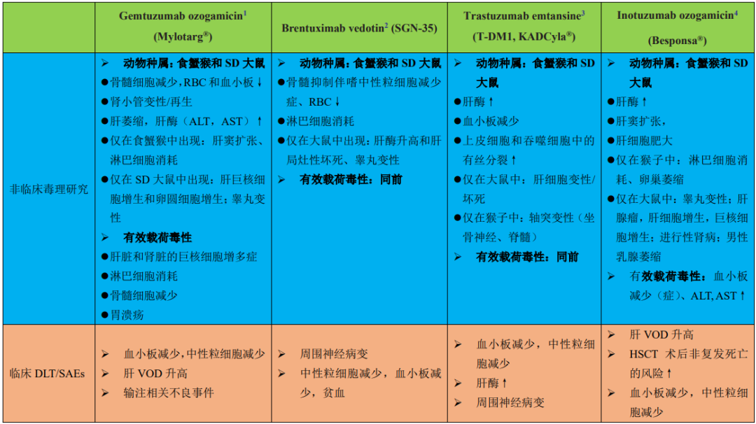
1: Anti-CD33 antibody conjugated via cleavable hydrazone linker (approved in 2000, Pfizer/Wyeth-Ayerst Laboratories)2: Anti-CD30 antibody conjugated to MMAE via a protease-cleavable valine-citrulline linker (approved in 2011, Seattle Genetics)3: Anti-HER2 antibody conjugated to DM1 via an uncleavable SMCC linker (approved in 2013, Genentech)4: Anti-CD22 antibody conjugated to calicheamicin via an acid-labile butyric acid linker (approved in 2017, Pfizer)Table 2: Clinical Toxicities or Adverse Events Associated with ADC Payloads
Although ADC toxicity is primarily believed to stem from the payload, the mechanisms of non-targeted uptake of ADCs in normal cells, delivering cytotoxic payloads, remain unclear. Only a small fraction of ADC accumulation occurs at the human target (tumor) site (~0.1% of the administered dose per gram of tumor). Most ADCs remain in circulation or are distributed in normal tissues, leading to toxicity in normal cells.The expression of ADC-targeted antigens in normal tissues (although at lower levels) may lead to target-dependent uptake of ADCs and subsequent toxicity. For example, the dose-limiting gastrointestinal toxicity (hemorrhagic gastritis) of BMS-182248-01 is associated with the expression of the Lewis-Y target antigen on normal gastric mucosal cells.It is also important to note that the expression of target antigens in normal cells does not typically predict ADC toxicity. For instance, the clinical toxicity of Trastuzumab emtansine (TDM1, KADCyla®), an ADC targeting HER2, is not associated with toxicity in important organs such as the heart and kidneys, despite high HER2 expression levels. Severe thrombocytopenia is a common DLT of T-DM1. Since HER2 is not expressed on platelets or megakaryocytes in circulation, this toxicity is largely considered a target-independent effect.Besides target antigen expression, factors such as the internalization rate of target antigens, circulation/transit kinetics, intrinsic sensitivity to the payload, and the in vivo distribution of ADCs to normal cells/tissues may determine ADC toxicity. Generally, tissues with high perfusion and vascular leakage (incomplete and/or absent basement membranes), such as the liver, bone marrow, and spleen, are expected to have higher IgG/ADC distribution and exposure compared to other normal tissues.Some ADCs share the same payload and linker but target different antigens, exhibiting similar maximum tolerated doses (MTDs) and showing similar toxicity in normal cells/tissues. The most common ADC toxicities are independent of target antigen expression. For example, neutropenia is commonly regarded as a DLT associated with most MMAE-based ADCs (with cleavable linkers). Similarly, ocular (corneal) toxicity is a DLT for multiple ADCs containing DM4.Moreover, ocular toxicity has been observed in multiple MMAF-based ADCs targeting different antigens. This suggests that ADC toxicity is largely an off-target effect, further indicating the potential effects of drug-linker combinations (ADC platforms) on specific off-target toxicity. Therefore, there is ample reason to believe that the three components of ADCs (i.e., monoclonal antibody, payload, and linker) can all contribute to toxicity in normal cells/tissues.The potential mechanisms of ADC or free payload uptake in normal cells are illustrated in Figure 1. Different receptor-dependent and non-receptor-dependent (non-specific endocytosis) mechanisms may contribute to the uptake of intact ADCs and/or the release of free payload in normal cells. Additionally, instability of the linker-payload in circulation leading to premature release of the payload may also result in target-independent toxicity. Understanding the mechanisms of target-independent ADC uptake and toxicity in healthy normal cells is crucial for improving ADC technology. 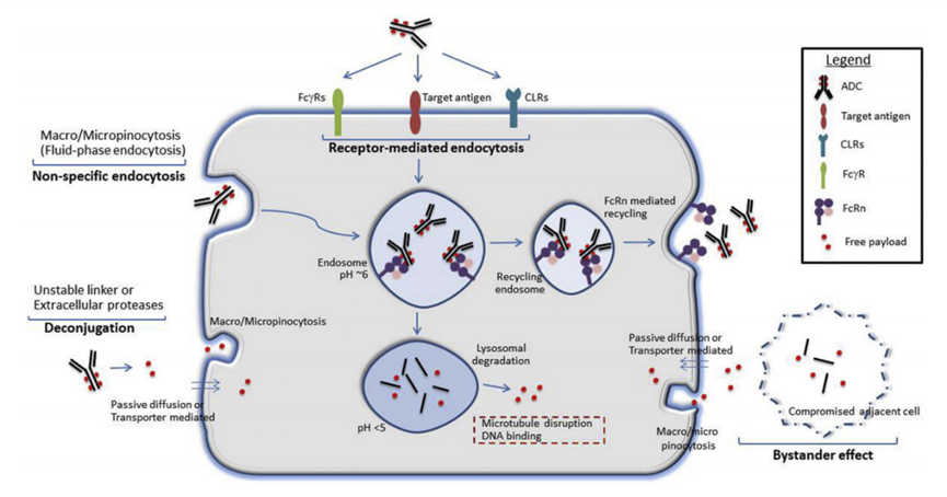 Figure 1: Potential mechanisms of ADC or free payload uptake in normal cells.Target antigens may be expressed on normal cells, facilitating target-dependent uptake of ADCs. Additionally, other receptors that bind to the conserved Fc region of IgG antibodies, such as FcγRs, neonatal Fc receptor (FcRn), and C-type lectin receptors (CLRs), may also contribute to the target internalization of ADCs in normal cells. Non-specific endocytosis mechanisms, such as phagocytosis or pinocytosis, may also facilitate the internalization of intact ADCs or free payloads (released extracellularly due to linker-payload instability or extracellular protease activity).Free payloads may also enter normal cells through other mechanisms, such as passive diffusion (if membrane-permeable), non-specific endocytosis, or uptake mediated by specific transporters (if they are substrates for membrane transporters). Furthermore, antigen-positive target cells may mediate toxicity by releasing free payloads into the local environment, which are subsequently absorbed by antigen-negative normal cells through passive diffusion, paracrine-mediated uptake, or other non-specific endocytosis mechanisms (bystander effect).
3
Non-Targeted Dependent ADC
Uptake and Potential Toxicity Mechanisms
1Linker-Payload Linkage Instability
Linker-payload instability can lead to premature release of the payload into the bloodstream, resulting in off-target toxicity of ADCs.The choice of linker is one of the main drivers behind the stability of ADCs. First-generation ADCs featured acid-labile bonds (e.g., hydrazone) that are stable at neutral pH in plasma but release in lysosomes at lower pH after ADC internalization.These early ADCs were often plagued by poor plasma stability, with free payloads detectable in circulation (Figure 1).The introduction of uncleavable linkers alleviated the cleavage issues in some cases, improving preclinical safety.The reduced toxicity of uncleavable linker types is believed to be due to decreased release of free cytotoxic payloads.However, not all targets are suitable for uncleavable ADCs, as complete antibody metabolism is needed to release the linker-payload.ADCs with cleavable linkers may also enhance efficacy through the bystander effect, making them preferable for low-copy, heterogeneous tumors with low expression or internalization rates of antigens.It is also important to note that, in addition to the cleavability of the linker, the membrane permeability of the released payload may also influence potential off-target cellular toxicity in normal cells, thereby impacting the TI.Conjugation sites may affect the stability and pharmacokinetic characteristics of ADCs. Traditional non-specific conjugation methods use surface-exposed amino acids, such as lysine or cysteine, resulting in highly heterogeneous ADCs (drug-to-antibody ratio [DAR], 0 to 8), increased aggregation, and decreased plasma stability. Thus, non-specifically conjugated ADCs may also contribute to increased target-independent uptake and toxicity in normal cells.Neutropenia is an important target-independent DLT of ADCs, associated with the instability of cleavable linkers in plasma and the systemic release of membrane-permeable free payloads. Neutropenia is a common toxicity associated with many adenylate cyclase-based DLTs (with cleavable valine-citrulline linkers), such as Brentuximab vedotin (ADCetris, Seattle Genetics), ASG-5ME (Agensys), Glembatumumab vedotin (Celldex Therapeutics), Indusatumab vedotin (Millennium Pharmaceuticals), Polatuzumab vedotin (Genentech), and PSMA ADC (Progenics Pharmaceuticals).According to the chemical nature of the linker, valine-citrulline linkers are cleaved by intracellular cysteine proteases in lysosomes. Local serine proteases secreted by neutrophils differentiating in the bone marrow contribute to the cleavage of extracellular VC linkers and the release of membrane-permeable MMAE, leading to cytotoxicity in neutrophil differentiation in the bone marrow.Similarly, peripheral neuropathy (PN) is another important target-independent toxicity associated with microtubule-inhibiting ADCs (regardless of target antigen). PN is believed to be driven by linker-payload instability, associated with premature release of membrane-permeable free payloads (microtubule inhibitors) in circulation. Microtubule inhibitors disrupt interphase microtubule function, which is crucial for the active transport of key essential proteins from neuronal cell bodies to distal synapses, ultimately leading to peripheral neuropathy. PN is a common adverse event for nearly all membrane-permeable ADCs (DM-1 and DM-4) bound to cleavable linkers.It is noteworthy that the PN observed clinically is not always predicted in preclinical animal models. For example, preclinical toxicology studies of VC-MMAE-based ADCs did not monitor PN. In contrast, PN was observed in preclinical species for other non-MMAE ADCs containing microtubule inhibitors, such as DM1 or DM4, demonstrating good clinical predictivity.Bystander Effect:Besides the direct cytotoxicity from ADC uptake by antigen-positive cells, the free payloads of ADCs may also exert cytotoxicity on adjacent antigen-negative cells through a phenomenon known as the bystander effect. In antigen-expressing target cells, the uptake and metabolism of ADC in lysosomes release free payloads into the cytoplasm. The free payloads can then passively enter the extracellular space (membrane-permeable, lipophilic payloads) or be released due to loss of membrane integrity (after target cell death). The released free payloads may enter antigen-negative cells through passive diffusion, transporter-mediated uptake, or other non-specific endocytosis mechanisms, leading to cytotoxicity (Figure 1).The bystander effect of ADCs is often associated with enhanced tumor killing, particularly for tumors with heterogeneous antigen expression. Its impact on the potency and efficacy of ADCs with membrane-permeable payloads has been demonstrated through in vitro colony sphere analyses, co-culture systems, and in vivo xenograft models. However, the increased cellular permeability required to achieve the bystander effect may also lead to off-target toxicity.Compared to uncleavable, less permeable payloads, released payloads can permeate normal tissues and increase toxicity. Recent advancements in ADC technology have enabled cytotoxic payloads to be metabolized into membrane-impermeable metabolites within tumor cells (e.g., Dolaflexin). This approach can control the bystander effect, preserving beneficial chemical properties to kill tumor cells while significantly reducing systemic toxicity to normal cells.
2Non-Specific Endocytosis
Endocytosis is a vital process for cellular uptake of nutrients, regulating transmembrane dynamics, and synaptic vesicle recycling. Endocytosis may also play an important role in the uptake and distribution of large molecules, including IgG/ADCs, in normal cells. Endocytosis can be broadly divided into phagocytosis (particle internalization) and pinocytosis (soluble molecule internalization, also known as liquid-phase endocytosis). Furthermore, depending on the size of the endocytic vesicles formed, endocytosis can be categorized into macro and micro endocytic processes. Table 3 lists the key characteristics of major endocytic mechanisms that may lead to non-specific uptake of IgG/ADCs.Table 3: Key Characteristics of Major Endocytic Mechanisms
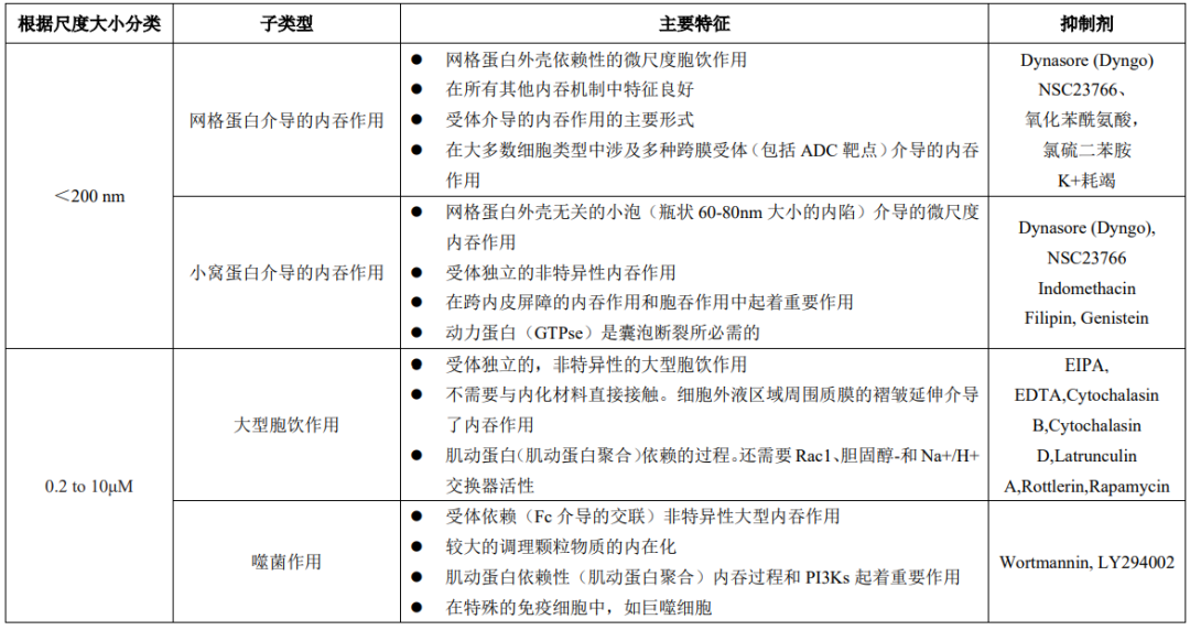
“Macroscopic” endocytosis includes phagocytosis and macropinocytosis, which involve the internalization of large particles and large volumes of fluid, respectively. Phagocytosis involves the uptake of larger particles, causing deformation of the cell membrane (local rearrangement of actin). Immune complexes containing ADCs or ADC aggregates may also be absorbed by this process.Similar to phagocytosis, macropinocytosis is also an actin-dependent process that involves the extension of membrane folds surrounding relatively large extracellular fluid (rather than particles) to mediate endocytosis. Microscopic endocytic processes involve the internalization of small vesicles smaller than 200 nm.These processes typically require specific coat proteins, such as clathrin or caveolin (Figure 2). The binding of ligands to specific membrane receptors initiates a series of signaling events that recruit specific adapter proteins to form clathrin-coated vesicles. These newly formed vesicles are cleaved by dynamin (GTPase) and released for further intracellular transport.Caveolin-mediated endocytosis involves the formation of flask-shaped structures (caveolae) by membrane coat proteins caveolin, which also relies on dynamin to cleave the vesicles. Caveolin-mediated uptake plays a major transport role in many cell types, particularly dominating in endothelial cells. 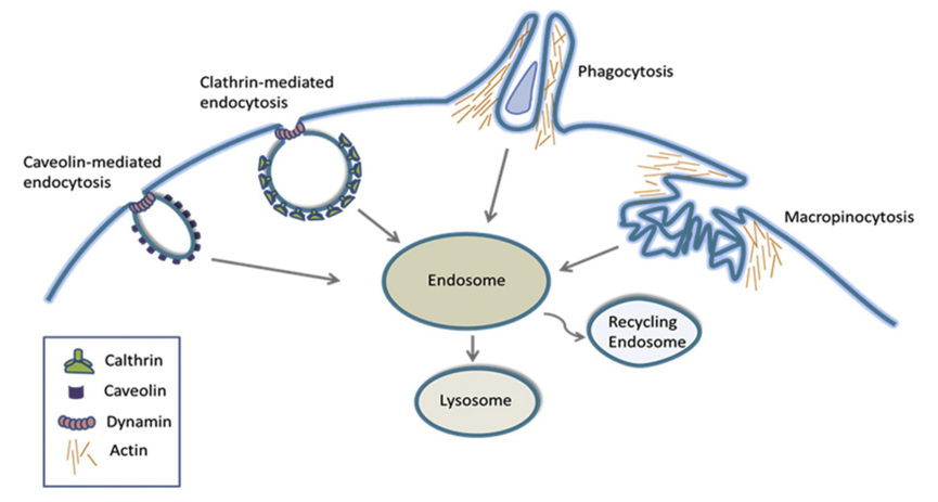 Figure 2: Schematic Diagram of Key Structural Features of Macroscopic and Microscopic Endocytic ProcessesOverall, the aforementioned macroscopic and microscopic endocytic processes may facilitate the entry of ADCs into normal cells. Regarding ADC toxicity in normal cells, non-specific endocytic mechanisms such as caveolin-dependent endocytosis, macropinocytosis, and phagocytosis are potential important mechanisms. It is also evident that endocytic mechanisms and overall endocytic rates differ among normal tissues and cell types.Many specialized immune cells (including macrophages and dendritic cells) exhibit higher endocytic rates. For example, Kupffer cells (resident macrophages in the liver) play a major role in the non-specific uptake and clearance of immune conjugates, including ADCs. Since endothelial cells are located at the interface between blood vessels and interstitial compartments, they also exhibit higher rates of macromolecular endocytosis. Understanding the endocytic rates of different normal cells/tissues is valuable for comprehending the role of non-specific endocytosis as a potential mechanism for ADC uptake and toxicity.Factors Affecting Non-Specific Endocytosis of IgG/ADCs: The physicochemical properties of macromolecules may influence endocytosis in normal cells/tissues. The molecular charge on the surface of IgG/ADCs is an important parameter among many that influence antibody tissue distribution and pharmacokinetics (PK). Positively charged molecules are attracted to negatively charged groups on mammalian cell membranes and extracellular matrices (heparan sulfate proteoglycans). This increases the local concentration of ADCs, leading to more non-specific endocytic uptake in normal tissues/cells.Overall, increasing the net positive charge of IgG antibodies leads to increased tissue distribution and plasma clearance rates, while decreasing net positive charge results in reduced tissue distribution. Importantly, a change in isoelectric point (pI) of at least one or more units is sufficient to produce measurable changes in tissue distribution and PK. These conclusions may also apply to ADCs, supporting the hypothesis that charge may influence non-specific endocytosis of normal cells. Therefore, charge modifications—by reducing positive charges or balancing the overall surface charge distribution—are a method to consider when designing ADCs.However, it is worth noting that, similar to normal tissues, charge modifications may also affect the target antigen-dependent uptake of ADCs required for tumor cell efficacy. Optimizing the surface charge of ADCs to reduce uptake in normal cells while retaining target-mediated uptake in tumor cells may benefit improving the TI.The hydrophobicity of ADCs may also play a role in their non-specific uptake by normal cells. Many drug-linker combinations used for ADCs are hydrophobic, imparting significant hydrophobicity to the antibody, especially for ADCs with high DAR. The increased hydrophobicity of high DAR ADCs can promote aggregation of ADCs and accelerate non-specific clearance. Similar to hepatocytes, high DAR ADCs may be rapidly cleared by other normal cells with high non-specific endocytic capabilities, leading to off-target toxicity.Non-specific endocytosis (particularly macropinocytosis) is considered a pathway for ADC uptake in normal corneal epithelial cells and megakaryocytes, leading to ocular toxicity and thrombocytopenia, respectively. Similarly, macropinocytosis-mediated internalization can mitigate the toxicity of ADC (AGS-16C3F) to megakaryocytes (thrombocytopenia).
3Receptor-Mediated Uptake Mechanisms
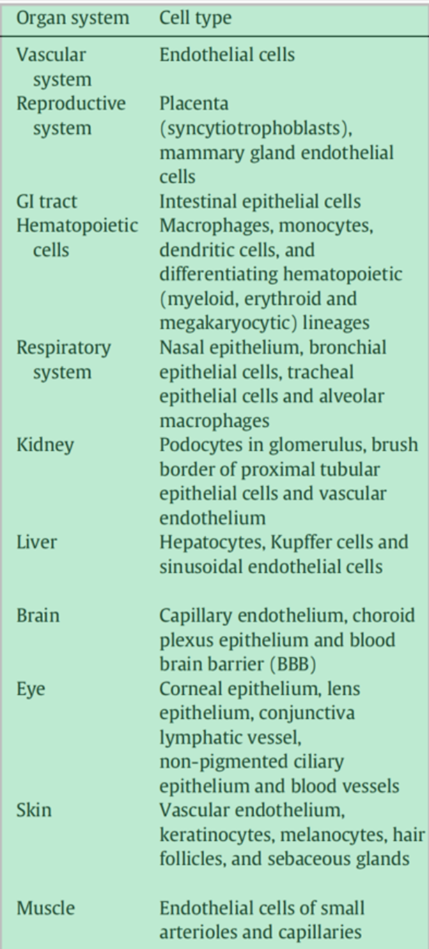
The target-dependent uptake and toxicity of ADCs can also be mediated by different receptors that recognize the Fc (fragment crystallizable) region of IgG in ADCs (Figure 3). The constant domain of IgG is structurally highly conserved, allowing interaction with other components of the immune system through Fc receptors, initiating effector immune functions. Although Fc-mediated effector functions are not typically required to achieve ADC efficacy, the recognition and binding of Fc receptors to the IgG component of ADCs may mediate the non-target internalization in normal cells. 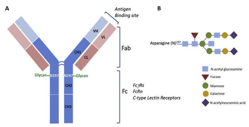 Figure 3: Schematic Diagram of the Structure of the IgG Portion of ADCsFcγ Receptors (FcγRs):FcγRs play a key role in mediating antibody-dependent effector functions (such as antibody-dependent cellular cytotoxicity (ADCC), complement-dependent cytotoxicity (CDC), phagocytosis, cytokines (IFN-γ and TNF-α), and other IgG immune complexes and released cytokines), connecting cellular and humoral immune responses. These effector functions play an important role in regulating the efficacy of several therapeutic IgG antibodies. FcγR-mediated effector functions are critical for target-related efficacy but may contribute to off-target uptake and toxicity in normal cells.Therefore, understanding the biology of FcγRs, their expression patterns in normal cells/tissues, and the physicochemical factors that facilitate FcγR binding to ADCs is essential for understanding potential off-target toxicity mechanisms. FcγRs can be primarily classified into activating (FcγRI, FcγRIIa, FcγRIIc, FcγRIIIa, and FcγIIIb) and inhibitory (FcγRIIb1/b2) receptors based on the type of signaling pathways initiated after receptor cross-linking.Activating FcγRs positively regulate effector functions through interactions with immune receptor tyrosine activation motifs (ITAMs), while inhibitory receptors negatively regulate IgG-mediated effector functions, including endocytosis/phagocytosis, through interactions with immune receptor tyrosine inhibitory motifs (ITIMs). In addition to immune cells, the expression of different FcγRs is also observed in various other normal cell types, including epidermal keratinocytes, sensory neurons, mesangial cells, osteoclasts, endothelial cells, fibroblasts, salivary gland epithelial cells, various cell types in the kidneys and eyes, megakaryocytes, platelets, and immature cells derived from bone marrow, including hematopoietic stem cells/progenitors.FcγR-Mediated IgG/ADC Internalization:FcγRs are not only important molecules mediating IgG antibody effector functions but also one of the most characteristic endocytic surface receptors, playing a role in the internalization/clearance of IgG-opsonized antigens in circulation. The binding of FcγRs to the Fc region induces the aggregation/cross-linking of IgGs on the cell surface and initiates downstream signaling events, leading to the phosphorylation and activation of kinases such as PI3K, p70S6K, and Akt. These are directly involved in the reorganization of the actin cytoskeleton and membrane remodeling for the formation of pseudopodia and phagosomes. Similar mechanisms may also apply to FcγR-mediated ADC internalization, contributing to non-target-dependent toxicity in normal cells.ADC toxicity in normal cells is associated with FcγR-mediated uptake of ADCs. Although the expression patterns of FcγRs in normal healthy cells/tissues correspond to several reported off-target toxicities of ADCs, the role of FcγRs in mediating ADC off-target toxicity is primarily considered a potential mechanism for hematologic toxicity (blood cell toxicity). Hematologic toxicity is the most common off-target toxicity of ADCs containing MAME (MMAF), calicheamicin, and maytansine (DM-1). In clinical studies, T-DM1-induced thrombocytopenia is a DLT.Neonatal Fc Receptor (FcRn):FcRn is a member of the MHC class I glycoprotein family that specifically binds to the Fc domain and plays a key role in extending the half-life (~21 days). Unlike other Fc receptors, FcRn interacts with ligands in a pH-dependent manner, exhibiting high-affinity binding at mildly acidic pH (~6.5). This pH-dependence is believed to be crucial for the mechanism of FcRn extending the half-life of IgG/ADCs. FcRn is widely expressed in many normal adult tissues/cell types (Table 4).In particular, vascular endothelial cells and myeloid-derived hematopoietic cells (antigen-presenting cells) play a dominant role in FcRn-mediated immune responses, which also affect the metabolism and PK of IgG/ADCs. FcRn expressed in polarized epithelial cells (such as intestinal epithelial cells and proximal tubular epithelial cells) also contributes to the bidirectional endocytosis of IgG or immune complexes.The expression patterns of FcRn may vary between different species, leading to differences in IgG binding affinity between humans and other preclinical species. While human FcRn only binds to human IgG, mouse FcRn is highly promiscuous and binds IgG from multiple species (including human) with a binding affinity approximately 10 times higher than that of mouse IgG. The binding affinity of human IgG to crab-eating macaque FcRn is also twice that of human FcRn.Table 4: Expression of FcRn in Different Normal Cells/Tissues
  FcRn Binding and Its Potential Role in ADC Toxicity:ADCs primarily undergo non-specific fluid-phase endocytosis, binding to FcRn in acidic early endosomes, and then, in the neutral pH extracellular space or circulation, the FcRn-ADC complex is transferred away from lysosomal degradation and recycled back to the cell surface to release ADCs. In each endocytic cycle, only unbound ADCs are transported to lysosomes for degradation and release of the cytotoxic payload (Figure 4).From a safety perspective, FcRn binding is also crucial for reducing the accumulation and metabolism of ADCs in normal cells to release cytotoxic payloads. Therefore, modifications that enhance FcRn binding in normal cells with significant FcRn expression may be a useful strategy to overcome adverse toxicity/adverse events.
 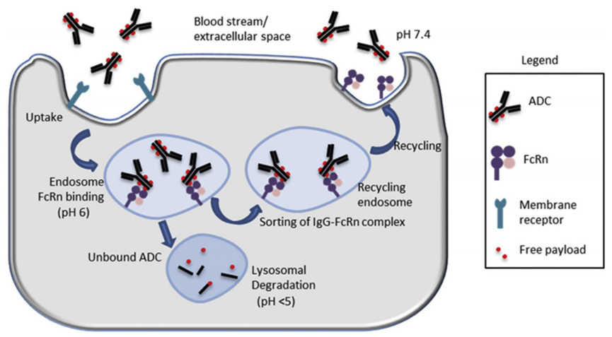 Figure 4: Role of FcRn in ADC CirculationC-Type Lectin Receptors (CLRs):CLRs are a large, highly conserved, and well-characterized family of endocytic receptors. Type I CLRs are calcium-dependent and have multiple (6 to 8) carbohydrate recognition domains (CRDs), as well as cysteine-rich and fibronectin domains. Their members include the macrophage mannose receptor (MR, MRC1, CD206), Endo180 (CD280, MRC2, uPAR-associated protein, uPARAP), DEC-205, PLA2R, and DCL-1. Type II CLRs contain one CRD and can be calcium-dependent (Dectin 2, Mincle, CLECSF8, DCIR, DCAR, BDCA-2, DC-SIGN, MGL) or calcium-independent (Dectin 1, CLEC5A, DNGR-1 (CLEC9A)).CLRs can be membrane-bound (mainly) or soluble/secreted, primarily found on myeloid cells. Increasing evidence also indicates functional expression of CLRs in various normal epithelial and endothelial cells, including dermal microvascular endothelial cells (DMECs), liver sinusoidal endothelial cells (LSECs), perivascular small glial cells, mesangial cells in the kidneys, and corneal epithelial cells.It is noteworthy that, although CLRs are expressed in various normal tissues, certain pathophysiological events, such as inflammation and infection (fungal, microbial), have been shown to significantly regulate the expression of these receptors. The important endocytic functions and structural expression of CLRs in some normal tissues, including common ADC target organs (liver, skin, and cornea), suggest that these receptors may mediate non-target internalization and toxicity of ADCs in these tissues.ADC toxicity is associated with CLR binding. Although there is no direct evidence that CLRs play a direct role in the off-target toxicity of ADCs, the uptake mediated by the mannose receptor (MR) is considered a potential mechanism for ADC hepatotoxicity. LSECs are a highly specialized type of endothelial cells. Compared to other liver cells, LSECs have significantly higher levels of lysosomal enzyme activity, which may further lead to the high degradation of their internalized ligands (including ADCs) and the release of degraded substances/cytotoxic payloads into surrounding compartments.LSECs rely on MR-mediated uptake of lysosomal enzymes (glycoproteins) to maintain their high degradation capacity. After the release of ligands in early endosomes, the rapid internalization of MR-ligand complexes and the rapid recycling of MRs back to the cell surface may further contribute to the high continuous endocytic capability of MR-expressing cells. MR-mediated uptake is also an important mechanism for clearing endogenous and therapeutic glycoproteins and immunoglobulins in LSECs.Additionally, due to the presence of large pores (approximately 50-150 nm) surrounded by microtubules (actin) without membranes and basement membranes, LSECs are the most permeable type of endothelial cells in vivo. Therefore, the diffusion of large molecules (including ADCs) through the clearance receptors of LSECs may also lead to hepatotoxicity.It is noteworthy that Kupffer cells also express MR and play a significant role in the non-specific uptake and processing of ADCs. Therefore, MR-mediated uptake of ADCs in Kupffer cells and the release of cytotoxic payloads into surrounding cells cannot be ruled out, leading to hepatotoxicity.
4
Conclusion
ADC toxicity primarily affects the bone marrow/hematological system, liver, eyes, peripheral nerves, kidneys, and serous effusions (vascular leakage syndrome). Both receptor-dependent and independent mechanisms may help understand the off-target toxicity of ADCs. These mechanisms may differ across different cell/tissue types, depending on the expression and function of key candidate receptors. Common DLTs include thrombocytopenia (FcγRIIa-mediated or macropinocytosis-mediated), ocular toxicity (macropinocytosis-mediated), neutropenia (extracellular protease-mediated), liver damage (mannose receptor-mediated), and peripheral neuropathy (circulation) associated with potential mechanisms of ADC/payload uptake.In summary, selectively targeting tumor cells expressing ADCs is far more complex than initially anticipated. Simply selecting antibody targets that are minimally or not expressed in normal tissues and highly expressed in tumors is insufficient for effectively delivering ADC payloads to tumor cells while minimizing toxicity to normal cells in vivo. The pathways for non-specific ADC entry into normal cells may vary by cell type and depend on the characteristics of the ADC itself. To date, not all parameters influencing non-specific ADC uptake have been identified, and the more we learn, the more the complexity seems to expand.References1Mahalingaiah PK, Ciurlionis R, Durbin KR, Yeager RL, Philip BK, Bawa B, Mantena SR, Enright BP, Liguori MJ, Van Vleet TR. Potential mechanisms of target-independent uptake and toxicity of antibody-drug conjugates. Pharmacol Ther. 2019 Aug;200:110-125.2Pretto F, FitzGerald RE. In vivo safety testing of Antibody Drug Conjugates. Regul Toxicol Pharmacol. 2021 Jun;122:104890. doi: 10.1016/j.yrtph.2021.104890. Epub 2021 Feb 13. PMID: 33587934.3Johns AC, Campbell MT. Toxicities From Antibody-Drug Conjugates. Cancer J. 2022 Nov-Dec 01;28(6):469-478. doi: 10.1097/PPO.0000000000000626. PMID: 36383910.
 Quality by Design (QbD) and Its Intersection with Drug DevelopmentOverview of Macrolide AntibioticsDesign and Development of Covalent Small Molecule TargetsResearch Progress Summary on Tumor and Autoimmunity Target—TNFR2International Rare Disease Day Special | Rare Does Not Equal Alone


Click “Read Original”, Stay Updated with Industry Trends