

1. Research Background
Ribonucleic acid (RNA) plays a complex and central role in cellular regulation, catalysis, and signaling. Visualizing RNA during isolation and analysis, as well as in its natural cellular environment, is an important tool for analyzing its function. The ability to image populations of RNA can monitor cellular transcription, track the mobility and dynamics of different classes of transcripts, and analyze RNA-rich subcellular compartments and biomolecular aggregates, such as nucleoli, P bodies, and stress granules. However, despite decades of effort, imaging of RNA populations in vitro and in cells and tissues remains a fundamental challenge in biomolecular visualization chemistry. There is an urgent need for labeling strategies that enable direct, sequence-independent but highly selective labeling of RNA molecules based on intrinsic chemical features rather than sequence. To this end, small molecule fluorescent dyes that non-covalently bind to RNA are being developed, leveraging their structural tunability (Figure 1a). These cationic push-pull fluorophores non-covalently bind to RNA through electrostatic attraction and intercalation, which increases the off-target background fluorescence from negatively charged and hydrophobic species and environments, leading to poor discrimination between RNA and DNA. Notably, cationic aromatic fluorophores, some of which have structures similar to reported RNA stains, are also widely used for mitochondrial labeling in cells, highlighting the background and specificity issues that may arise from these low-specificity interactions.
Recent studies have elucidated a key chemical distinction of RNA as a biomacromolecule: the presence of a 2′-hydroxyl (2′-OH) in ribose. This functional group, which is absent in DNA, constitutes an inherent chemical handle for selective RNA targeting. The pKa of the 2′-OH group in RNA (approximately 12.5) is lower than that of reference alcohols (ethanol pKa=15.9) and exhibits unusually high reactivity towards electrophiles. Examples of functional electrophiles that react with the 2′-OH with high yields include acyl, sulfonyl, and aryl electrophiles. These reactions can be effectively conducted under mild aqueous conditions, and in some cases, their reactivity can rival that of protein thiol modifications in terms of rate and specificity. Covalent reagents developed for 2′-OH modification have been applied to structural probing (e.g., SHAPE), labeling, RNA protection and delivery, RNA purification, and interaction analysis, while preserving the potential native structure of RNA. Acyl derivatives on RNA can be designed to be reversible through chemical triggers or catalysts, releasing unmodified RNA for further analysis. Reagents targeting the 2′-OH can exhibit tunable reactivity, solubility, and cellular compatibility, enabling emerging applications from transcriptome-wide structural mapping to live-cell RNA imaging and RNA targeting chemical tool development.
2. Results Discussion
To address the limitations of existing general RNA labeling and staining strategies, a molecular approach utilizing this 2′-OH reactivity has been developed (Figure 1b). A class of previously unexplored fluorescent acylation probes that utilize covalent reactions with RNA has been described. These tunable reagents, termed RiboLight (RL) fluorophores, are capable of highly selective sequence-independent labeling of RNA, while enhancing the signal (Figure 1c). Notably, RL fluorophores are not cationic, thus do not rely on non-specific electrostatic attraction for interaction with their targets. Each RL fluorophore is designed to emit in different spectral windows, enabling the possibility of multicolor imaging and ratio analysis. By integrating fluorescence activation with covalent bond formation in a single step, the RL platform overcomes key limitations of non-covalent fluorescent probes for RNA, such as low specificity, transient binding, moderate signal enhancement, and the need for washing steps. These probes are compatible with gel- and cell-based RNA imaging and work efficiently across different RNA sequences and structural contexts. Notably, the absence of the 2′-OH group in DNA ensures minimal cross-reactivity of RL fluorophores, resulting in high levels of RNA:DNA selectivity. Finally, these fluorophores can be removed from their targets under mild conditions, restoring unmodified RNA for further analysis. Collectively, these features establish RL fluorophores as useful chemical tools for RNA imaging and analysis in vitro and in cells.
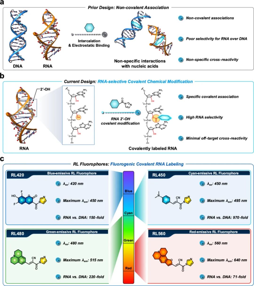
Figure1. Development of RNA-selective fluorescent covalent acylation probes (RiboLight, RL)
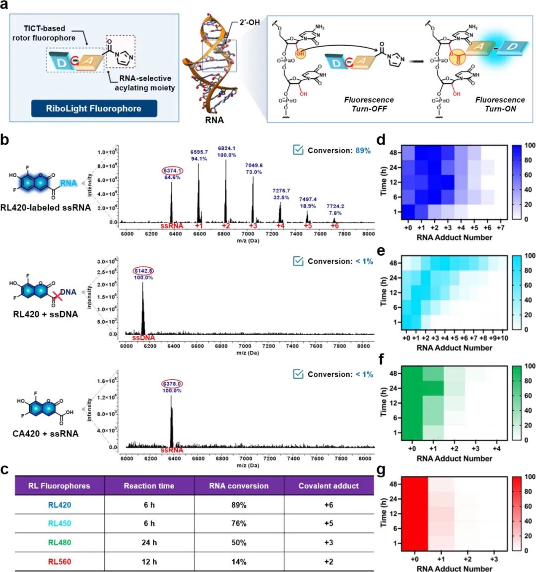
Figure2. Selective acylation of RNA via RL fluorophores’ 2′-OH reactivity for covalent labeling
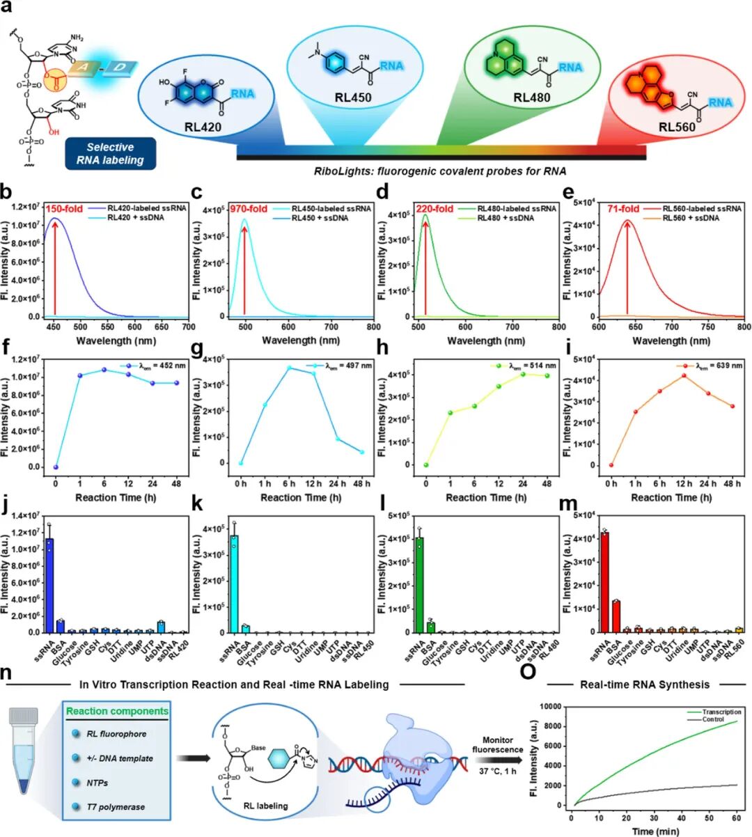
Figure3.. Multicolor fluorescence RNA labeling via selective acylation with RL fluorophores’ 2′-OH reactivity
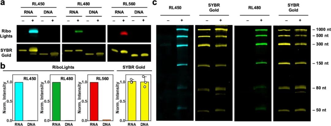
Figure4.. RNA selective gel imaging using RL fluorophores
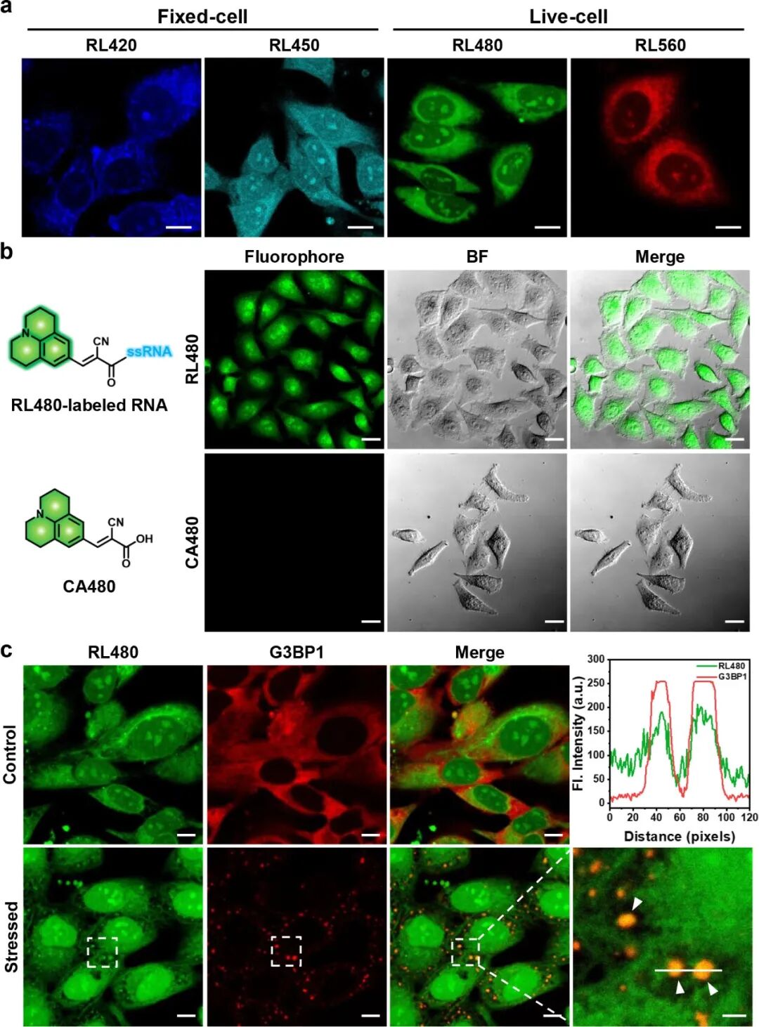
Figure5.. Cell imaging of RNA using RL fluorophores
DOI: 10.1021/jacs.5c14938
Click to read the original article for more details