AI Summary
- This study investigates the reasons for resistance to treatment with 8201 in breast cancer patients, finding that it primarily stems from HER2 gene mutations or loss of HER2 protein.
- In experiments with HER2 mutations (e.g., V597M), normal cancer cells showed a 53.75% decrease in survival after exposure to 8201, while mutated cancer cells showed no significant change in survival, and the efficiency of drug entry into mutated cells was only one-tenth that of normal cells (19-fold increase vs. 200-fold increase).
- A retrospective analysis of 102 breast cancer patients treated with 8201 revealed that 50 patients (49%) experienced a decrease in HER2 expression levels at disease progression.
- Among these 50 patients with decreased HER2 expression, 26 (52%) completely lost HER2 expression (transformed to HER2 0), indicating that half of the patients with decreased expression ultimately lost their treatment target.
- In contrast, among 20 patients with metastatic breast cancer treated with sacituzumab govitecan, only 1 patient (5%) experienced HER2 loss, suggesting that 8201 induces HER2 loss more readily than sacituzumab.
- Cell experiments precisely measured that when HER2 expression dropped from high levels (induction concentration 1 µg/mL) to zero, the amount of 8201 entering cancer cells decreased by 99.43%.
- Loss of HER2 led to a dramatic increase in resistance: when HER2 expression dropped to low levels (simulating HER2 1+), the half-maximal inhibitory concentration (IC50) increased 60-fold; when HER2 was completely lost (HER2 0), IC50 increased 700-fold (i.e., 700 times the drug amount was needed to achieve the same lethal effect).
- Although cancer cells became resistant to 8201 due to HER2 loss, they remained sensitive to the chemotherapy payload carried by the drug (DXd). Therefore, the study proposed a combination strategy with Dato-DXd (targeting TROP2 protein), as TROP2 is widely expressed in breast cancer.
- Cell experiments showed that the combined use of low-dose drugs (1 µg/mL each of 8201 and Dato-DXd) had a killing effect equivalent to the high-dose (10 µg/mL) use of either drug alone.
- In a breast cancer mouse model (ZR-75-1), the use of 3 mg/kg of 8201 or Dato-DXd alone only delayed tumor growth, while the combined use of half-dose drugs (1.5 mg/kg each) successfully reduced tumor volume (tumor regression).
- Drug distribution tests confirmed that the concentration ratio of chemotherapy payload in tumor tissue to plasma concentration in the combination treatment group was significantly higher than that in the 3 mg/kg 8201 monotherapy group (p<0.05), and the weight loss in mice receiving low-dose combination treatment was less than that in the high-dose (10 mg/kg) monotherapy group.
- In summary, for breast cancer patients resistant to 8201 with decreased HER2 expression, the combined use of drugs carrying the same payload but targeting TROP2 is an effective strategy to overcome resistance caused by the loss of a single target.
“8201 Resistance Due to Loss of HER2 Expression and Binding“
8201 has clinical benefits in HER2-positive and low HER2-expressing metastatic breast cancer. However, most patients develop treatment resistance over time, and the mechanisms remain unclear. Molecular characterization analysis of paired patient samples before and after 8201 treatment revealed that 49% of cases exhibited significant decreases in HER2 expression during progression, with 52% completely losing HER2 expression. Using a homologous gene model system, it was confirmed that decreased HER2 expression was associated with reduced internalization of 8201 and significantly increased IC50 for tumor growth inhibition drugs. Further identification and validation of HER2 mutations affecting trastuzumab binding (V597M and P593R) were found to lead to 8201 resistance. To overcome impaired binding and internalization of 8201, a low-dose combination scheme of 8201 with TROP2-targeting ADC was tested, which was found to deliver DXd payload more uniformly, thereby overcoming HER2 loss-mediated resistance.
Significance Statement The mechanisms of clinical resistance to 8201 remain unclear. This article reports two resistance mechanisms due to loss of target binding and proposes a combination strategy using different antibodies but sharing effective payloads of ADCs to overcome resistance through enhanced intratumoral delivery.
Introduction
Antibody-drug conjugates (ADCs) are a class of breakthrough therapeutic agents designed to combine the specificity of antibodies with the potency of chemotherapy drugs. These complex molecules consist of monoclonal antibodies (mAbs) targeting tumor-specific antigens, cytotoxic payloads that kill cancer cells, and linkers connecting the two. Their unique design aims to achieve selective delivery of potent chemotherapy drugs to cancer cells while minimizing off-target effects, thus providing better efficacy and safety than traditional cytotoxic therapies. However, the actual mechanisms of action of ADCs may be more complex than initially envisioned [1,2].
HER2-targeting ADCs are among the most transformative representatives, as this oncogenic protein is overexpressed in 15-20% of breast cancers [3]. Anti-HER2 ADCs (such as trastuzumab emtansine (T-DM1) and 8201) have dramatically changed the treatment landscape for HER2-positive breast cancer, significantly improving clinical outcomes compared to single antibodies or chemotherapy regimens [4-6]. 8201 is composed of the anti-HER2 monoclonal antibody trastuzumab conjugated to a potent topoisomerase I inhibitor payload (DXd) via a cleavable peptide linker at a 1:8 ratio [7,8]. Recently, 8201 has shown significant clinical benefits in breast cancer with HER2 IHC 2+/FISH negative or IHC 1+ status, and responses have even been observed in cases previously classified as HER2 0 [9,10]. This breakthrough has prompted a reevaluation of the classical IHC-based HER2 classification in breast cancer, introducing new categories such as low and ultra-low HER2 expression, and expanding the drug indications beyond traditionally defined HER2-positive breast cancer [11]. These advances raise critical questions about the role of target expression in the primary and acquired resistance to 8201.
Loss or downregulation of target antigens is considered a resistance mechanism for ADCs [10,12,13]. While several studies have shown that ADC target expression correlates with response and/or monoclonal antibody uptake [14-20], systematic studies on the relationship between target binding and ADC resistance are limited. In this study, by assessing HER2 abundance and its binding to 8201, we found: 1) HER2 expression downregulation or loss [21,22]; 2) mutations interfering with 8201 binding sites significantly weakened the anti-cancer efficacy of 8201 by reducing drug internalization. Given that these cells remain sensitive to the naked DXd payload, we confirmed that DXd can be delivered via alternative targets (TROP2) to overcome target-mediated 8201 resistance, and low-dose multi-target ADC combinations are a promising therapeutic strategy.
Results
ERBB2 Extracellular Domain Mutations Can Lead to 8201 Resistance:
Activating somatic mutations in the ERBB2 kinase activity are associated with enhanced internalization of 8201 and response [23,24], but mutations that may hinder 8201 binding have not been studied. We conducted clinical genomic analysis on HER2 low-expressing and HER2-positive MBC patients (n=451) who received 8201 treatment and underwent next-generation sequencing (NGS) between 2018 and 2024 to identify new mutations affecting the trastuzumab binding domain (557-561, 570-575, 593-603) [25]. Three potential relevant and previously unreported ERBB2 mutations were found in the cohort. Two mutations (V597M and p.Q213* (c637C>T)) were found in pre-treatment samples from patients with clinical HER2-positive disease who rapidly progressed after receiving 8201 (defined as progression-free survival (PFS) <3 months), suggesting primary resistance (Figure 1A). The third mutation (P593R) was found in a patient with an unusually prolonged response (approximately 32 months) to 8201, whose post-progression biopsy detected the P593R mutation, indicating acquired resistance.
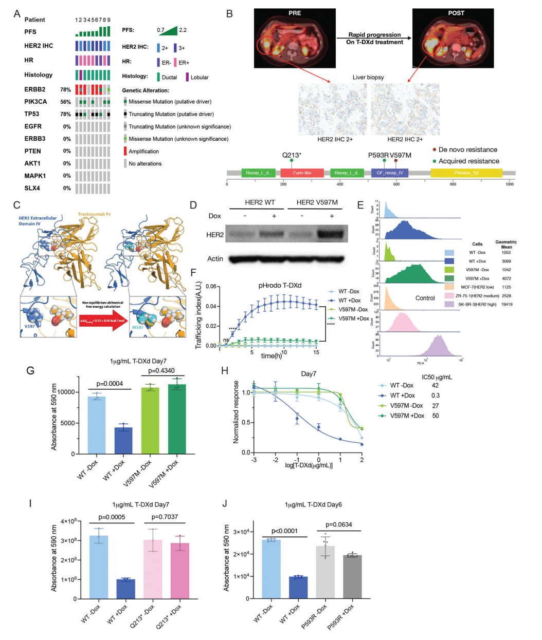
Figure 1: ERBB2 Extracellular Domain Mutations and 8201 Resistance
(A) Clinical genomic data of 9 HER2+ MBC patients who rapidly progressed (PFS<3 months) after 8201 treatment displayed in an oncoprint, with each column representing a different patient. Two patients were found to have target ERBB2 mutations: Patient #2 had a Q213* mutation, and Patient #9 had a V597M variant of uncertain significance (VUS). No mutations in EGFR, ERBB3, PTEN, AKT1, MAPK1, or SLX4 were observed.
(B) (Top) Imaging and pathological data of the ERBB2 V597M mutation patient (rapid progression after 8201 treatment). Interval PET CT showed rapid progression of known liver lesions between the initiation of 8201 treatment and two months into treatment. (Middle) HER2 IHC staining showed no change in HER2 expression. (Bottom) Lollipop plot shows that P593R and V597M mutations are located in one of the trastuzumab binding regions, while the Q213* mutation is located in the furin-like cysteine-rich region.
(C) Free energy calculations predict that the binding affinity of trastuzumab to the HER2 extracellular domain IV with the V597M mutation decreases (red arrow). (Top) Cartoon representation of the trastuzumab variable region (orange) in complex with wild-type V597 (left) and mutant M597 (right) HER2 extracellular domain IV. (Bottom) Magnified view of wild-type V597 and mutant M597 residues, with side chain atoms represented as deep blue and cyan spheres, respectively. The red arrow indicates the predicted relative binding free energy (ΔΔGbinding) of trastuzumab to HER2 after the V597M mutation.
(D) Western blot showing HER2 protein levels in MCF10A cells overexpressing HER2 WT and HER2 V597M after incubation with 0.2 µg/mL tetracycline (abbreviated as Dox) or solvent control for 48 hours. Representative results are shown, independently repeated three times.
(E) Flow cytometry analysis showing surface HER2 levels in MCF10A cells overexpressing HER2 WT and HER2 V597M after incubation with 0.2 µg/mL tetracycline or solvent control for 48 hours. The geometric mean calculated by FCS Express is shown. MCF7 (HER2 low), ZR-75-1 (HER2 medium), and SK-BR-3 (HER2 high) were used as standard controls. Repeated twice.
(F) Internalization of 5 µg/mL pHrodo Red-labeled 8201 in MCF10A cells overexpressing HER2 WT and V597M (incubated with 0.2 µg/mL tetracycline or solvent control). Data represent the mean transport index ± SD of 12 images at each time point. Representative results from three independent experiments are shown. Statistical significance was determined using two-way ANOVA with multiple comparisons. ****, p<0.0001; ns, no significance (p>0.05).
(G) Assessment of cell viability in MCF10A cells overexpressing HER2 WT and V597M after incubation with 0.2 µg/mL tetracycline or solvent control on day 7 (1 µg/mL 8201 treatment). Data are mean ± SD, n=3. Statistical significance was determined using unpaired two-tailed t-test. Repeated twice.
(H) Percentage of live cells in MCF10A HER2 WT and V597M overexpressing cells after treatment with different concentrations of 8201 following incubation with 0.2 µg/mL tetracycline or solvent control. IC50 values were calculated based on cell growth curves using Prism software on day 7. Data are mean ± SD, n=3. Repeated twice.
(I) Assessment of cell viability in MCF10A overexpressing HER2 WT (incubated with 0.2 µg/mL tetracycline or solvent control) and P593R (incubated with 0.01 µg/mL tetracycline or solvent control) on day 6 (1 µg/mL 8201 treatment). Data are mean ± SD, n=6. Statistical significance was determined using unpaired two-tailed t-test. Repeated twice.
ERBB2 V597M
Patient #1 had ER-positive HER2-positive MBC (HER2 2+, FISH positive) (Figure 1B, Supplementary Figure 1A) and experienced rapid progression of liver metastasis after only two cycles of first-line treatment with 8201 for metastatic disease. Hybrid capture panel NGS analysis of the liver metastasis was performed using MSK-IMPACT [24], revealing the presence of the ERBB2 V597M mutation in both pre-treatment and post-8201 treatment samples (this mutation has not been reported in clinical databases such as OncoKB [25]).
Given that this mutation is located in the trastuzumab binding domain, it is hypothesized that it may impair 8201 binding and thus affect drug efficacy. First, molecular dynamics simulations were guided by previously reported co-crystal structures [23]. The V597M mutation simulation showed a ΔΔG change of 0.75 ± 0.10 kcal/mol, suggesting weakened binding of trastuzumab to mutant HER2 (Figure 1C). To experimentally verify the impact of this mutation, MCF10A cell lines overexpressing wild-type (WT) or mutant ERBB2 were constructed under a tetracycline-inducible promoter. MCF10A, as a breast epithelial cell line with negligible endogenous HER2 expression, allows for direct comparison of transgenic effects. A wide characterization of breast cancer cell lines with three different HER2 amplification levels and IHC scores was included as controls: MCF-7 (HER2 low), ZR-75-1 (HER2 medium), SK-BR-3 (HER2 high).
Without tetracycline induction, both MCF10A WT and V597M cells exhibited low baseline HER2 levels (corresponding to IHC HER2 low expression). After tetracycline induction, HER2 protein expression levels increased equivalently in both (Figure 1D). Flow cytometry analysis showed that overexpressed HER2 protein localized to the cell membrane (Figure 1E), with mean fluorescence intensity (MFI) values between ZR-75-1 and SK-BR-3, indicating that HER2 levels were in the medium-high expression range. Droplet digital PCR (ddPCR) further confirmed the presence of the V597M mutation (Supplementary Figure 1B).
To assess whether the mutation affects 8201 internalization, pH-sensitive fluorescent dye (pHrodo) was conjugated to 8201 to monitor 8201-HER2 endocytosis (Supplementary Figure 1C) [22]. Tetracycline-induced HER2 WT increased surface HER2 by 2.9-fold (calculated from FACS histograms) and promoted rapid internalization of 8201 (200-fold increase at 15 hours, Figure 1F). In contrast, surface HER2 V597M increased by 3.9-fold, resulting in only a 19-fold increase in 8201 internalization. To evaluate the impact of internalization changes on tumor growth, cell viability was assessed in both models. Continuous exposure to 1 µg/mL (6.66 nM) 8201 for seven days led to a 53.75% decrease in viability of MCF10A cells overexpressing WT HER2 compared to control (p = 0.0004), with a corresponding decrease in IC50; whereas cells overexpressing HER2 V597M showed no significant change in viability or IC50 compared to control (Figure 1G-H).
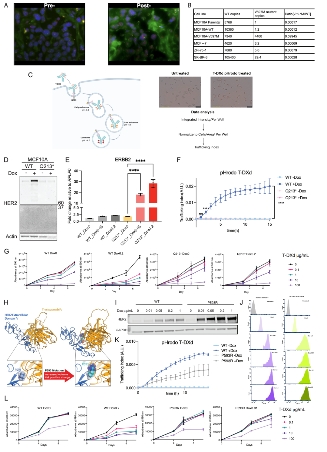
Supplementary Figure 1 (A) FISH images of liver tissue from the V597M mutation patient before (left) and after (right) 8201 treatment, with ratios of 2.4 and 2.18, respectively. (B) ddPCR analysis of parental MCF10A, overexpressing HER2 WT and V597M cells (HER2 WT and V597M copy numbers). MCF7 (HER2 low), ZR-75-1 (HER2 medium), SK-BR-3 (HER2 high) were used as controls. (C) (Left) Schematic of pHrodo labeling (generated by BioRender) and Incucyte image acquisition process: after internalization of the 8201-HER2 complex, pHrodo fluoresces in acidic environments. (Right) Representative images of MCF10A HER2 OE cells treated with 5 µg/mL pHrodo-8201 for 9 hours under 0.2 µg/mL tetracycline induction (vs untreated control). (D) Western blot: HER2 protein levels in MCF10A overexpressing HER2 WT/Q213 after 48 hours of induction with 0.2 µg/mL tetracycline (Q213 not detectable). (E) qPCR: ERBB2 RNA levels in MCF10A HER2 WT/Q213 after 48 hours of treatment with different concentrations of tetracycline (one-way ANOVA: **, p<0.0001). (F) Internalization curves of pHrodo-8201 in MCF10A overexpressing HER2 WT/Q213 cells induced with 0.2 µg/mL tetracycline (two-way ANOVA: ****, p<0.0001; ns, p>0.05). (G) Cell growth curves under different concentrations of 8201 treatment (mean ± SD, n=3). (H) (Top) Cartoon representation of trastuzumab variable region (orange) in complex with wild-type P593 (left) / mutant R593 (right) HER2. (Bottom) Magnified view of P593/R593 residues (side chain atoms blue/cyan spheres), with mutations leading to increased volume and net positive charge. (I) Western blot: total protein levels in MCF10A overexpressing HER2 WT/P593R after 48 hours of induction with different concentrations of tetracycline. (J) Flow cytometry analysis: surface HER2 levels after 48 hours of induction with different concentrations of tetracycline. (K) Internalization curves of pHrodo-8201 in HER2 WT (0.2 µg/mL tetracycline induction) and P593R (0.01 µg/mL tetracycline induction) cells. (L) Cell growth curves under different concentrations of 8201 treatment (mean ± SD, n=6).
ERBB2 Q213* (c637C>T)
Patient #2 had a poor response to 8201 and was found to have the ERBB2 Q213* mutation. This patient had ER+ HER2 0 breast cancer and received standard first-line endocrine therapy combined with CDK4/6 inhibitors until rapid progression of liver metastasis prompted a biopsy showing newly developed HER2 3+ disease, leading to a switch to paclitaxel-carboplatin-trastuzumab-perjeta (TCHP) with sustained complete remission for 17 months. Upon further progression, T-DM1 was switched, achieving an additional 10 months of disease control until progression. At this time, NGS of the biopsy confirmed clonal homology with the initial HER2-positive disease (both samples had the same TP53 and PIK3CA mutations). However, the post-T-DM1 biopsy showed acquired ERBB2 Q213* mutation. Copy number analysis revealed sustained 8-fold local copy number amplification at the ERBB2 locus. The early appearance of a stop codon truncated the protein at the 5′ end of the trastuzumab binding domain, leading to reduced HER2 protein expression (IHC = 2+). Subsequently, the disease rapidly progressed under various subsequent regimens, including capecitabine/trastuzumab/tucatinib (PFS=5 months) and abemaciclib/fulvestrant/trastuzumab (PFS=4 months), with 8201 ultimately leading to disease progression and death in less than 2 months.
Interestingly, mutation feature analysis of the post-T-DM1 sample showed that 81.7% of mutations were associated with APOBEC3 activity, with the Q213* (GTC>TAG) mutation occurring at a classic APOBEC3 background site [26]. This case highlights the potential role of APOBEC3-induced mutations (a common mutational process driving breast cancer evolution [27]) in promoting acquired resistance.
To validate the results, the Q213* mutation was introduced into the aforementioned MCF10A HER2 overexpression (OE) system. However, Western blot did not detect the truncated HER2 protein (Supplementary Figure 1D). qPCR analysis confirmed that HER2 mRNA levels significantly increased under 0.05 and 2 µg/mL tetracycline treatment compared to the no tetracycline control (p<0.0001) (Supplementary Figure 1E). Despite the elevated transcription levels, the truncated HER2 could not internalize 8201 (Supplementary Figure 1F), nor did it enhance the response to 8201 treatment (Figure 1I, Supplementary Figure 1G).
ERBB2 P593R
In contrast to the previous two patients, Patient #3 was found to have a newly developed ERBB2 P593R mutation in a biopsy taken during progression after a long response to 8201. This patient was initially diagnosed with primary metastatic ER+ HER2 0 breast cancer, and the disease rapidly progressed during first-line treatment with fulvestrant + abemaciclib. At this time, NGS of the liver metastasis detected a clear activating mutation in the ERBB2 kinase domain (L755S) and another pathogenic ERBB3 E928G mutation. Given the ERBB2 L755S mutation, the patient subsequently received a clinical trial with fulvestrant, neratinib, and trastuzumab, achieving nearly 2 years of good disease control. The patient ultimately experienced liver progression, at which point the biopsy still showed HER2 0 disease. However, considering the known ERBB2 activating mutation and good response to anti-HER2 treatment, 8201 was switched and achieved approximately 32 months of excellent disease control. Eventually, liver progression was again observed. At this time, the somatic NGS of the liver tumor re-detected the previously known ERBB2 L755S and ERBB3 E928G mutations, along with the newly identified ERBB2 P593R mutation (with a mutation allele frequency comparable to the known ERBB2 L755S) (Figure 1B). This mutation changes a hydrophobic proline to a hydrophilic arginine (located at the start of the AA 593-603 trastuzumab binding domain), leading to increased volume and net positive charge (Supplementary Figure 1H). Data suggest this is an acquired clonal mutation associated with 8201 progression. Overall, although such events are rare, the findings emphasize that mutations affecting trastuzumab binding to HER2 can significantly reduce the efficacy of 8201.
Similarly, a HER2 P593R OE system was constructed, and total HER2 and surface HER2 levels under different tetracycline concentrations were assessed via Western blot (Supplementary Figure 1I) and flow cytometry (Supplementary Figure 1J) compared to the WT model. It was found that the HER2 levels induced by 0.01 µg/mL tetracycline expression of P593R were comparable to those induced by 0.2 µg/mL tetracycline in the WT model. Therefore, subsequent functional studies were conducted under this condition.
Despite higher HER2 expression levels, the P593R mutant still showed reduced internalization of 8201 (Supplementary Figure 1K) and impaired response to 8201 treatment (Figure 1J, Figure 1L).
Loss of HER2 Levels Leads to 8201 Resistance
To explore whether patients receiving 8201 treatment experience a decrease or loss of HER2 expression independent of ERBB2 mutation status during progression, a retrospective analysis was conducted on 102 breast cancer patients treated with standard 8201 therapy, all of whom had paired pre-treatment and post-treatment samples, with the most recent pre-treatment samples showing detectable HER2 expression by IHC [28]. Among these, 50 patients (49%) exhibited a decrease in HER2 levels measured by IHC, with 26 patients (52%) showing loss of HER2 expression down to IHC 0 (Figure 2A). This finding has also been reported in previous small-scale cohorts [10,19]. Given that IHC results may be influenced by pre-analytical and analytical variables such as tissue fixation and processing [29,30], we employed an independent method to further validate the observed decrease in HER2 expression. Immunofluorescence (IF) analysis was performed on 15 pre-treatment and 16 post-treatment patient samples (including 9 paired samples). Representative images are shown in Figure 2B. Quantitative analysis showed a significant reduction in HER2 staining intensity after 8201 treatment (p < 0.01, Figure 2B’), supporting the observed loss of HER2 expression by IHC. As a control, 20 patients with metastatic breast cancer treated with the TROP2-targeting ADC sacituzumab govitecan, who had detectable HER2 expression (1-2+) at baseline and paired post-treatment biopsy samples without 8201 treatment, showed only 1 case (5%) with HER2 loss after sacituzumab treatment (IHC 0). This supports the hypothesis that changes in HER2 expression arise from the pressure of HER2-targeted therapy (rather than TOP1 inhibition or broader/non-specific evolutionary changes).
Given that 8201 still has activity in HER2 low-expressing breast cancer, we sought to determine whether the loss of HER2 expression is sufficient to lead to 8201 resistance. To this end, homologous gene models were established to simulate the observed decrease in HER2 expression levels across cellular environments in patients. In a tetracycline-inducible ERBB2 cDNA transgene model as the sole source of HER2 protein in MCF10A (Figure 2C,D), reducing tetracycline concentration could downregulate HER2 expression to levels corresponding to clinical HER2 IHC scores (e.g., 1+ and 0) (Figure 2E). To promote universality, similar methods were used to construct other models, including CAMA-1 (ER+/PR+/HER2 0 luminal BC cell line), 18cS1 (ER+/PR-/HER2 1+ primary cells derived from patient-derived xenografts (PDX)), T47D (ER+/PR+/HER2 2+ luminal BC cell line), and 293T (ER+/PR+/HER2+ human embryonic kidney cells) (Supplementary Figure 2A-B).
Using pHrodo-labeled 8201, it was found that HER2 expression levels were strongly correlated with 8201 internalization: when tetracycline concentration was reduced from 1 µg/mL to 0.05 µg/mL (p<0.0001) or from 0.05 µg/mL to 0.01 µg/mL (p<0.0001), internalization decreased by nearly 50% (Figure 2F). In the absence of HER2 expression (0 µg/mL tetracycline), 8201 internalization was negligible. In contrast, the pHrodo-labeled human IgG-DXd conjugate showed no internalization even at the highest tetracycline concentration (1 µg/mL). Similar patterns were observed in the other four models (Supplementary Figure 2C), confirming that 8201 internalization is dependent on HER2 expression.
To validate the correlation between 8201 internalization and HER2 expression levels, intracellular and extracellular trastuzumab levels were quantitatively detected by ELISA (Figure 2G, Supplementary Figure 2D,E). It was found that intracellular trastuzumab levels significantly decreased when tetracycline was reduced from 1 µg/mL to 0.2 µg/mL (p=0.0389). The decrease was even more significant between 1 µg/mL and 0.05 µg/mL (p=0.0017), and further decreased when tetracycline was reduced to 0.01 µg/mL or 0 µg/mL (p=0.0002 or p=0.0001). There was no significant difference in extracellular trastuzumab levels between 0.2 µg/mL and 0.05 µg/mL tetracycline (compared to 1 µg/mL). However, significant increases were detected when tetracycline concentrations were reduced to 0.01 µg/mL or 0 µg/mL (p=0.0105 or p=0.0366). These results confirm that lower HER2 expression levels lead to reduced 8201 internalization, and are associated with extracellular trastuzumab levels. Finally, to verify the internalization results using a non-pH-dependent method, 8201 was labeled with Alexa488 and tracked for uptake after exposure in live cells (Figure 2H,H’). When tetracycline was reduced from 1 µg/mL to 0 µg/mL, intracellular 8201 signal decreased by 99.43% (p<0.0001), further supporting the correlation between expression levels and drug internalization.
Subsequently, the impact of different 8201 doses and tetracycline concentrations on cell viability in the models was assessed (Figure 2I and Supplementary Figure 2F). When tetracycline was reduced from 1 µg/mL to 0.05 µg/mL, the number of live cells under 1 µg/mL 8201 treatment significantly increased (p=0.0136), and further increased when reduced to 0 µg/mL (p=0.0005) (Figure 2I). The IC50 value increased 5-fold when tetracycline concentration was reduced from 1 µg/mL to 0.2 µg/mL, increased 60-fold when reduced to 0.05 µg/mL, and increased 700-fold when reduced to no tetracycline conditions (reflecting HER2 0 parental MCF10A cells) (Figure 2J). Similar patterns of viability changes were observed in other tested models (Supplementary Figure 2G-I).
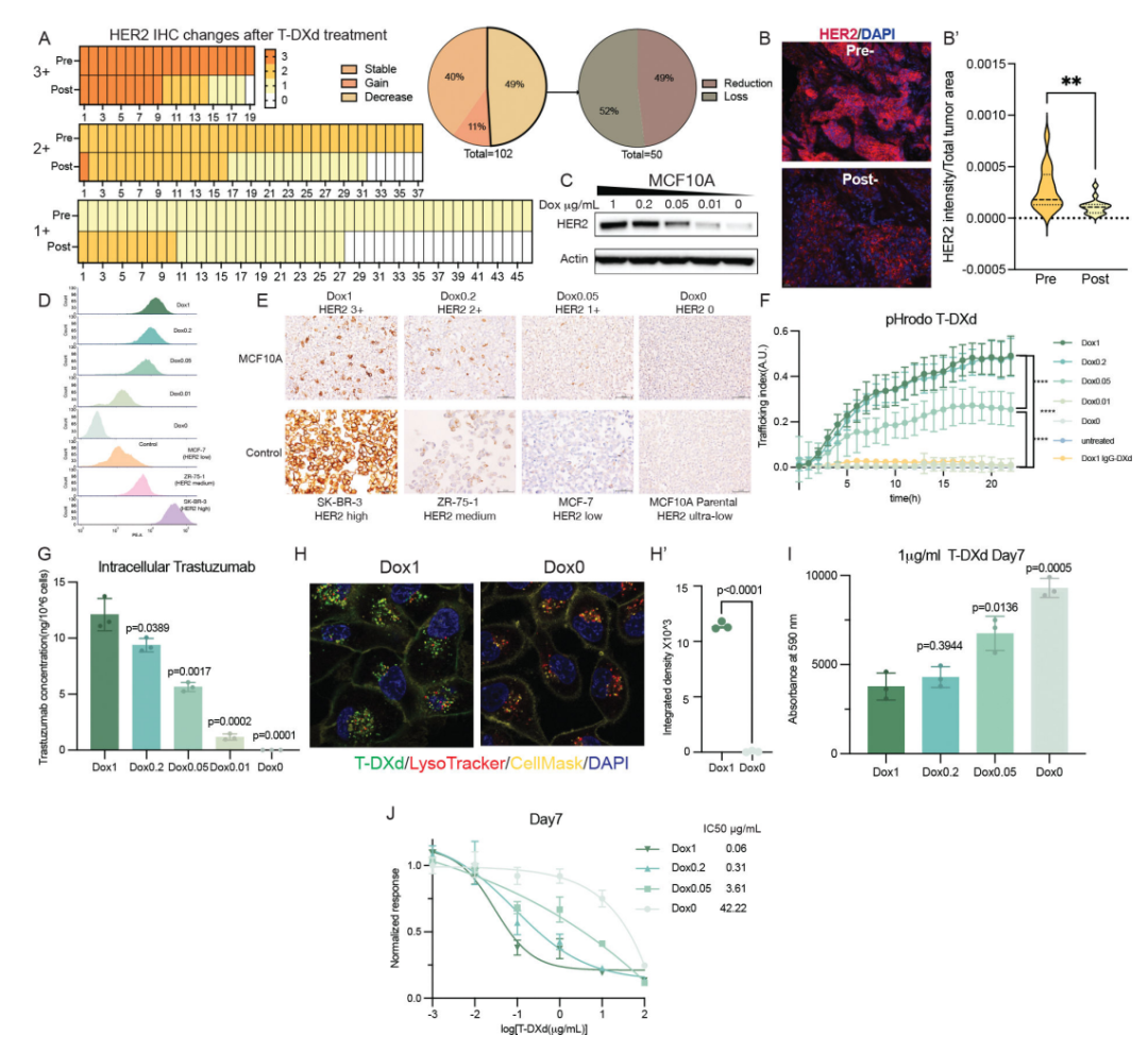
Figure 2: Loss of HER2 Expression and 8201 Resistance
(A) Our team conducted a retrospective study on 102 breast cancer patients receiving standard 8201 treatment with varying baseline HER2 expression levels, most of whom exhibited varying degrees of HER2 expression downregulation when comparing real-world pre-treatment and post-progression biopsies (based on HER2 IHC and next-generation sequencing [24]).(B) (Left) Representative images of HER2 IF in paired pre- and post-treatment patient biopsy samples. (Right) Quantification of HER2 staining time changes measured by IF in 15 pre-treatment and 16 post-treatment samples (including 9 paired samples). p-values were determined using unpaired two-tailed t-tests. **, p<0.01.(C) Western blot analysis of total HER2 expression levels in MCF10A cells after 48 hours of induction with different concentrations of tetracycline. Representative results are shown, independently repeated three times.(D) Flow cytometry analysis showing surface HER2 levels in MCF10A cells after 48 hours of induction with different concentrations of tetracycline. MCF-7 (HER2 low), ZR-75-1 (HER2 medium), SK-BR-3 (HER2 high) were used as internal controls. Representative results are shown, independently repeated three times.(E) IHC showing membrane HER2 levels in MCF10A cells after 48 hours of induction with different concentrations of tetracycline (pathologist classified IHC scores). Parental MCF10A (HER2 ultra-low), MCF-7 (HER2 low), ZR-75-1 (HER2 medium), SK-BR-3 (HER2 high) were used as internal controls.(F) Internalization of 5 µg/mL pHrodo Red-labeled 8201 in MCF10A cells treated with different concentrations of tetracycline. Human IgG-DXd was used as a positive control. Data represent the mean transport index ± SD, n=12. Representative results are shown, independently repeated three times. p-values for the final time point (22h) between Dox1 and Dox0.05, Dox0.05 and Dox0, Dox1 and Dox0 are shown (Dox1 = 1 µg/mL, Dox0.2 = 0.2 µg/mL, Dox0.05 = 0.05 µg/mL, Dox0.01 = 0.01 µg/mL, Dox0 = no Dox). ****, p<0.0001.(G) ELISA quantification of intracellular trastuzumab concentrations in MCF10A cells after treatment with 1 µg/mL 8201 for 24 hours. Data are mean ± SD, n=3. p-values compared to Dox1 conditions were determined using unpaired two-tailed t-tests.(H) Confocal imaging showing intracellular Alexa488-labeled 8201 in MCF10A cells under Dox1 and Dox0 conditions. LysoTracker labeled lysosome localization. Right side shows statistical analysis. The integral density of Alexa488-labeled 8201 was quantified using Fiji, n=3. p-values were determined using unpaired two-tailed t-tests. ****, p<0.0001.(I) Cell viability on day 7 of MCF10A cells under different concentrations of tetracycline (1 µg/mL 8201 treatment). Data are mean ± SD, n=3. p-values compared to Dox1 conditions were determined using unpaired two-tailed t-tests. Representative results are shown, independently repeated three times.(J) IC50 curves and values derived from cell growth curve data on day 7. Data are mean ± SD, n=3. Representative results are shown, independently repeated three times.
In summary, the observed downregulation of HER2 expression in the subgroup of patients with 8201 progression is sufficient to reduce 8201 internalization to clinically relevant levels of treatment resistance.
Combination of HER2- and TROP2-targeting DXd-based ADCs Synergistically Overcomes 8201 Resistance
Given that HER2 loss or mutation leads to reduced 8201 internalization, it is hypothesized that enhanced alternative strategies for DXd internalization may be effective in this context. Cells with low HER2 levels remain sensitive to the naked DXd payload: adding 0.01 µM DXd to a base of 1 µg/mL 8201 significantly improved (p<0.0001) the cell growth inhibition effect compared to high HER2 level cells treated with only 1 µg/mL 8201 (Figure 3A). Based on this preliminary result, it is speculated that increasing the delivery mechanism of DXd independent of HER2 could maximize the delivery of cytotoxic effective payloads and anti-cancer potential.
The TROP2-targeting ADC datopotamab deruxtecan (Dato-DXd) delivers the same DXd payload as 8201 (but with a lower drug-antibody ratio (DAR) of 4), showing monotherapy efficacy in MBC [31-33]. Using an inducible HER2 model library, it was found that TROP2 is highly expressed in breast cancer cell lines and MCF10A cells (Supplementary Figure 3A). Such expression can also be detected by IHC staining (Supplementary Figure 3B). Comparing the efficacy of Dato-DXd in MCF10A cells with high and low HER2 levels, it was found that HER2 expression did not affect (p>0.05) Dato-DXd efficacy (Figure 3B). These data also suggest that when HER2 is lost, alternative DXd delivery mechanisms may still be effective. Given the theoretical possibility that TROP2 may be lost due to Dato-DXd resistance, the combination of 8201 and Dato-DXd at different drug concentrations was tested to prevent target-mediated resistance. Notably, the low-dose combination of 8201 (1 µg/mL) and Dato-DXd (1 µg/mL) was equivalent in efficacy to high-dose monotherapy (10 µg/mL) (Figure 3C). Similar results were observed in multiple models, including MCF-7 HER2 OE cell line models, 18cS1 HER2 OE primary cell line models, and ZR-75-1 (HER2 medium expressing breast cancer cell line) (Supplementary Figure 3C).
After labeling both 8201 and Dato-DXd with pHrodo, the total transport index was quantified as an indirect measure of intracellular DXd levels (Figure 3D). The combination of 1 µg/mL 8201 and 1 µg/mL Dato-DXd resulted in significantly higher intracellular DXd levels than either 10 µg/mL 8201 (p<0.01) or 10 µg/mL Dato-DXd (p<0.0001) monotherapy (Figure 3D’). There were no significant differences in intracellular DXd levels between the 1, 5, or 10 µg/mL 8201 treatment groups (p>0.05), nor between the 1 and 5 µg/mL Dato-DXd treatment groups (p>0.05).
To evaluate the in vivo efficacy of the combination scheme, mice bearing 18cS1 xenografts were randomly grouped to receive either 8201, Dato-DXd monotherapy, or combination treatment (Supplementary Figure 3E). Monotherapy only led to slight tumor growth delays, while combination treatment significantly inhibited tumor growth (despite both ADCs sharing the same DXd payload).
To assess potential toxicity, weight changes were monitored. Mice receiving low-dose 8201 (3 mg/kg) and Dato-DXd (3 mg/kg) combination treatment experienced less weight loss than those receiving high-dose 8201 (10 mg/kg) monotherapy (Supplementary Figure 3E’), suggesting improved tolerability.
Given concerns that the combination of ADCs sharing the same effective payload may exacerbate toxicity and reduce patient tolerability, further testing was conducted to determine whether low-dose combinations could achieve comparable efficacy. In the ZR-75-1 cell line-derived xenograft (CDX) model with HER2 expression, the tumor growth inhibition effect of 1.5 mg/kg 8201 + 1.5 mg/kg Dato-DXd was superior to that of 3 mg/kg 8201 or 3 mg/kg Dato-DXd monotherapy, with only the combination group showing tumor regression (Figure 3E). Additionally, there were no significant differences in body weight between groups (p > 0.05), indicating good tolerability of the combination.
To explore whether the enhanced therapeutic effect of the combination scheme was due to increased delivery of effective payloads to the tumor, LC-MS/MS [10,34-36] was used to measure tumor and plasma DXd concentrations 24 hours post-treatment. The tumor/plasma DXd ratio in the combination group was significantly higher than that in the 3 mg/kg 8201 monotherapy group (p<0.05), comparable to the 3 mg/kg Dato-DXd group (p>0.05) (Figure 3F). These findings support that ADC combinations can enhance intratumoral effective payload delivery and potentially circumvent target loss-mediated resistance.
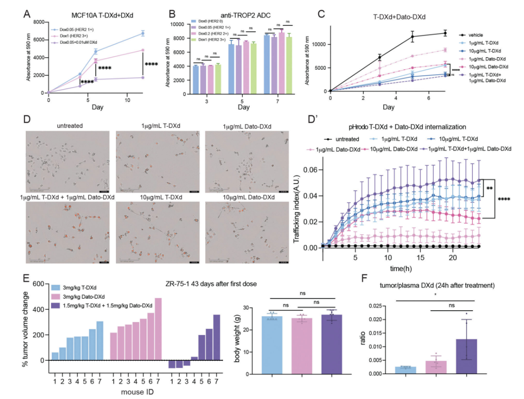
Figure 3: Effects of Combination of 8201 and Dato-DXd
(A) MCF10A cells treated with 1 µg/mL 8201 (0.05 µg/mL Dox induction (Group 1) (representing HER2 1+) or 1 µg/mL Dox induction (Group 2) (representing HER2 3+)), or adding 0.01 µM effective payload DXd under 0.05 µg/mL Dox induction (Group 3). Data are mean ± SD, n=3. p-values between Group 2 and Group 3 were determined using unpaired two-tailed t-tests. ****, p<0.0001.(B) The number of cells treated with Dato-DXd after treatment with different concentrations of tetracycline (representing different HER2 levels). p-values between groups were determined using unpaired two-tailed t-tests: ns, no significance (p>0.05).(C) Cell growth curves of MCF10A HER2 OE cells induced with 0.2 µg/mL tetracycline treated with 8201, Dato-DXd, and their combination. Data are mean ± SD, n=3. p-values on day 7 (final time point) between 1 µg/mL 8201 + 1 µg/mL Dato-DXd combination and 10 µg/mL 8201 or 10 µg/mL Dato-DXd combination are shown in the figure. ****, p<0.0001; ns, no significance (p>0.05).(D) Incucyte images showing internalization of pHrodo-labeled 8201 or Dato-DXd or their combination in MCF10A HER2 OE cells induced with 0.2 µg/mL tetracycline for 24 hours. E’ shows statistical analysis. Data represent the mean transport index ± SD, n=12. Representative results are shown, independently repeated three times. p-values for the final time point (24h) between 1 µg/mL 8201 + 1 µg/mL Dato-DXd combination and 10 µg/mL 8201 or 10 µg/mL Dato-DXd combination are shown in the figure. **, p<0.01; ****, p<0.0001.(E) (Left) Waterfall plot showing the percentage change in tumor volume after 43 days of treatment (3 mg/kg 8201, 3 mg/kg Dato-DXd, or 1.5 mg/kg 8201 + 1.5 mg/kg Dato-DXd combination (IV injection every 3 weeks)). (Right) Body weight of mice in different treatment groups. Data are mean ± SD, n=7. p-values between groups were determined using unpaired two-tailed t-tests: ns, no significance (p>0.05).(F) Tumor/plasma DXd ratios 24 hours post-treatment in ZR-75-1 CDX model (3 mg/kg 8201, 3 mg/kg Dato-DXd, or 1.5 mg/kg 8201 + 1.5 mg/kg Dato-DXd combination). Data are mean ± SD, n=5. p-values between groups were determined using unpaired two-tailed t-tests: ns, no significance (p>0.05); *, p<0.05.
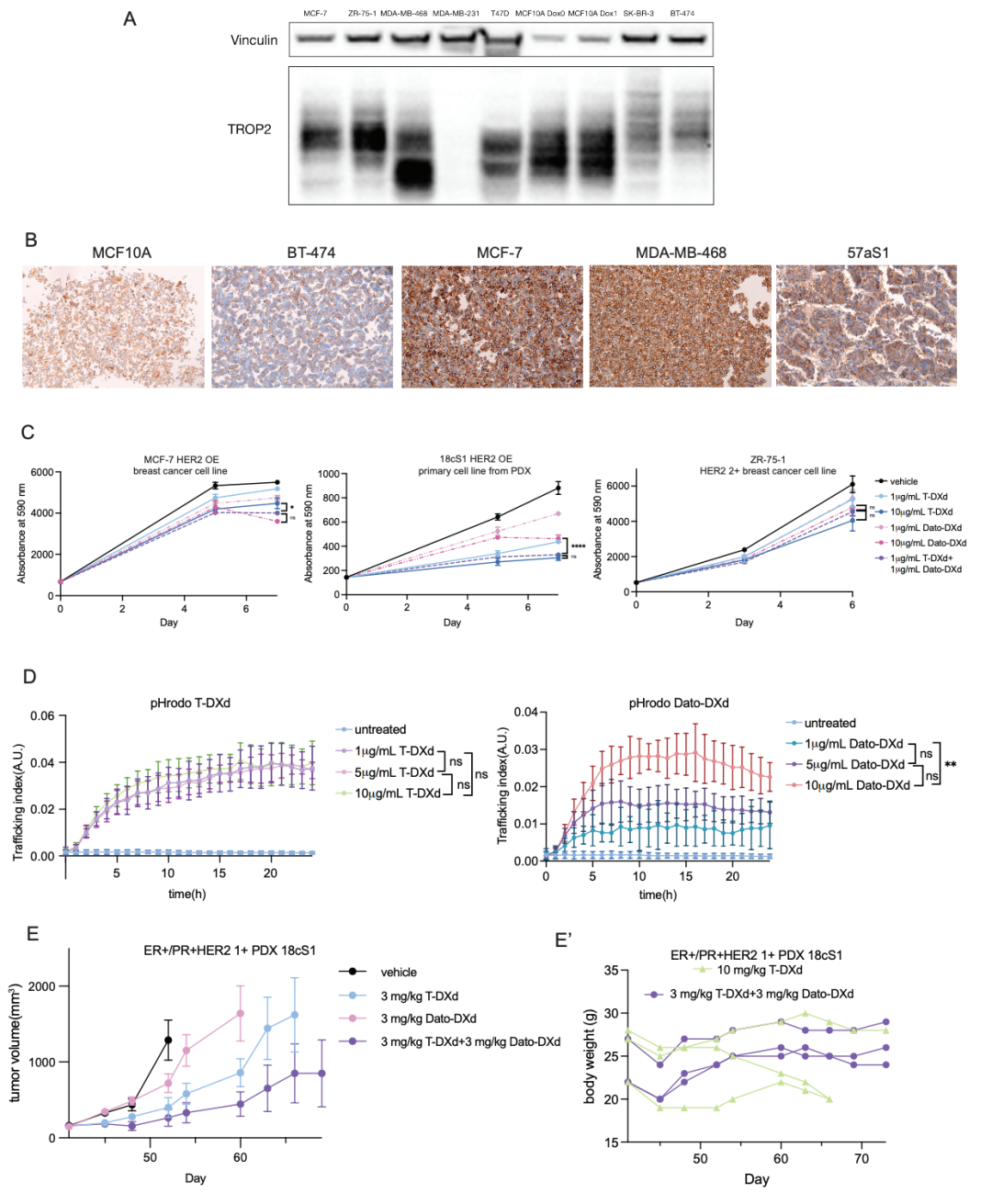
Supplementary Figure 3 (A) Western blot: TROP2 expression levels in different breast cancer/non-cancer cell lines. (B) IHC staining of TROP2 in cell lines and PDX model 57aS1. (C) Cell growth curves of 8201, Dato-DXd, and their combination in different models (MCF-7 HER2 OE, 18cS1 HER2 OE primary cells, ZR-75-1) (two-way ANOVA: *, p<0.05; ****, p<0.0001; ns, p>0.05). (D) Internalization curves of different concentrations of pHrodo-8201 (left)/Dato-DXd (right) in MCF10A HER2 OE cells induced with 0.2 µg/mL tetracycline (two-way ANOVA: **, p<0.01; ns, p>0.05). (E) In vivo efficacy of ER+/PR+/HER2 1+ PDX model 18cS1: solvent, 3 mg/kg 8201, 3 mg/kg Dato-DXd, or combination (IV injection every 3 weeks) (mean ± SEM, n=3).(E’) Body weight of mice receiving 10 mg/kg 8201 monotherapy vs. 3 mg/kg 8201 + 3 mg/kg Dato-DXd combination.
Discussion
8201’s significant activity in HER2-amplified and non-amplified breast cancer reveals the multifaceted anti-tumor potential of ADCs. On one hand, its outstanding activity in HER2-overexpressing cancers validates the concept of utilizing tumor-specific antigens to drive selective uptake of chemotherapy drugs. On the other hand, the relatively limited activity observed in HER2 non-amplified breast cancer reveals the potential for broader uptake, which may underpin the anti-tumor activity of ADCs in the absence of clonal high-level target expression. Although multiple studies emphasize the correlation between target expression and ADC response, the role of target expression and ADC-target binding in treatment resistance has not been fully characterized [10,37-39]. This study reveals various mechanisms of 8201 resistance through preclinical models and clinical cohorts, including intrinsic/acquired HER2 mutations and loss of HER2 expression.
First, the study reveals that some patients develop resistance to 8201 due to mutations in the ERBB2 extracellular domain. Two of these mutations directly disrupt trastuzumab binding, while the third mutation eliminates HER2 expression (impairing drug internalization and efficacy). Although such events are rare, the results elucidate the importance of target binding for ADC efficacy and have implications for sequential therapy and potential cross-resistance of ADCs sharing the same monoclonal antibody backbone [40]. We note that ERBB2 activating mutations (as seen in Patient #3) seem to sensitize tumors to HER2-targeting ADCs (due to increased drug internalization and effective payload delivery), potentially leading to durable responses even in tumors with extremely low HER2 expression [22].
Secondly, we report that loss of HER2 protein expression is a resistance mechanism for 8201. While true HER2 loss is rare in HER2-amplified breast cancer patients (likely because HER2 is a key driver in the development of this subtype of tumor [41,42]), the likelihood of HER2 loss is greater in low HER2-expressing cases (where HER2 is less critical for tumor growth). This is reflected in our cohort: among 19 HER2 3+ patients, only 1 (5.3%) showed HER2 loss, among 37 HER2 2+ patients, 6 (16.2%), and among 46 HER2 1+ patients, 19 (41.3%). The DAISY trial also confirmed this phenomenon: 3 patients dropped from IHC 2+ to 1+, 3 from IHC 2+ to 0, and 4 from IHC 1+ to 0; while only 3 IHC 3+ patients showed decreased HER2 expression after 8201 treatment [10]. Similar reports have been noted after treatment with trastuzumab, T-DM1, and next-generation anti-HER2 ADCs (such as ARX788 and XMT-1522) [16,43-46]. We note that in a parallel cohort of MBC patients receiving sacituzumab treatment, only 1/20 (5%) observed HER2 loss, supporting the hypothesis that changes in HER2 expression arise from the pressure of HER2-targeted therapy (rather than TOP1 inhibition or broader/non-specific evolutionary changes). We acknowledge the inherent limitations of IHC as a measure of HER2 expression (which is influenced by pre-analytical and post-analytical variables—especially at low expression levels and when using formalin-fixed tissues). It remains unclear whether such HER2 expression changes stem from a global downregulation of all cancer cells or selective elimination of HER2-expressing cells [47,48]. Therefore, using IF staining as an independent method, we confirmed that surface HER2 expression decreased after 8201 treatment. However, it should be acknowledged that due to sample accessibility and the technical challenges associated with manual staining and quantification, interpretations are limited.
Through a series of homologous gene cell line models, we demonstrated that such HER2 loss can directly lead to reduced 8201 internalization. The models showed that this internalization reduction was significantly associated with altered drug efficacy. This is consistent with previous studies indicating that enhanced transport of 8201 is associated with improved treatment response [22]. Given the specific changes in drug internalization, it is speculated that these cells ultimately remain sensitive to the effective payload DXd. In fact, adding low levels of DXd in models with reduced HER2 expression proved effective. This prompted exploration of the possibility of using alternative DXd delivery mechanisms to overcome resistance.
In models with low HER2 expression levels, the addition of the TROP2-targeting ADC (Dato-DXd) was highly effective. Surprisingly, the data indicated that even low levels of Dato-DXd combined with 8201 could achieve anti-tumor effects comparable to high-dose monotherapy. This finding suggests that the saturation dose of certain ADCs may not be necessary for optimal tumor uptake. It indicates that clinical combination regimens of such drugs may not require maximum target doses to achieve efficacy, providing a pathway to minimize drug toxicity. Overall, this work suggests that combination regimens of ADCs with different antibodies but similar effective payloads should be validated in clinical trials as a potential strategy to reduce toxicity and enhance efficacy.
This study has several limitations that warrant further exploration. While resistance mechanisms related to antigen targets have been identified, other potential biological processes associated with 8201 resistance remain. The role of the tumor microenvironment, tumor cell plasticity, or DNA damage response/effective payload resistance was not emphasized in this work. Regarding ADC combinations, the study is limited to conceptual progress related to internalization and does not address potential barriers to translating these findings to clinical practice. Further research (especially in dose optimization and administration regimens) is needed to maximize the therapeutic potential of this strategy.
Materials and Methods omitted
References omitted
Source
Chen W, Gupta A, Mai N, Nag S, Lau JS, Singh S, Chodera JD, Liu B, de Stanchina E, Pareja F, Hashmi AA, Lieberman MM, Modi S, Bromberg J, Razavi P, Drago JZ, Chandarlapaty S. Trastuzumab Deruxtecan (T-DXd) Resistance via Loss of HER2 Expression and Binding. Cancer Discov. 2025 Nov 10:10.1158/2159-8290.CD-25-0647. doi: 10.1158/2159-8290.CD-25-0647. Epub ahead of print. PMID: 41212147; PMCID: PMC12631751.