Little Ma’s mother has been quite distressed lately.
She noticed that her originally lively and cheerful son
suddenly became less talkative
and insisted on wearing long sleeves even in hot weather.
This has worried her greatly.
After inquiring, she learned that
Little Ma discovered that his arm
looked different from other children, it had“bent”.
As a result, he was often teased at school,
which led him to become self-conscious and withdrawn.

A year ago,
Little Ma fell and landed on his elbow,
resulting inelbow pain, swelling, and deformity.
His movement was restricted, and he was treated at an external hospital,
where he was diagnosed with a fracture of the left humeral condyle.
He was put in a cast for conservative treatment.
After the cast was removed,
over time,
Little Ma’s left arm gradually bent.
Realizing that his arm looked different from other children,
he became increasingly self-conscious and withdrawn.
Worried, his mother
took Little Ma to our hospital’s orthopedic department.
After a detailed explanation from our experts,
they finally understood that
Little Ma’s bent arm is called cubitus varus deformity,
which is generally a complication of improper treatment of pediatric humeral condyle fractures.
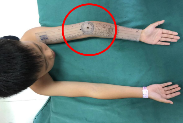
It manifests as inward deviation of the forearm.
Further examination revealed
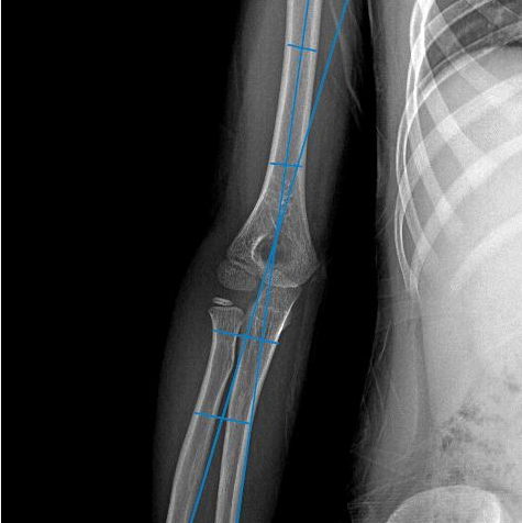
significant cubitus varus at Little Ma’s left elbow,
with hyperextension and rotational deformities, and limited flexion and extension.
If not treated promptly,
the varus deformity will worsen,
affecting elbow function
and increasing the difficulty of future treatment.
 X-ray Results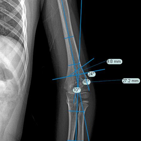

Considering that Little Ma is already 8 years old this year,
which is a critical period for physical and mental development,
to help him quickly restore his appearance and regain confidence,
after admission, under the guidance of department head Tu Yumin,
Deputy Director Wang Xiaoping and Dr. Wei Zhantu
agreed that Little Ma should undergo corrective surgery as soon as possible,
and that the results would be more satisfactory with the assistance of 3D printing technology,
which is more conducive to restoring appearance and function.
Through detailed preoperative planning, assessment, and computer-aided design,
the doctors used 3D printing technology to develop
a precise individualized surgical plan for Little Ma.
 Examination Results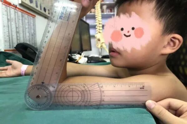

First,
preoperative measurements and osteotomy design were performed.
Based on Little Ma’s preoperative imaging data,
the modeling and design of the osteotomy correction guide were done using specialized software on the computer.
A 1:1 scale 3D model was printed,
allowing the doctors to rehearse the osteotomy procedure on the model.
This way, they could perform the operation more accurately during surgery.
 Digital Precision Design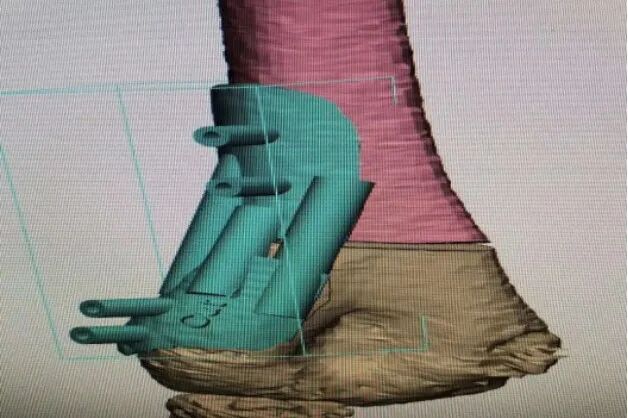
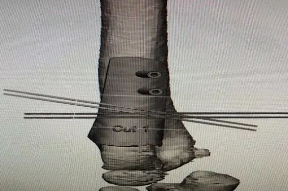

During the surgery,
the doctor exposed the osteotomy site and protected important blood vessels and nerve tissues.
Using an advanced ultrasonic bone scalpel under the guidance of the osteotomy correction guide,
the three-dimensional osteotomy correction for cubitus varus was successfully completed.
Good correction results were achieved during the surgery.
Compared to previous osteotomy surgeries,
the osteotomy was not only precise, but the surgery time was significantly reduced,
and the amount of bleeding was also noticeably less.
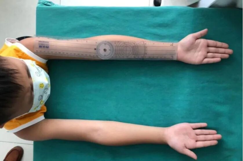
Three weeks post-surgery, it was found that
Little Ma’s elbow appearance and function had basically recovered,
achieving correction of varus, hyperextension, and rotational deformities in one step,
maximizing the recovery of elbow function
and preventing joint contracture and stiffness,
resulting in very good outcomes.
 Postoperative Situation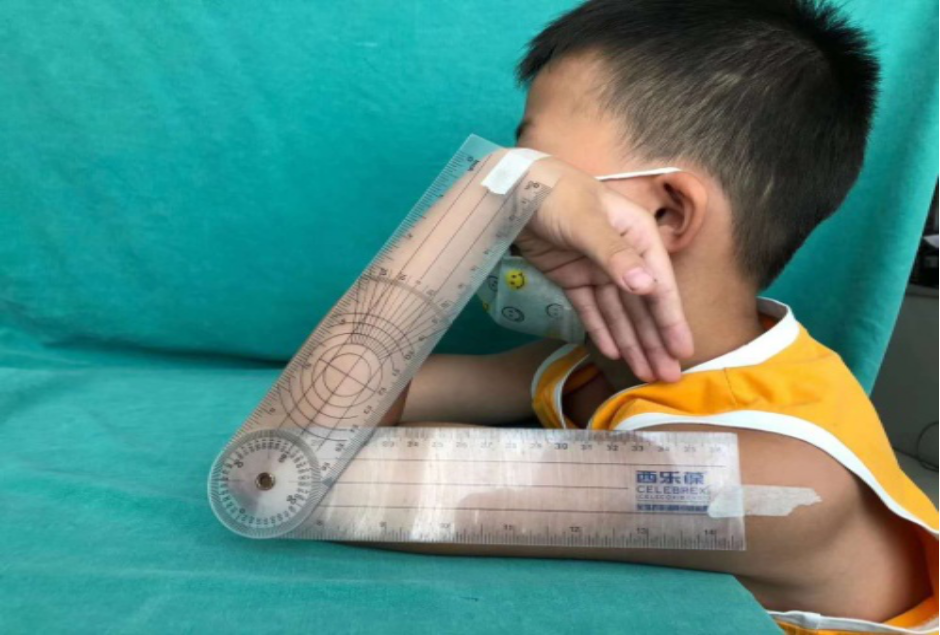

According to Deputy Director Wang Xiaoping,
the application of 3D printing technology in clinical medicine
mainly reflects in personalized treatment.
3D printing technology uses precise three-dimensional physical models
to visually present complex fractures, deformities, tumors, and other pathological conditions
to clinical doctors,
allowing for better assessment of bony lesions,
more accurate preoperative diagnosis,
and deeper understanding of the pathological site by clinical physicians.
Science Popularization Time
3D Printing
3D printing, also known as additive manufacturing technology, is a type of rapid prototyping technology that constructs objects based on digital model files, using powdery metals or plastics and other adhesive materials, through a layer-by-layer printing method.
Application of 3D Printing in Orthopedics
1. Preoperative Planning
For fracture patients, especially those with complex fractures, having a solid anatomical model for analysis or simulating surgical operations is more instructive than relying solely on two-dimensional MRI or CT scans.
2. Creation of Surgical Navigation Templates
These help achieve precise reduction of fractures, assist screws and other implants or instruments to reach the desired position, improving the convenience of surgical operations while reducing the rate of screw perforation and directional errors, significantly enhancing the accuracy of surgical procedures.
3. Customization of Personalized Prosthetics and Implants
In the repair of bone and joint injuries, prosthetics and implants are often required. Traditional prosthetics and implants have fixed specifications, requiring adjustments during surgery with knives and drills to achieve the desired shape and size, while 3D printing can provide “tailor-made” personalized materials for patients.
4.Custom External Fixation Rehabilitation Braces
3D printing technology can fully simulate the characteristics of the patient’s rehabilitation site, and the individualized rehabilitation braces made through 3D printing increase the precision of the braces, providing patients with a “customized” rehabilitation experience.
END
Department Introduction
Orthopedic Department One
Established in 1999, the Orthopedic Department One was approved in October 2010 as the clinical research base for trauma treatment in Guangdong Province. It integrates medical care, teaching, and research.
The department has 45 beds, 8 doctors, and 16 nurses, including 5 senior titles, 3 intermediate titles, and 3 graduate students.
Orthopedic Department One specializes in spinal surgery, with features in digital orthopedics, minimally invasive spinal surgery, and microsurgery, while also focusing on trauma orthopedics. It was the first in the Zhongshan area to carry out related microsurgery procedures. After years of effort, the department’s business scope covers limb trauma, spinal trauma, degenerative spinal diseases, spinal deformities, tuberculosis, tumors, foot and ankle surgery, pediatric orthopedics, and sports medicine, and has conducted almost all spinal surgical techniques.
Contact Number: 88662120 ext. 6321 Editor:Liang JiahaoFirst Review: Liu Benjian, Ren WeiSecond Review: Xu YuanwenThird Review: Office