Tissue engineering and 3D printing of biological inks provide new ideas for tissue regeneration. However, current biological inks face issues such as limited functionality and insufficient compatibility, making it difficult to meet the challenges of defect repair in pathological microenvironments. Developing drug delivery biological inks may allow for targeted treatment in different pathological microenvironments, but physical blending of drugs with delivery materials can lead to drug burst release and cellular stimulation, while chemical grafting may damage the functional groups of the drugs, reducing their pharmacological activity; self-assembled nanoparticles and microspheres often face the risk of being difficult to degrade in vivo.
To address this issue, Professor Liu Hairong and his team from Hunan University developed a cellular capsule delivery strategy, using joint cartilage injury as an experimental model and oxidative stress environment as a pathological model, to develop a punicalagin-loaded cellular capsule multifunctional biological ink. This biological ink has functions such as accelerating cartilage regeneration, antioxidant properties, and antibacterial effects, and has great application potential in the fields of tissue engineering and clinical applications. This research was published in the international journal Advanced Functional Materials under the title “Cellular Capsule Delivery Bioink with Punicalagin-Loaded Chondrocyte Membrane Vesicles for Tissue Engineering Therapy,” with master’s student Yu Mengyi as the first author. Repairing damaged cartilage in joints is a major challenge in clinical treatment, as pathological conditions hinder cartilage regeneration caused by inflammatory factors and reactive oxygen species (ROS). To solve this problem, the authors developed a cellular capsule delivery bioink. By modifying chondrocyte membrane vesicles (CMVs) with acrylate-poly(ethylene glycol)-succinimidyl (AC-PEG-NHS) and loading multiple reactive hydroxyl groups of punicalagin inside, cellular capsules were prepared. These capsules were combined with methacrylated silk fibroin (SerMA) to form a photocurable bioink precursor, which not only exhibited excellent antioxidant properties but also demonstrated significant antibacterial activity and strong cartilage protective effects. All structures printed using the precision micro-projection micro-stereolithography (PμSL) technology maintained high shape fidelity and preserved good cell viability and activity.
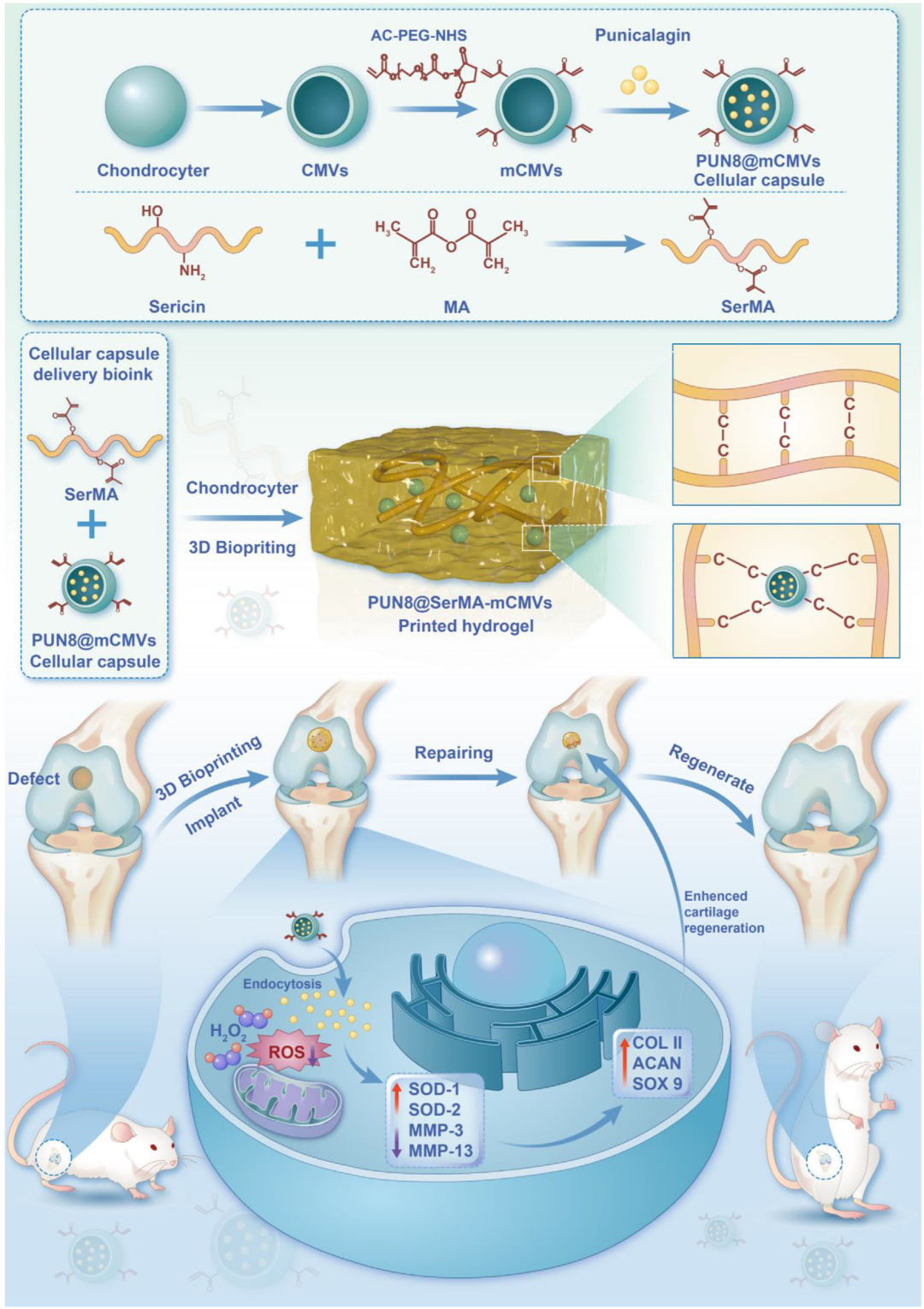 Figure 1. Schematic diagram of the preparation and application of cellular capsule delivery bioink. Negative staining TEM images show that the vesicles exhibit characteristic bilayer cup-shaped structures, with a size of approximately 100 nm. The characteristic peaks in the nuclear magnetic resonance hydrogen spectrum indicate the successful preparation of AC─PEG─NHS modified mCMVs. By loading the natural antioxidant punicalagin into the mCMVs, PUN@mCMVs nanoparticles were prepared. TEM images show that the cup-shaped structure of the mCMVs is sealed, and the drug is successfully loaded into the mCMVs. Immunofluorescence staining observed specific proteins related to cell integration on the surface of chondrocytes, including CD44, integrin α1, integrin β1, and E-cadherin. Furthermore, cells can uptake PUN@mCMVs, indicating successful preparation of the cellular capsules.
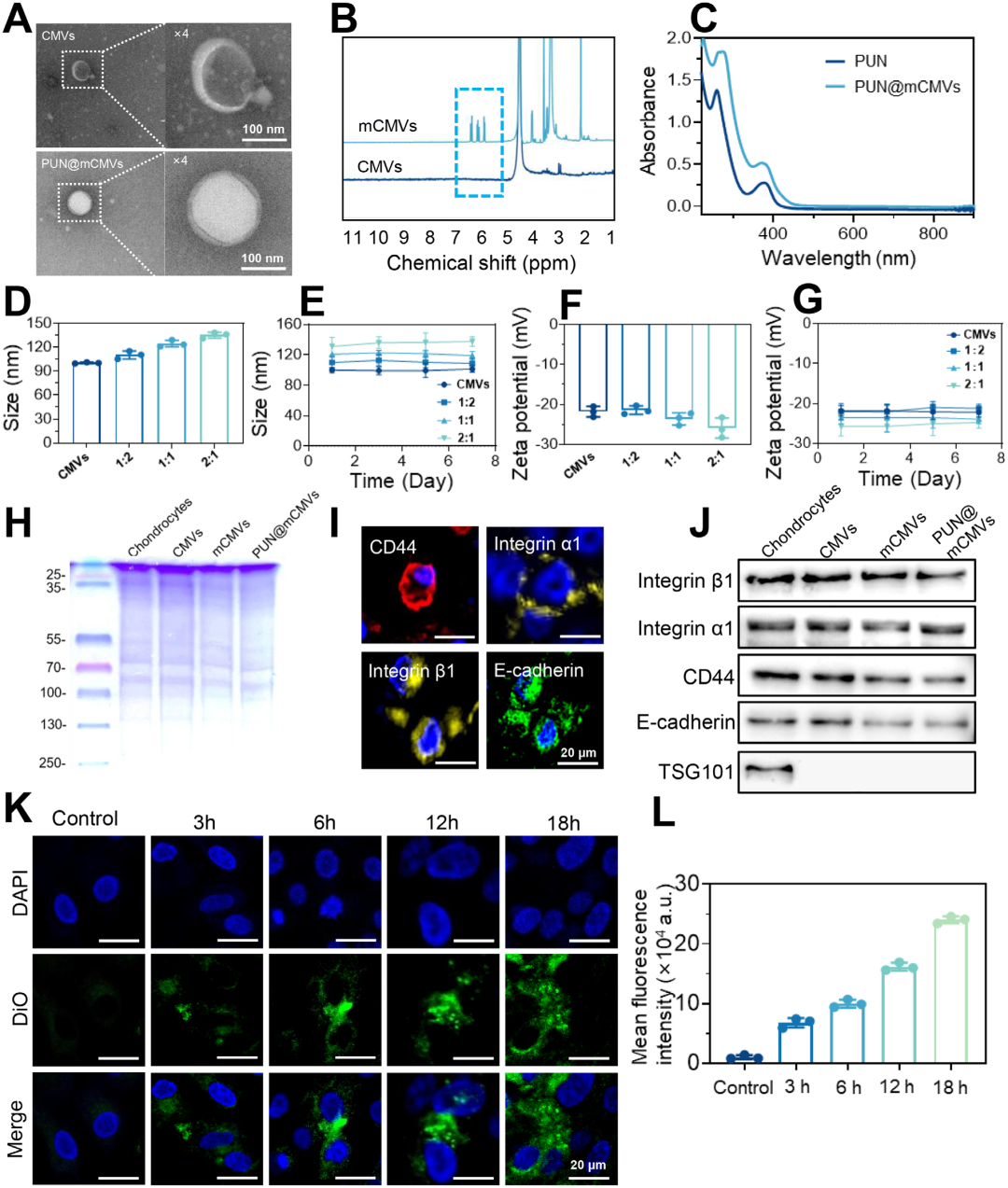 Figure 2. Preparation and characterization of PUN@mCMVs cellular capsules. Among them, the PUN8@SerMA-mCMVs hydrogel exhibited the highest cell proliferation-promoting effect. Additionally, the PUN8@SerMA-mCMV group displayed more typical cartilage lacuna structures and more significant extracellular matrix secretion, with enhanced proteoglycan deposition and increased secretion of COL II. The excessive accumulation of ROS at the site of cartilage injury often leads to oxidative stress, inhibiting cell proliferation, degrading the extracellular matrix, and disrupting the endogenous tissue repair process. The 17 active phenolic hydroxyl groups in punicalagin act as hydrogen atom donors, neutralizing free radicals and forming stable non-radical substances. This process terminates the free radical chain reaction, thereby slowing down or preventing oxidative damage to cells and tissues. Therefore, the antioxidant activity of PUN8@SerMA-mCMVs may support cell proliferation and cartilage regeneration. Experimental results indicate that the cellular capsule hydrogel exhibits significant free radical scavenging activity, effectively removing excess reactive oxygen species from cells, significantly upregulating the expression of antioxidant metal enzymes SOD1 and SOD2 and downregulating the expression of matrix metalloproteinases MMP-3 and MMP-13, thereby protecting chondrocytes from oxidative stress induced by cellular inflammation.PUN8@SerMA-mCMV cellular capsule hydrogel exhibited antibacterial activity, significantly inhibiting the activity of Escherichia coli and Staphylococcus aureus, which may benefit the entire process of 3D bioprinting and tissue engineering.Using the precision nanoArch®S140 (precision: 10 μm) 3D printer, the printing capability of the cellular capsule delivery bioink was tested, and chondrocytes were loaded into the PUN8@SerMA-mCMV bioink. Experimental results showed that the hydrogel products printed from the bioink exhibited excellent printability and shape fidelity. After 3D bioprinting, the chondrocytes encapsulated in the printed hydrogel exhibited high cell viability. These results indicate that the cellular capsule delivery bioink PUN8@SerMA-mCMVs is suitable for 3D bioprinting and tissue engineering applications.
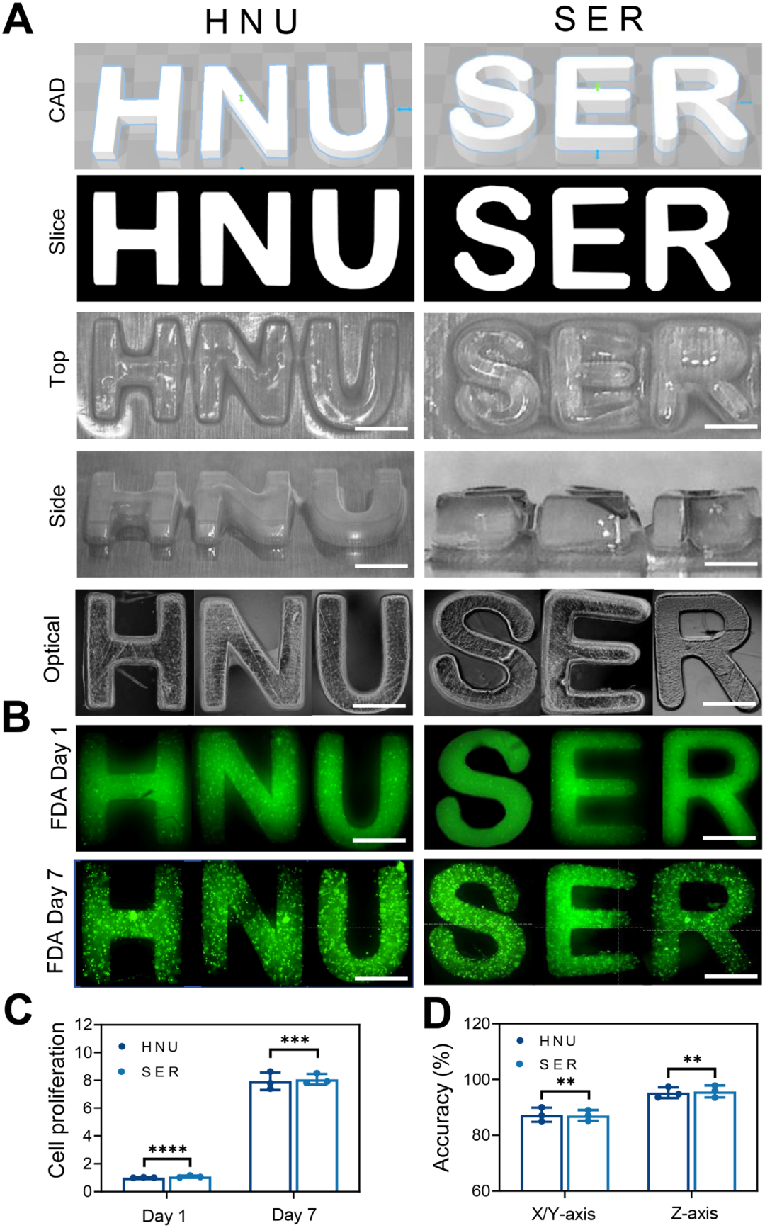 Figure 3. PUN@SerMA-mCMVs bioink DLP bioprinting.PUN8@SerMA-mCMV bioink promoted cartilage defect repair in SD rats. Results showed that compared to the blank group, the experimental group completely covered the original defect area with new cartilage, highly fused with the surrounding normal cartilage tissue, achieving the highest histological score. Micro-CT indicated that the newly formed cartilage had filled the defect, and the surface of the repair area was relatively flat. Histological staining experiments showed that in the PUN8@SerMA-mCMVs group, chondrocytes proliferated and were neatly arranged, exhibiting characteristic cartilage lacunae and layered structures, with minimal incomplete repair tissue. Furthermore, the newly formed cartilage presented a clear and complete tide line, secreting more uniform extracellular matrix, upregulating the expression of COL II and downregulating the expression of MMP-13, indicating that this bioink has effective cartilage defect repair capabilities.
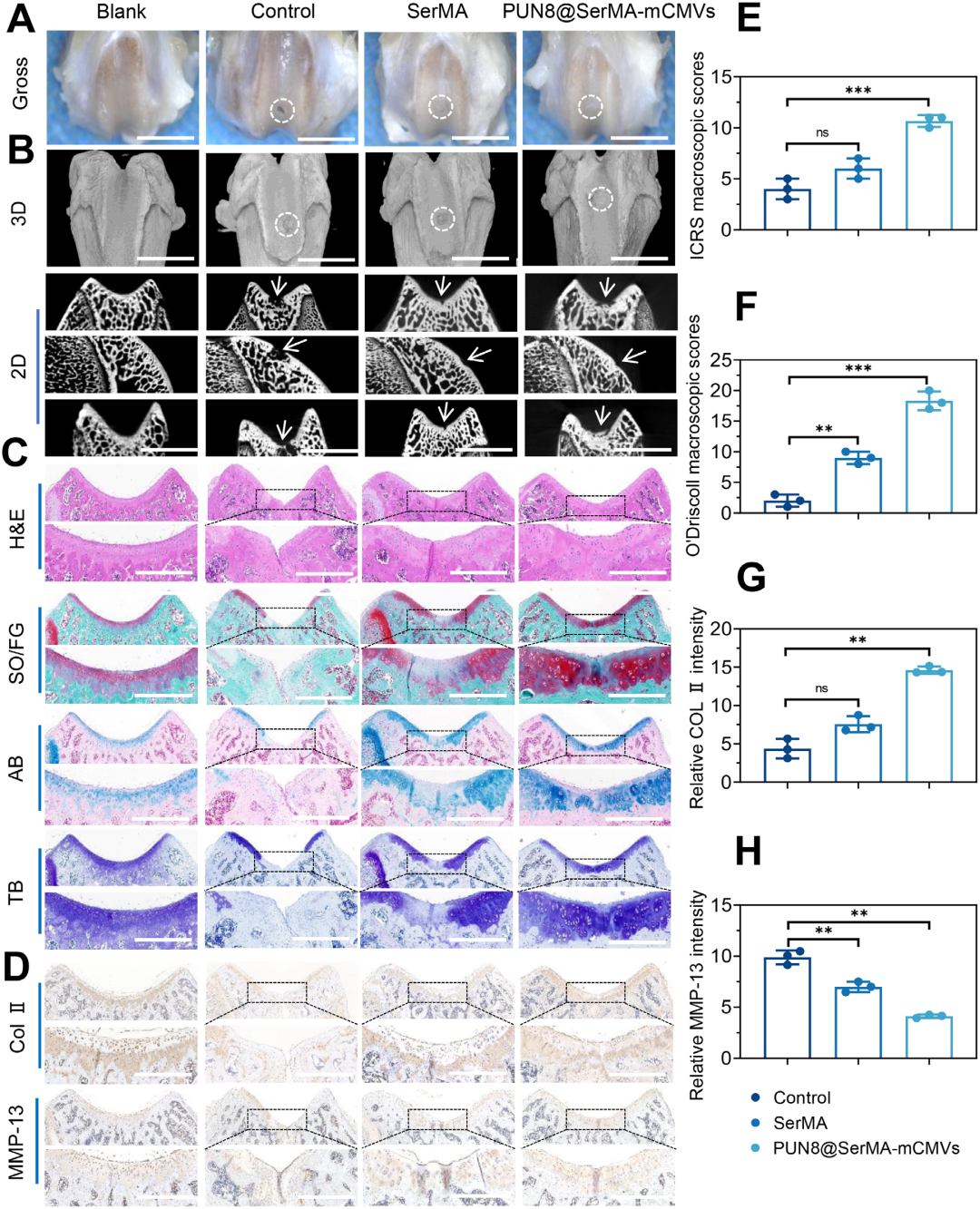 Figure 4. Macroscopic, histological, and immunohistochemical evaluation of joint cartilage repair.Conclusion and Outlook:In this study, a strategy for developing cellular capsule delivery bioink was developed, which combines conventional bioink materials (such as SerMA) and mCMVs into cellular capsules loaded with a certain amount of antioxidant agents, thus producing bioink. Compared to bioinks using alternative drug loading methods, this strategy not only extends the half-life of the drug but also enhances the biocompatibility, printability, and utilization of the bioink. The cartilage defect repair test results in SD rats indicated that compared to SerMA bioink and the control group, PUN8@SerMA─mCMV bioink exhibited the highest tissue regeneration efficiency. Considering the antioxidant activity and antibacterial capability of PUN8@SerMA-mCMV bioink, it may be applied in tissue engineering in the future, especially in specific pathological microenvironments, such as under oxidative stress.Original link: https://doi.org/10.1002/adfm.202504180Source: Polymer Science Frontiers
Experts and scholars are welcome to submit manuscripts (related to micro-nano 3D printing research results, cutting-edge technologies, academic exchanges). Submission email: [email protected].
The purpose of this article is to convey more information. If there are any issues regarding the content, copyright, or other matters, please contact our company, and we will delete the content promptly!
Most Viewed Articles TOP 5
-
Guo Chuanfei from Southern University of Science and Technology, Wang Liu from University of Science and Technology of China, and Chen Yingchun from Commercial Aircraft Corporation of China, published in Nat. Commun.: Ultrafast Response Capacitive Electronic Skin
-
Academician Huang Wei from Northwestern Polytechnical University and Professor Yu Tao’s research group published in Nat. Commun.: Adjustable Afterglow for Mechanical Self-Monitoring 3D Printed Structures
-
Professor Fan Zhiyong’s team from Hong Kong University of Science and Technology published a cover article in Science Robotics: Ultra-Wide Field Pinhole Compound Eye Based on Hemispherical Nanowire Arrays for Machine Vision Applications
-
Professor Zang Jianfeng/Jiang Xiaobing from Huazhong University of Science and Technology and Chen Xiaodong’s research group from Nanyang Technological University published in Nature: Injectable Ultrasound Sensors for Intracranial Signal Monitoring
Professor Shao Jinyou’s research group from Xi’an Jiaotong University published in Adv. Mater: Wearable Flexible Electrodes with High Water Permeability, Stable Adhesion, and Long-Term Durability Inspired by Tree Frog Feet