Recently, a study by Revathi Sukesan and her team from New York University published in Scientific Reports showcased an innovative 3D printed multimodal optogenetic neural probe. This research successfully addresses the challenge of multiple surgeries required in traditional optogenetic studies by integrating optical stimulation with microfluidic drug/virus delivery functions into a single device, providing an efficient, customizable, and minimally invasive solution for the field of neuroscience. Optogenetic technology allows for precise spatiotemporal control of neuronal activity by introducing light-sensitive proteins (such as Channelrhodopsin-2, ChR2) into specific neurons via viral vectors and then illuminating them with specific wavelengths of light. This has become a core tool in the study of neural circuits and behavior. However, traditional procedures typically involve two separate surgical operations: first, delivering the viral vector to the target brain region using stereotactic injection techniques; after several weeks of waiting for the light-sensitive protein to express adequately, a second surgery is performed to implant optical fibers or probes for light stimulation. This two-step process not only increases the complexity of the surgeries and the suffering of the animals but also risks misalignment between the viral infection area and the illumination area due to positional deviations in the two operations, potentially affecting the reliability of experimental results. Additionally, repeated surgical trauma can exacerbate tissue inflammatory responses and glial proliferation, possibly leading to glial scarring that hinders light penetration and reduces the efficiency of neural modulation.
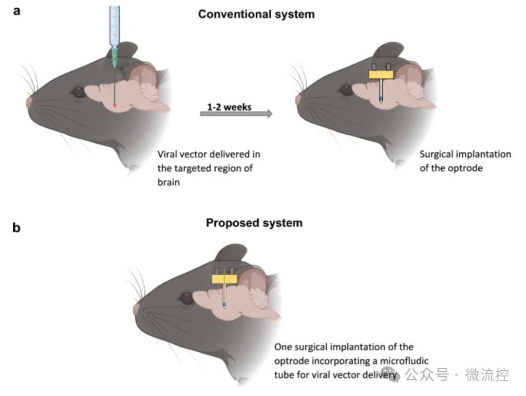 To address these challenges, the research team proposed and manufactured a 3D printed optogenetic probe integrated with microfluidic channels (MIO). The core design of the device lies in its multifunctional integration. The probe’s main structure is fabricated using two-photon polymerization 3D printing technology (utilizing Nanoscribe’s Photonic Professional GT system), which enables micron-level high-precision processing, ensuring the accuracy and reproducibility of the probe structure.
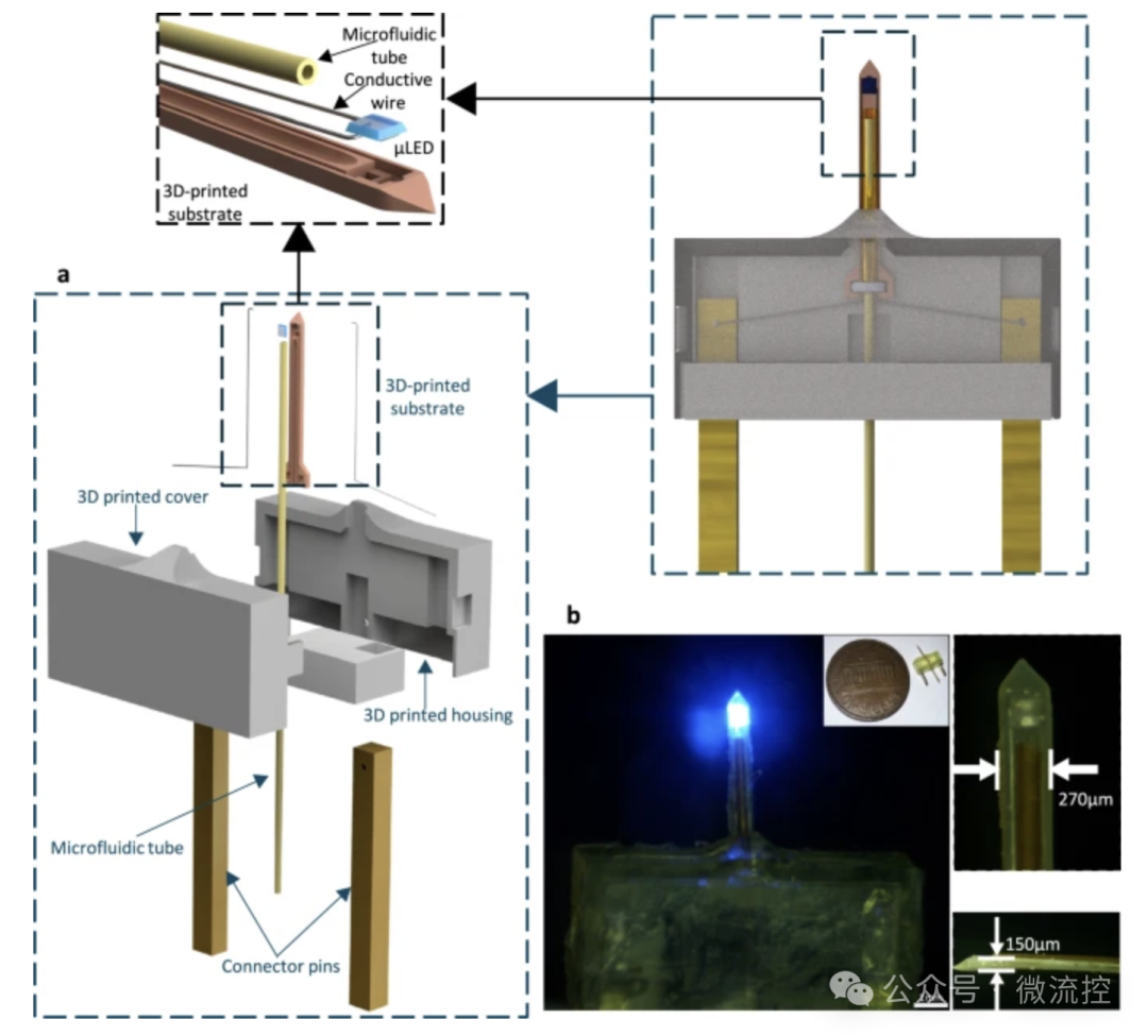 The probe integrates a micro LED (µLED) for optical stimulation, alongside a microfluidic tube with a diameter of 150 micrometers for delivering viral vectors or drugs. This clever design combines viral injection with probe implantation into a single surgical operation, fundamentally simplifying the experimental process. The researchers successfully implanted this probe in the subthalamic nucleus (STN) of mice—a key brain region closely related to motor control—completing both viral vector injection and device fixation in one surgery.The study conducted a series of rigorous performance characterizations on the MIO probe. Electrical and optical test results confirmed that the device operates safely at a working voltage (e.g., approximately 12 mA at 3.0 V), with the blue light emitted by the µLED (peak wavelength 465 nm) being strong enough to exceed the threshold required to activate ChR2 (1 mW/mm²), and the spectrum highly matching ChR2’s response spectrum.
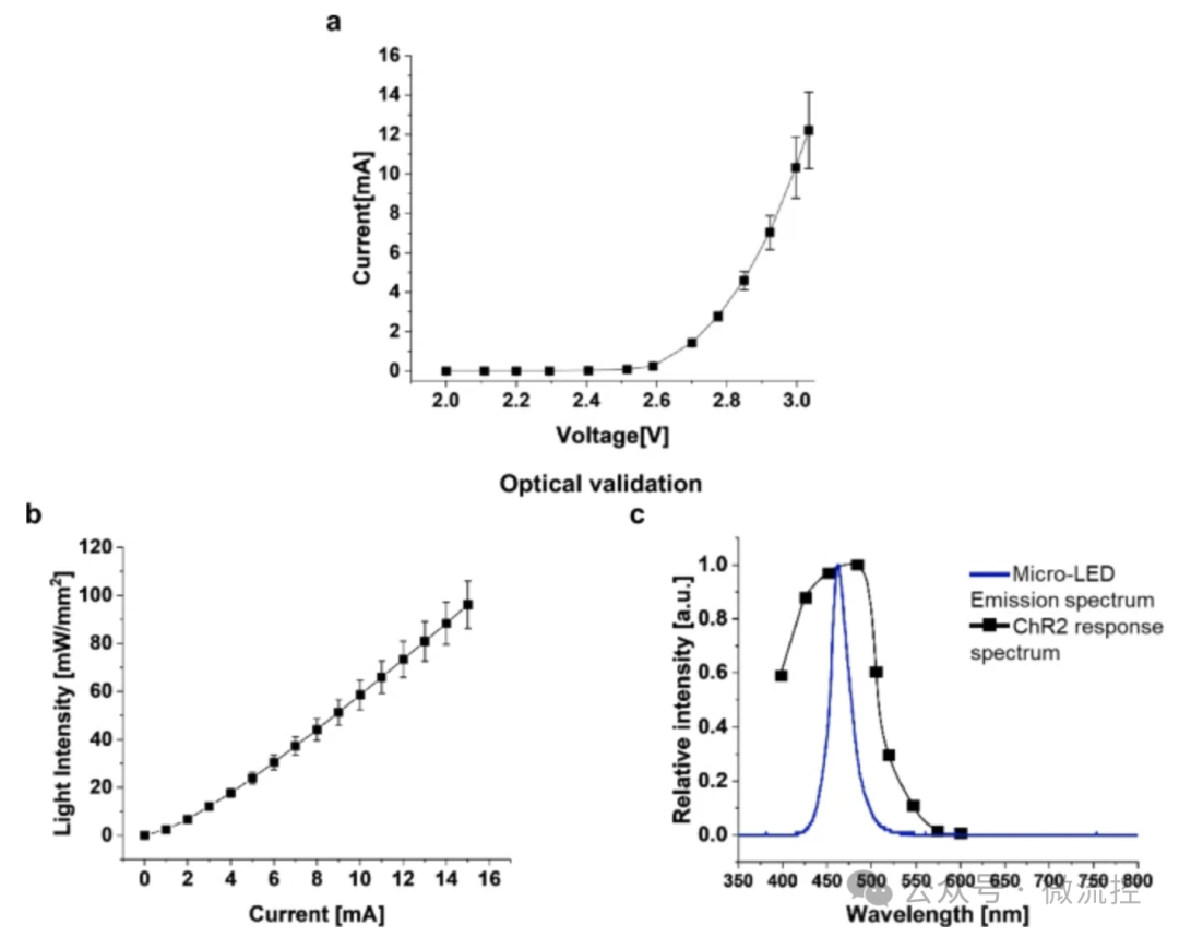 Thermal validation showed that under simulated in vivo experimental conditions, even at higher stimulation frequencies and pulse widths, the temperature rise at the probe tip could be controlled within a safe range of 2°C, avoiding thermal damage to surrounding neural tissue. These results indicate that the 3D printed probe meets the requirements for long-term, stable in vivo neural modulation in key physical performance aspects.In behavioral experiments, the research team compared the experimental group implanted with the MIO probe to a control group implanted with traditional optical fiber probes (after two surgeries). The results showed that during a seven-week observation period, both groups of mice exhibited significant increases in movement distance and speed when receiving optical stimulation, demonstrating that both devices effectively activated STN neurons and elicited corresponding motor behaviors. Notably, the MIO device group exhibited more stable and consistent behavioral effects throughout the experimental period, suggesting that the single-surgery integrated implantation approach may provide better long-term stability and modulation effects. Immunohistochemical analysis further confirmed the biocompatibility and effectiveness of the device. Through mCherry fluorescent labeling, researchers observed that the MIO probe successfully delivered the virus and achieved effective expression of ChR2 protein in the target neurons.
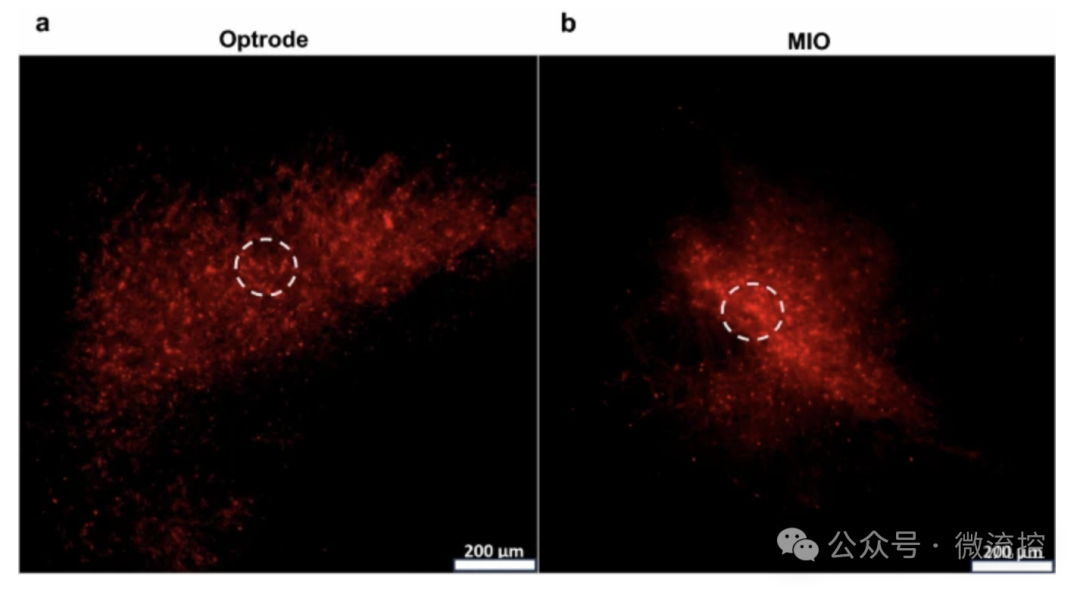 More importantly, compared to the control group, the density of neurons around the MIO probe implantation site was higher, and the activation levels of astrocytes (GFAP labeled) and activated microglia (ED1 labeled) were significantly reduced. This result strongly demonstrates that the single-surgery strategy can significantly alleviate mechanical damage and inflammatory responses caused by the implant, better protecting neurons and reducing the formation of glial scars, providing histological support for the long-term functional stability of the device.In summary, this study successfully developed a multifunctional optogenetic probe based on 3D printing technology, integrating optical stimulation with liquid delivery functions, achieving virus injection and device implantation in a single surgery, significantly reducing surgical trauma and immune responses, and enhancing the efficiency and reliability of experiments. The emergence of this technology not only provides a more powerful and convenient tool for basic neuroscience research but also opens new avenues for the future development of neural modulation devices for clinical treatment, such as deep brain stimulation for Parkinson’s disease. With further advancements in materials science and micro-nano manufacturing technology, such integrated and customizable neural interface devices are expected to play an increasingly important role in the treatment of neurological diseases and human-machine interaction in the future.Paper link:https://doi.org/10.1038/s41598-025-13654-4
Further Reading:
“Printing and Flexible Sensor Technology and Market – 2024 Edition”“3D Electronics and Additive Manufacturing Electronics Technology and Market – 2024 Edition”
“Brain-Computer Interface Technology and Market – 2025 Edition”
Disclaimer:This article is a reprint, and the purpose of reprinting is to convey more information. The copyright belongs to the original author. If there are any copyright issues with the videos, images, or text used in this article, please contact us for modification or deletion. We also welcome submissions, recommendations, and collaborations. Phone: 17898818163.