Sun, X., Quan, S., Wang, B., Wang, Q., Li, W., & Xiao, J. (2023). Peptide-triggered self-assembly of collagen mimetic peptides into nanospheres by electrostatic interaction and π–π stacking.
Journal of Materials Chemistry B , 11(21), 4677–4683.
https://doi.org/10.1039/D3TB00088E
Abstract
Collagen is the most abundant protein in various connective tissues, providing mechanical integrity and regulating cellular activities. Self-assembling peptides have been widely explored to develop collagen mimetic materials due to their attractive features such as ease of synthesis, selective sequences, and low immunogenicity. Recently, metal ion-triggered self-assembly of collagen mimetic peptides has gained increasing attention, as the addition of external stimuli provides programmable control over the self-assembly process. Here, we report for the first time the self-assembly of peptide-triggered collagen mimetic peptides into nanospheres via electrostatic interaction and π–π stacking. We serendipitously discovered that the positively charged, FAM-modified, triple helical peptide FAM-PRG is highly soluble, and the addition of the negatively charged, single-stranded peptide EOG-10 effectively drives its self-assembly into ordered spherical nanomaterials. The peptide EOG-10 has been shown to mediate the self-assembly of TPE-modified triple helical peptide TPE-PRG into luminescent delicate nanospheres, consistently demonstrating the robustness of this peptide-triggered strategy. Fluorescence monitoring of the interaction between EOG-10 and TPE-PRG at different ratios indicates that EOG-10 specifically binds to TPE-PRG to form a 3:1 complex. High salt concentrations were found to inhibit the self-assembly of TPE-PRG with EOG-10, indicating that their self-assembly is controlled by electrostatic interactions. The self-assembly of TPE-PRG with EOG-10 was further revealed to require precise lengths of both peptides and complementary sequences without mutations, indicating a paired “side-by-side” binding mode. Notably, the identity of the N-terminal residue of X-PRG was found to play a decisive role in self-assembly, while the loss of aromatic residues resulted in the loss of self-assembly capability, indicating that π–π stacking and electrostatic interactions jointly regulate the self-assembly of X-PRG and EOG-10. In summary, we have developed a highly biocompatible and programmably controlled peptide-triggered self-assembly method to create novel collagen mimetic nanomaterials, which may have great potential in advanced functional materials.
1. Introduction
Collagen is the main structural protein in the extracellular matrix and serves as a scaffold that contributes to the structural integrity of connective tissues such as bone, skin, and tendons. The three polypeptide chains of collagen bundle together to form a rope-like triple helix, which further self-assembles into fibers with a D-periodic pattern. Collagen fibers interact with cell surface receptors to mediate cell adhesion, migration, and proliferation. Additionally, collagen exhibits excellent biocompatibility, minimal immunogenicity, and natural biodegradability, making collagen-based biomaterials highly attractive in tissue engineering and regenerative medicine.
Collagen is primarily extracted from animal tissues, exhibiting various biological properties. Collagen extracted from bovine cornea shows effective promotion of cell growth. Cod skin is a source of collagen fragments that exhibit accelerated skin wound healing capabilities. However, animal-derived collagen raises increasing concerns in clinical applications due to its insolubility in water, uncontrollable molecular weight, and pathogen transmission.
Due to controllable structures, facile chemical modifications, and zero risk of infectious diseases, peptides have attracted widespread attention in creating collagen mimetic nanomaterials. Significant efforts have been made to construct collagen peptides that can self-assemble into higher-order structures. Collagen mimetic peptide H-(byp)2 modified with two bipyridine ligands self-assembles into nanodisks triggered by Fe(II). Collagen-related peptides containing phenylalanine (F5-Phe) and Phe residues located at the N and C termini self-assemble into fibrils and fibers through π–π stacking. C16-assisted collagen mimetic amphiphilic peptides self-assemble to form nanofibers. These strategies require the introduction of non-natural amino acids, synthetic polymers, or metal ions to trigger the self-assembly of collagen mimetic peptides into ordered nanomaterials, which may severely limit their clinical applications due to potential toxicity. Therefore, collagen mimetic peptides composed solely of natural amino acids have garnered increasing attention, and the inclusion of selectively positioned charged amino acids has been shown to trigger the self-assembly of collagen mimetic peptides into distinct nanofibers.
We serendipitously discovered that collagen mimetic peptides can self-assemble into delicate spherical nanomaterials under the trigger of single-stranded peptides. The self-assembly system consists of two peptides, one being a triple helical peptide with an N-terminal aromatic group and the sequence G(PRGPOG)5, and the other being the single-stranded peptide EOG-10. When simply mixed at room temperature, the two peptides self-assemble into ordered nanospheres mediated by electrostatic interactions and π–π stacking. This self-assembly system is triggered by peptides that exhibit biocompatibility, while it provides a new platform for the design of self-assembling collagen peptides.
2. Experimental Section
2.1. Materials
5(6)-Carboxyfluorescein (FAM) was purchased from Aladdin Biochemical Technology Co., Ltd. (Shanghai, China). 1-(4-Carboxyphenyl)-1,2,2-triphenylethylene (TPE) was purchased from Diba Biotechnology Co., Ltd. (Shanghai, China). Trifluoroacetic acid (TFA) was purchased from Jinkeide Science Co., Ltd. (Beijing, China). Triisopropylsilane (TIS) was purchased from Tokyo Chemical Industry Co., Ltd. (Tokyo, Japan). All Fmoc-amino acids, Rink Amide resin, 2-chlorotrityl chloride resin, O-benzotriazole-N,N,N’,N’-tetramethyluronium hexafluorophosphate (HBTU), and hydroxybenzotriazole (HOBt) were obtained from GL Biochem Ltd. (Shanghai, China). All commercially sourced reagents were analytical grade and used without further purification. All solutions were prepared with ultrapure water.
2.2. Peptide Synthesis
All peptides were synthesized using standard Fmoc solid-phase peptide synthesis methods with Rink Amide resin or 2-chlorotrityl chloride resin. The stepwise coupling of amino acids was performed according to the double coupling method, using Fmoc-amino acids (4 equivalents), DIEA (6 equivalents), and activating reagents (HBTU + HOBt 0.66 mmol mL-1, 4 equivalents). DMF (3 × 5 mL) and DCM (3 × 5 mL) were used to wash the reaction mixture after each coupling step, and the Fmoc protecting group was removed with 20% piperidine. Test reagents (2% acetaldehyde DMF, 2% tetrachlorobenzoquinone DMF) were used to check the completion of each coupling reaction and Fmoc deprotection. TPE-PRG/FAM-PRG was synthesized by conjugating the dye to the N-terminus of peptide G(PRGPOG)5 at room temperature for 24 hours using TPE/FAM (12 equivalents), DIEA (6 equivalents), and activating reagent PyAOP (12 equivalents). TFA/TIS/H2O (95:2.5:2.5) was used to treat the resin for 3 hours to remove the tBu groups and cleave the peptide from the resin. Cold ether was used to precipitate the peptide, and the precipitate was resuspended in cold ether, sonicated, and centrifuged again. The crude product was repeatedly dialyzed against 10 mM PB buffer, and these peptides were confirmed by mass spectrometry.
2.3. Fluorescence Spectroscopy
Fluorescence spectra were obtained on an RF-5301PC fluorescence spectrophotometer (Shimadzu Corporation, Japan), using a xenon lamp as the excitation source. Peptides TPE-PRG and EOG-10 were dissolved in 20 mM PB buffer at pH 7.4. All peptide solutions were heated at 90°C for 20 minutes and then re-equilibrated at 4°C for at least 24 hours. Standard solutions of peptide EOG-10 were prepared by continuous dilution. Fluorescence emission spectra were recorded for different concentrations of TPE-PRG and EOG-10 mixtures at an excitation wavelength of 327 nm.
The “premixing” strategy was used to allow sufficient interaction between peptide TPE-PRG and the single-stranded peptide. Peptide TPE-PRG (300 µM) was mixed with single-stranded peptides (EOG-6, EOG-8, EOG-10[G16-A], and EOG-10[E14-A]) (100 µM) and incubated at 25°C for 24 hours. The mixture solution of peptides was diluted 300 times to obtain a final concentration of 1 µM for TPE-PRG for fluorescence measurement. All fluorescence measurements were repeated three times.
2.4. Scanning Electron Microscopy
SEM images of the self-assembled nanomaterials were obtained on a Hitachi S-4800 scanning electron microscope (Hitachi, Japan) at an operating voltage of 5.1–5.3 kV. The triple helical peptide and single-stranded peptide were mixed in a 2:1 molar ratio and then incubated at 25°C for 24 hours. The supernatant was removed by centrifugation at 8000 rpm for 3 minutes. The precipitate was washed three times with the same volume of ultrapure water and then resuspended in anhydrous ethanol. A 10 µL resuspended solution was dropped onto the center of a silicon wafer. Dried samples were treated with AuPd for 2 minutes before characterization.
3. Results and Discussion
3.1. EOG-10 Triggered Self-Assembly of Collagen Mimetic Peptide FAM-PRG
The amino acid sequences of all collagen mimetic peptides are presented in Table 1. We previously reported that the peptide mixture 2FAM-PRG:1EOG-10 formed heterotrimers after preheating at 90°C and then incubating at 4°C. Here, we found that an unexpected precipitate formed when the two peptides were directly mixed at room temperature. The solutions of FAM-PRG and EOG-10 remained clear when alone (Figure 1A and B). When the peptide FAM-PRG was mixed with EOG-10 in solution at 90°C and then incubated at 25°C for 24 hours, they still remained clear (Figure 1C). However, the solution of peptide FAM-PRG immediately became turbid after the addition of peptide EOG-10 at 25°C and remained turbid after incubation at 25°C for 24 hours (Figure 1D). It has been reported that collagen mimetic peptide FAM-PRG possesses a unique triple helical structure at room temperature, while EOG-10 remains in a single-stranded state. These results indicate that maintaining the triple helical structure of FAM-PRG is a prerequisite for EOG-10-triggered self-assembly.
Table 1 Design of Collagen Mimetic Peptides

The morphology of the nanomaterials self-assembled from peptide FAM-PRG triggered by EOG-10 was evaluated by scanning electron microscopy (SEM) (Figure 1E and F). The SEM images of FAM-PRG aggregates at a 2:1 molar ratio with EOG-10 show delicate nanospheres with an average diameter of approximately 1 µm (Figure 1E and F). The particle size of the nanomaterials self-assembled from peptide FAM-PRG triggered by EOG-10 was characterized by dynamic light scattering (DLS) (Figure S1A, ESI†). DLS indicated that EOG-10 mediates the formation of FAM-PRG nanospheres with an average diameter of approximately 1 µm. This finding is consistent with the SEM results. These results indicate that EOG-10 triggers the self-assembly of peptide FAM-PRG to form ordered nanomaterials.
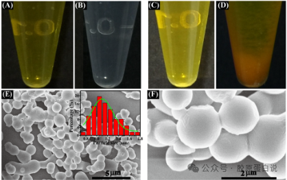
Figure 1 Self-assembly of Collagen Mimetic Peptide FAM-PRG Triggered by EOG-10. The photos show the visual changes of peptide solutions FAM-PRG (A) and EOG-10 (B) incubated separately at 25°C, the peptide mixture re-equilibrated at 25°C (C), and mixed at 25°C (D). SEM images of the assembled nanomaterials of FAM-PRG and EOG-10 (E and F). Illustration (E): Particle size distribution of the assembled nanomaterials. The self-assembled nanomaterials were prepared in 20 mM PB, pH 7.4.
3.2. EOG-10 Triggered Self-Assembly of Collagen Mimetic Peptide TPE-PRG
As a fluorescent dye with aggregation-induced emission, TPE endows materials with excellent optical properties. FAM was replaced with TPE to construct a new triple helical collagen mimetic peptide TPE-PRG (Table 1). The self-assembly of collagen mimetic peptide TPE-PRG triggered by EOG-10 was evaluated through a series of measurements (Figure 2). The solutions of TPE-PRG and EOG-10 remained transparent when alone (Figure 2A and B). When TPE-PRG and EOG-10 were mixed at 90°C and then incubated at 25°C for 24 hours, the mixed peptide solution still remained clear (Figure 2C). However, the mixture of TPE-PRG and EOG-10 became turbid immediately at 25°C and remained turbid after 24 hours (Figure 2D). SEM images show that FAM-PRG self-assembles into nanospheres with an average diameter of approximately 950 nm under the trigger of EOG-10 (Figure 2E and F). The particle size of the nanomaterials self-assembled from peptide FAM-PRG triggered by EOG-10 was characterized by DLS (Figure S1B, ESI†). DLS results showed that EOG-10 mediates the formation of FAM-PRG nanospheres with an average diameter of approximately 950 nm. This result is consistent with the SEM data. It demonstrates that EOG-10 triggers the self-assembly of peptide TPE-PRG to form ordered nanomaterials, exhibiting the same morphology as the assembled FAM-PRG and EOG-10 materials.
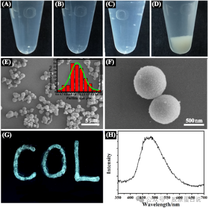
Figure 2 Self-assembly of Collagen Mimetic Peptide TPE-PRG Triggered by EOG-10. The photos show the visual changes of peptide solutions TPE-PRG (A) and EOG-10 (B) incubated separately at 25°C, the peptide mixture re-equilibrated at 25°C (C), and mixed at 25°C (D). SEM images of the assembled nanomaterials of TPE-PRG and EOG-10 (E and F). Illustration (E): Particle size distribution of the assembled nanomaterials. Luminescent photos of TPE-PRG/EOG-10 nanomaterials under 330 nm light on black paper (G). Fluorescence emission spectra of TPE-PRG/EOG-10 nanomaterials obtained at an excitation wavelength of 300 nm (H).
The photoluminescent characteristics of the TPE-PRG and EOG-10 assembled nanomaterials were characterized by solid-state fluorescence. The nanomaterials emitted blue-green light under UV irradiation (λex = 330 nm) (Figure 2G). The fluorescence emission spectrum showed that the maximum fluorescence emission wavelength (472 nm) is consistent with that of TPE-PRG in solution at an excitation wavelength of 300 nm (Figure 2H). Our results demonstrate that the self-assembly of peptide TPE-PRG triggered by EOG-10 shows great potential in the development of luminescent materials.
3.3. Binding Ratio of Collagen Mimetic Peptide TPE-PRG and EOG-10
The fluorescence response of peptide TPE-PRG with different concentrations of EOG-10 was studied to obtain the binding ratio of TPE-PRG and EOG-10 (Figure 3). TPE-PRG showed relatively low fluorescence intensity in the absence of EOG-10, and the fluorescence intensity significantly increased after the addition of EOG-10 (Figure 3A). As the concentration of EOG-10 increased from 0 to 3.0 µM, the fluorescence intensity of TPE-PRG at 472 nm gradually increased and plateaued at EOG-10 concentrations of 3.0 µM or higher (Figure 3B). The enhancement of fluorescence intensity after the addition of EOG-10 demonstrates that EOG-10 induces the aggregation of TPE-PRG.
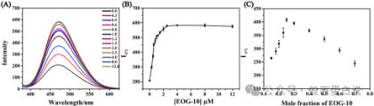
Figure 3 Fluorescence Characterization of Collagen Mimetic Peptide TPE-PRG with Different Concentrations of EOG-10. Fluorescence emission spectra of TPE-PRG in the presence of different concentrations of EOG-10 (0.0, 0.3, 0.5, 0.6, 0.8, 1.0, 1.2, 1.5, 2.0, 2.5, 3.0, 4.0, 8.0, and 12.0 µM) (A). Monitoring the fluorescence intensity at 472 nm (I472) as a function of EOG-10 concentration (B). Job plot of peptide TPE-PRG with EOG-10 in 20 mM PB, pH 7.4 (C).
Job analysis was used to further determine the binding ratio of peptide TPE-PRG and EOG-10 (Figure 3C). The total concentration of TPE-PRG and EOG-10 was maintained at 1.0 µM, and the fluorescence intensity at 472 nm was measured at different molar fractions of EOG-10. The maximum fluorescence intensity was observed at an EOG-10 molar fraction of approximately 0.25. The results indicate that peptide TPE-PRG and EOG-10 bind in a 3:1 molar ratio.
3.4. EOG-10 Triggered Self-Assembly of TPE-PRG via Electrostatic Interactions
The electrostatic interactions triggering the self-assembly of TPE-PRG by EOG-10 were studied through fluorescence assays and SEM at different salt concentrations (Figure 4). As the salt concentration in the mixed solution of TPE-PRG and EOG-10 increased from 50 mM to 200 mM, the fluorescence intensity of TPE-PRG gradually decreased and remained constant at salt concentrations of 200 mM and 300 mM (Figure 4A). This indicates that the tendency of TPE-PRG to aggregate decreases during the process. According to SEM images, collagen mimetic peptide TPE-PRG self-assembled into spherical nanomaterials under the mediation of EOG-10 at a salt concentration of 50 mM (Figure 4B). As the salt concentration increased to 100 mM and 150 mM, the aggregates of EOG-10-triggered TPE-PRG self-assembly became disrupted and more disordered (Figure 4C and D). The mixed solution remained clear when the salt concentration further increased to 200 mM and 300 mM (Figure 4E and F). The electrostatic interactions triggering the self-assembly of TPE-PRG by EOG-10 were further studied at different pH levels through SEM (Figure S2A–D, ESI†). Under acidic (pH 5 and pH 6) and alkaline (pH 9 and pH 12) conditions, the results showed that EOG-10-induced self-assembly of TPE-PRG was disrupted and more disordered. These results significantly inhibit the EOG-10-triggered self-assembly of TPE-PRG, indicating that electrostatic interactions play a key role in the self-assembly of collagen mimetic peptide TPE-PRG triggered by EOG-10.
3.5. Influence of Peptide EOG-10 Sequence Variants on TPE-PRG Self-Assembly
Peptides EOG-6 and EOG-8, designed with 6 and 8 repeats of the EOG tripeptide sequence, respectively, were studied for their influence on TPE-PRG self-assembly through fluorescence assays. The fluorescence intensity of TPE-PRG (black) was recorded in the presence of other peptides EOG-10 (red), EOG-8 (blue), and EOG-6 (pink) (Figure 5A). Compared to peptide EOG-10, peptides EOG-6 and EOG-8 showed much lower fluorescence intensity. This result demonstrates that EOG-6/EOG-8 lack the ability to bind with TPE-PRG, proving that TPE-PRG and EOG-10 bind in a paired “side-by-side” arrangement.
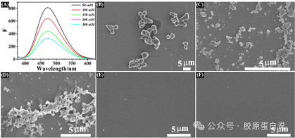
Figure 4 Effect of Different Salt Concentrations on EOG-10 Triggered Self-Assembly of Collagen Mimetic Peptide TPE-PRG. Fluorescence emission spectra of TPE-PRG with EOG-10 at different salt concentrations (50, 100, 150, 200, and 300 mM) (A). SEM images of EOG-10 triggered self-assembly of TPE-PRG nanomaterials at different salt concentrations: 50 mM (B), 100 mM (C), 150 mM (D), 200 mM (E), and 300 mM (F).
Peptides EOG-10[G16-A] and EOG-10[E14-A] were designed to further investigate the effects of different mutations on TPE-PRG self-assembly (Table 1). The fluorescence intensity of TPE-PRG (black) was recorded in the presence of other peptides EOG-10 (red), EOG-10[G16-A], and EOG-10[E14-A] (Figure 5B). Compared to EOG-10, EOG-10[E14-A] showed much less enhancement in fluorescence intensity. Furthermore, EOG-10[G16-A] showed almost no enhancement in TPE-PRG fluorescence intensity. The results indicate that single amino acid mutations in the EOG-10 sequence can inhibit TPE-PRG self-assembly, proving that peptide-triggered self-assembly of collagen mimetic peptides strictly requires sequence complementarity. Moreover, the inhibition caused by the Gly-Ala mutation is significantly more pronounced than that caused by the Glu-Ala replacement, demonstrating the close binding of TPE-PRG and EOG-10.
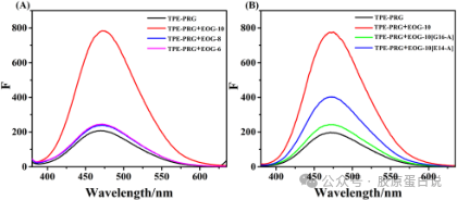
Figure 5 Influence of Peptide EOG-10 Sequence Variants on TPE-PRG Self-Assembly. (A) Fluorescence emission spectra of peptide TPE-PRG alone (black), in the presence of EOG-10 (red), EOG-8 (blue), and EOG-6 (pink). (B) Fluorescence emission spectra of peptide TPE-PRG alone (black), in the presence of EOG-10 (red), EOG-10[G16-A] (green), and EOG-10[E14-A] (blue).
3.6. EOG-10 Triggered Self-Assembly of X-PRG
Seven types of triple helical peptides were designed to study the role of the N-terminal residues of X-PRG in self-assembly triggered by EOG-10. The peptides W-PRG, Y-PRG, G-PRG, Q-PRG, L-PRG, P-PRG, and PRG were designed (Table 1). Turbidity experiments were conducted using a UV-Vis spectrophotometer to measure the turbidity of EOG-10 in the presence of peptide X-PRG (Figure 6A). Compared to peptides W-PRG and Y-PRG, the other five peptides showed much lower absorbance at OD313nm. SEM images showed that W-PRG and Y-PRG self-assemble into spherical nanoparticles mediated by EOG-10 (Figure 6B and C). However, no supramolecular structures were observed in the SEM images of G-PRG, Q-PRG, L-PRG, P-PRG, and PRG mixed with EOG-10. These results reveal that π–π stacking plays a decisive role in the self-assembly of X-PRG and EOG-10.
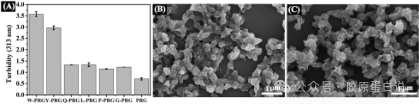
Figure 6 Self-assembly of X-PRG Triggered by EOG-10 (W-PRG, Y-PRG, Q-PRG, L-PRG, P-PRG, G-PRG, and PRG). The turbidity of the solution of the three helical peptides PRG after the addition of EOG-10 (A). SEM images of collagen mimetic peptides W-PRG (B) and Y-PRG (C) with EOG-10.
3.7. Biocompatibility of Peptide-Triggered Collagen Mimetic Peptide Self-Assembly
The in vitro cytotoxicity of peptide-triggered collagen mimetic peptide self-assembly was examined using CCK-8 assay with HFF-1 cells. HFF-1 cells showed similar high cell viability at different concentrations (0, 0.1, 0.5, 1, 2, 5, 10, 50, and 100 µg mL-1) demonstrating that the nanomaterials exhibit almost no cytotoxicity and good biocompatibility (Figure 7).
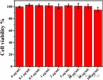
Figure 7 CCK-8 Assay of Peptide-Triggered Collagen Mimetic Peptide Nanomaterial Self-Assembly.
3.8. Possible Mechanism of Peptide-Triggered Collagen Mimetic Peptide Self-Assembly
The mechanism of peptide-triggered collagen mimetic peptide self-assembly is hypothesized from the experimental results. The single-stranded peptide EOG-10 triggers the self-assembly of the triple helical peptide in a paired “side-by-side” binding mode through electrostatic interactions, and the formation of delicate nanospheres is further facilitated by electrostatic interactions and π–π stacking (Figure 8).
4. Conclusion
Collagen is not only a major component of connective tissues but also an essential biomolecule that regulates various cellular functions. Significant progress has been made in constructing self-assembling systems of collagen mimetic peptides that can form supramolecular structures. The incorporation of metal ions has been an effective strategy to promote the self-assembly of collagen peptides into advanced nanomaterials. However, their biomedical applications are also limited due to potential toxicity.
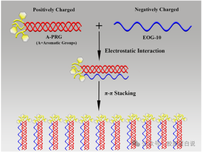
Figure 8 Possible Mechanism of Peptide-Triggered Collagen Mimetic Peptide Self-Assembly.
We report for the first time a self-assembly system of collagen mimetic peptides triggered by peptides to form delicate nanospheres through electrostatic interactions and π–π stacking. We serendipitously discovered that the single-stranded peptide EOG-10 can trigger the self-assembly of the triple helical peptide FAM-PRG into delicate spherical nanomaterials. The peptide TPE-PRG also forms nanospheres mediated by EOG-10, while TPE endows the self-assembled materials with excellent luminescent properties.
In summary, this study investigates the mechanism of peptide-triggered self-assembly of collagen mimetic peptides. The results indicate that the electrostatic interactions between TPE-PRG and EOG-10 are responsible for the formation of a 3TPE-PRG:1EOG complex. The self-assembly process is influenced by salt concentration and pH, and requires complementary sequences for the close packing of TPE-PRG and EOG-10. Shortened sequence peptides EOG-8 and EOG-6 cannot trigger self-assembly, indicating the importance of pairing between TPE-PRG and EOG-10. Moreover, the N-terminal aromatic amino acids of peptide X-PRG contribute to self-assembly through π–π stacking. Overall, this biocompatible peptide-triggered collagen mimetic peptide self-assembly system has potential applications in tissue regeneration and other biomedical fields.
This article is for personal learning and research purposes only, aimed at promoting academic exchange and knowledge dissemination in related fields, and cites the original literature source and author information. This behavior strictly follows the provisions of the Copyright Law of the People’s Republic of China regarding fair use, non-commercial purposes, and does not infringe upon the copyright and other legal rights of the original authors and related parties.