Abstract:
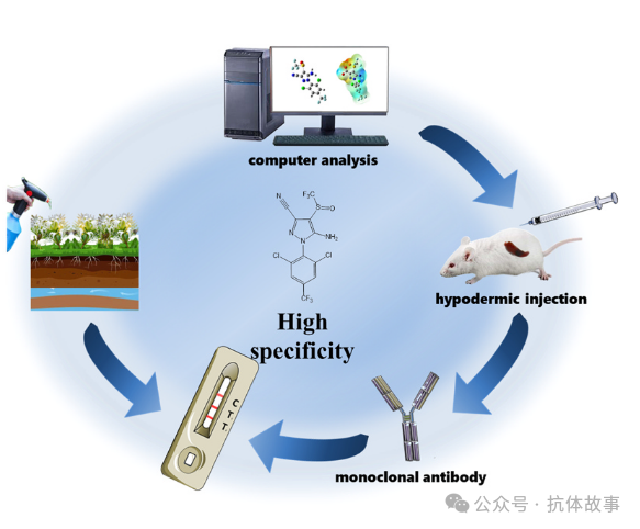
Fipronil (FPN) is one of the widely used pesticides in plant cultivation and animal husbandry, causing extensive pollution to various environmental media such as water, soil, and plants. To address the challenge of trace detection of fipronil (FPN) in complex environmental matrices, this study designed a semi-antigen through computer simulation, modified carrier proteins, and synthesized a novel FPN-immunogen, successfully preparing high-specificity monoclonal antibodies. Based on a double T-line AuNP-labeled immunosensor, a visual quantitative test strip was developed. The method achieved a detection limit (cLOD) of 1.23 μg/kg for FPN in water (6.46 μg/kg in soil, 13.7 μg/kg in honeysuckle), with a recovery rate of 92.3–108.5%. Verified by LC-MS/MS, it demonstrated excellent stability and accuracy, making it suitable for rapid on-site screening and quantitative analysis of FPN contaminants in complex environments.
Introduction
Fipronil (FPN), a phenylpyrazole insecticide, exhibits biological activity by acting on GABA receptors, and its residues are widely present in environmental media (e.g., 65 μg/kg in Pakistani rice, 0.13 mg/kg in Chinese vegetables exceeding standards) and in the food chain (honey from bees, poultry products). FPN persists and accumulates in water and soil. FPN and its metabolites exhibit multiple toxicities: causing neurofunctional disorders in aquatic organisms, DNA damage in mammals, and thyroid abnormalities, and are classified as Class C carcinogens in the United States. China has set the maximum residue limit (MRL) for vegetables at 0.02 mg/kg (GB 2763–2021). Current detection relies on chromatography/mass spectrometry techniques (e.g., HPLC, LC-MS/MS) or emerging aptamer sensors (sensitivity of 0.033 μg/kg), while immunoassays are still in exploratory stages due to limitations in semi-antigen design. This study breakthrough selected semi-antigens retaining the FPN characteristic SOCF3 motif, synthesized a novel immunogen through carrier protein modification, and successfully prepared high-specificity monoclonal antibodies (mAbs) sensitive only to FPN, subsequently developing a double T-line immunosensor based on AuNP labeling. This method can rapidly identify FPN in complex environmental and food samples, significantly reducing detection costs and time, providing an efficient template for on-site screening.
Research Content
1. Joint Computer-Aided Design to Predict Semi-Antigens
A semi-antigen, as an incomplete antigen, has no immunogenicity but can bind to antibodies. For the small molecule fipronil (FPN), the design of its semi-antigen must meet the following criteria: contain active groups for protein coupling, have a structure highly fitting to FPN, and possess exposed sites to achieve high specificity in ic-ELISA detection. Although broad-spectrum antibodies can detect FPN and its metabolites, the preparation of high-specificity antibodies remains challenging. This study confirmed that the SOCF3 radical is key to producing high-specificity FPN antibodies (Figure 1B,C), thus retaining this group in the semi-antigen design; simultaneously constructing a heterologous coated antigen H3FPN (with a different coupling site than H2FPN, Figure 1A) to enhance sensitivity. By optimizing the three-dimensional structures of FPN and semi-antigens to their lowest energy states through computer simulation, the surface charge distribution was calculated using the DET formula (Figure 1B,C), revealing that HFPN2/HFPN3 pairs are closer to the FPN conformation. Further analysis of physicochemical properties indicated that HFPN1 exhibited weak immunogenicity due to its small dipole moment and poor charge matching (experimental validation showed a decrease in its titer), while computer simulation provided effective analytical tools for catalyst screening and immunogenicity research by analyzing electronic configurations and spatial structures.
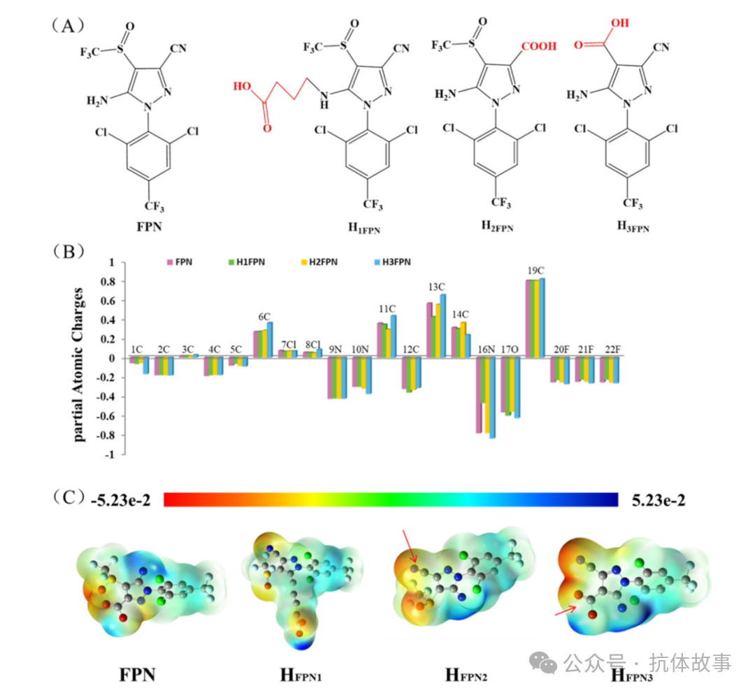
Figure 1. (A) Structures of FPN and semi-antigens reported in literature; (B) Calculated partial atomic charges of FPN and semi-antigens; (C) ESP of FPN and semi-antigens.
2. Identification of FPN-Semi-Antigens and Complete Antigens
HFPN2 was characterized by MS (ESI) m/z = 362.9 [M-H]- and 1H NMR (δ 8.22(s,2H), 6.57(s,2H)), and synthesized into a complete antigen with cationized carrier proteins; UV-Vis spectroscopy confirmed the successful cationization of the carrier protein, with more -NH2 used for coupling with semi-antigen -COOH to obtain immunogenicity. UV-Vis data showed changes in absorption peaks confirming effective coupling. To validate this hypothesis, three semi-antigens (H1FPN, H2FPN, H3FPN) were subjected to immunization experiments (Figure 2A), where the immunogen used was semi-antigen coupled cationized proteins (HFPN1-HAD-KLH, HFPN2-HAD-KLH, HFPN3-HAD-KLH) (Figure 2B), and the coated antigens used were semi-antigens directly coupled to proteins (HFPN1-BSA, HFPN2-BSA, HFPN3-BSA). UV-Vis further confirmed the changes in absorption peaks of the synthesized products supporting successful coupling.
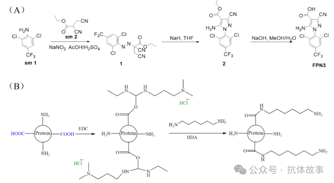
Figure 2. (A) Synthesis route of HFPN3 semi-antigen; (B) Modification of cationized proteins.
3. Characterization of Different mAb Properties
During the evaluation of immune responses, serum from the sixth immunized mouse was analyzed with different coated antigens (Figure 3A). Through ic-ELISA, it was found that among mice with titers of 1.2-1.8, the combination of HFPN2-KLH immunogen and HFPN3-BSA coated antigen exhibited the best inhibition effect, thus this mouse was selected for cell fusion to prepare high-specificity monoclonal antibodies 3B1 and 2B12 that recognize FPN. Subtype analysis showed that 3B1 is IgG2a heavy chain λ light chain, while 2B12 is IgG1 heavy chain λ light chain (Figure 3B). Affinity testing (Figure 3C) indicated that under a gradient of coated antigen concentrations (1, 0.3, 0.1 mg/mL), the affinity constant of 3B1 reached 1.35×10¹⁰ L/mol, significantly higher than 2B12’s 2.72×10⁹ L/mol, confirming that 3B1 has stronger affinity and activity, thus it was selected as the antibody for subsequent development of the AuNP immunosensor.
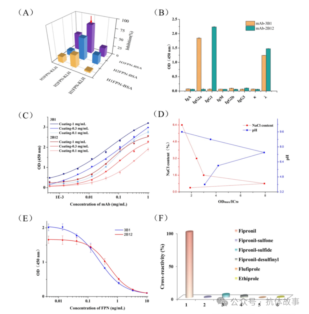
Figure 3. (A) Determination of mouse serum; (B) Isotype identification of mAb – 3B1 and mAb – 2B12; (C) Affinity of mAb – 3B1 and mAb – 2B12; (D) Optimization of working buffer, different pH PBS (4.0, 6.0, 7.4, 8.8, and 9.6), NaCl content (0.4%, 0.8%, 1.6%, 3.2%, 6.4%); (E) Standard inhibition curves of mAb – 3B1 and mAb – 2B12; (F) Cross-reactivity.
4. Optimization of Monoclonal Antibodies
In establishing ic-ELISA for detecting FPN, the NaCl content and pH of the solution affected antibody binding, thus optimizing NaCl (0.4%, 0.8%, 1.6%, 3.2%, 6.4%) and pH (4.0, 6.0, 7.4, 8.8, 9.6) parameters (Figure 3D). The higher the OD Max/IC50 ratio, the higher the sensitivity; analysis showed the optimal conditions were pH 7.4 and NaCl 0.7%-1.0%. Under optimal conditions, antibody performance was evaluated (Figure 3E), with an IC50 of 0.236 ng/mL, a linear range of 0.06-0.87 ng/mL, and an LOD of 0.0083 ng/mL, indicating high sensitivity and a wide detection range. In LFIA test strip determinations, cross-experiments (Figure 3F) showed negligible cross-reactivity with similar substances, proving its highly specific recognition of FPN, meeting the development needs of the AuNP immunosensor.
5. Optimization of LFIA
In the electrostatic adsorption of proteins by AuNP, adsorption is enhanced when the pH exceeds the isoelectric point. The experiment optimized the K2CO3 content (1-2 μL solution turns purple and OD value increases, stabilizing after >2.5 μL), with 2 μL selected as optimal to ensure solution stability without precipitation. The particle size and uniformity of AuNP affect color development; TEM showed uniformity, and UV-Vis confirmed successful synthesis, with AuNPs labeled monoclonal antibodies synthesized (Figure 4A). To enhance the accuracy of the immunosensor, a double T-line system was designed: at T2 line, AuNPs labeled mAb bind to low-concentration antigens, while at T1 line, they bind to higher-concentration antigens (T1 line slightly deeper than T2 line), followed by non-specific binding at the C line (Figure 4B). This design expands the linear range, allowing detection of low concentrations (e.g., in water samples) and high concentrations (e.g., in soil and honeysuckle) of FPN. Surfactant optimization tested eight types (5% BSA, PVP, PEG, On-870, Span-20, Trito X-100, Brij-35, Tween-20), where Span-20 caused rapid flow inhibiting binding, Trito X-100 and Brij-35 reduced hydrophilicity worsening inhibition, while BSA/PVP/PEG/Tween-20 lightened the C line, and 5% On-870 provided moderate flow rate, optimal inhibition effect, and positive differentiation, thus selected as the best.
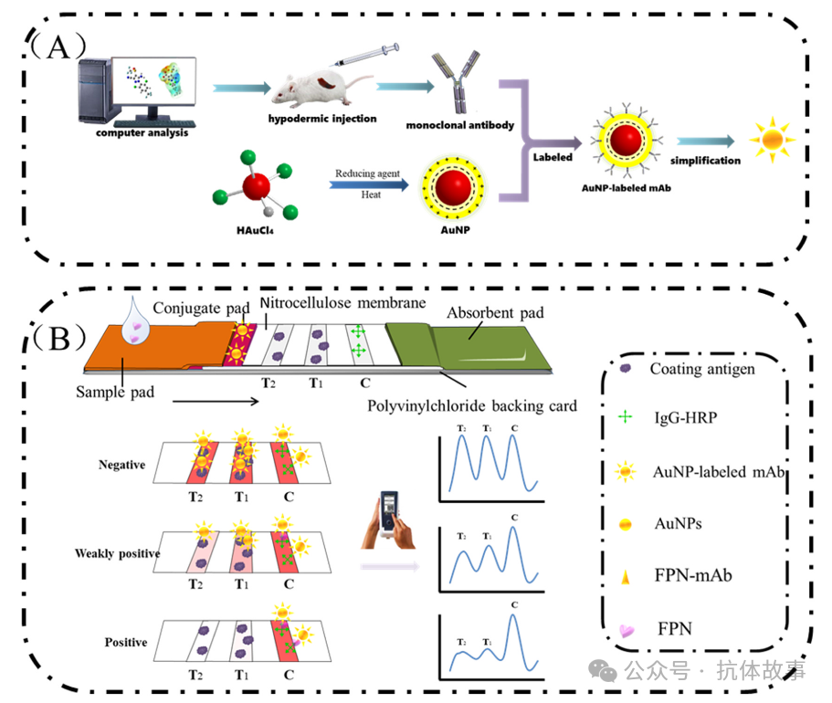
Figure 4. (A) Schematic diagram of AuNP labeled mAb synthesis; (B) Schematic diagram of AuNP labeled immunosensor.
6. Optimization of Sample Pretreatment Methods
When detecting FPN in honeysuckle, soil, and water, directly testing the sample extraction solution interfered with LFIA performance, thus a 5% On-870 + PBS gradient dilution was adopted. Experimental results showed that at a 10-fold dilution, matrix interference caused abnormal C line/T line; as the dilution factor increased, the C line and double T line color deepened; at a 30-fold dilution, the C line stabilized and showed the best color, thus determined as the optimal dilution. Additionally, four organic solvents (acetonitrile, methanol, acetone, DMSO) were screened as extraction solutions, with the blank sample interference intensity ranking as acetonitrile > methanol > acetone > DMSO, with acetonitrile selected to reduce background interference.
7. Validation of AuNP-Based Immunosensor
In the performance evaluation of the optimized AuNP immunosensor, FPN spiked detection was conducted on water, soil, and honeysuckle samples. Qualitative analysis showed that at an FPN concentration of 5 μg/kg in water, the T2 line weakened, and at 200 μg/kg, it disappeared (detection limit 5 μg/kg, critical value 200 μg/kg, Figure 5A); in soil, at 20 μg/kg, the T2 line weakened, and at 500 μg/kg, it disappeared (detection limit 20 μg/kg, critical value 500 μg/kg, Figure 5B); in honeysuckle, at 20 μg/kg, the T2 line weakened, and at 1000 μg/kg, it disappeared (detection limit 20 μg/kg, critical value 1000 μg/kg, Figure 5C). Quantitative analysis fitted standard curves through (T1+T2)/C values: for water samples y=3.21–1.4lgx (R²=0.989, LOD=1.23 μg/kg); for soil y=7.85–3.64lgx (R²=0.975, LOD=6.46 μg/kg); for honeysuckle y=5.356–1.76lgx (R²=0.993, LOD=13.7 μg/kg). The double T-line design significantly broadened the detection range compared to a single T-line, meeting China’s MRL requirements. Verification in cabbage matrix confirmed LOD=8.35 μg/kg, suitable for food detection.
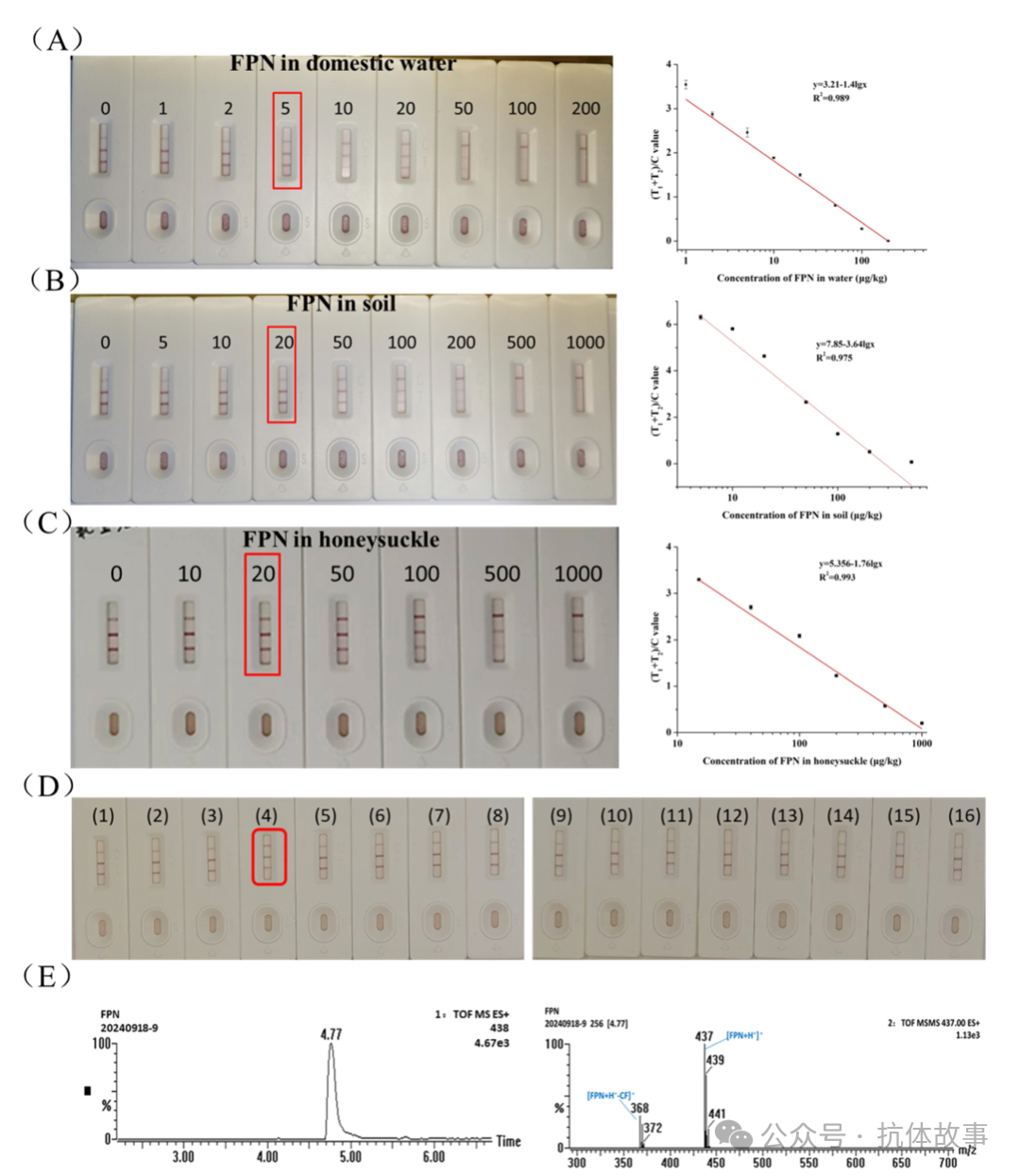
Figure 5. Performance of the immunosensor (A-C) Analysis results of the immunosensor in water, soil, and honeysuckle; (D) Results of 16 actual samples; (E) Positive sample LC-MS/MS detection results.
In assessing the practical application stability of the immunosensor, accelerated aging tests at 37°C (0, 3, 5, 7, 14, 28 days) were conducted on water/soil/honeysuckle samples (containing 10 μg/kg and 50 μg/kg FPN). Results showed that during aging, the (T1+T2)/C values changed insignificantly within 28 days (water samples ±5.2%, soil ±7.1%, honeysuckle ±8.3%), proving the stability of antibody-antigen interactions on the AuNP-based immunosensor membrane, supporting long-term rapid detection of FPN in complex matrices.
8. Detection of FPN in Real Samples
To evaluate the accuracy of the AuNP immunosensor compared to LC-MS/MS methods: FPN was added to water/soil/honeysuckle samples at 0, 5/20/20 μg/kg to 200/500/1000 μg/kg (Table 1), with sensor recovery rates of 92.3%-108.5% (coefficient of variation CV=1.7%), highly consistent with LC-MS/MS recovery rates (95.3%-110.4%, CV=1.6%). Further detection of 16 actual samples showed that sample #4 indicated a deepening of the C line and weakening of T1 and T2 lines by the immunosensor (Figure 5D,E), qualitatively identified as weak positive (46.8 μg/kg), confirmed to contain FPN residues by LC-MS/MS. Results indicate that this sensor possesses excellent accuracy and stability in environmental and plant samples, enabling rapid on-site detection of FPN.
Table 1. Recovery rates of AuNP labeled immunosensor and LC-MS/MS (n = 3) for FPN in honeysuckle samples.
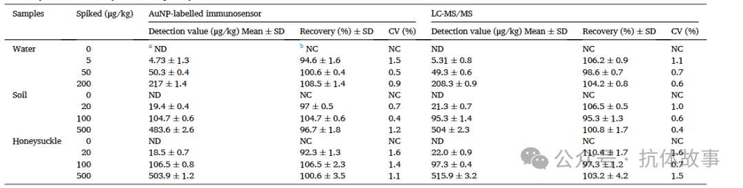
a. Nd, not detected; b. Nc, not calculated.
Conclusion
This study screened three structures suitable for FPN through computer simulation to synthesize a novel immunogen modified by carrier proteins, successfully preparing high-specificity monoclonal antibodies only for FPN. Based on this, a double T-line immunosensor labeled with AuNPs was developed, capable of rapidly identifying FPN in complex environmental and food samples, effectively addressing matrix interference issues, complying with China’s maximum residue limit standards (MRL), and achieving efficient screening of water, soil, and herbal samples. The current challenge lies in the lack of specific antibodies targeting FPN metabolites (fipronil-sulfone/sulfide/desulfonyl), necessitating the development of multi-band immunosensors capable of separately detecting FPN and its metabolites.
Original Source
Kou S, Qiu G, Liu L, et al. Visual immunosensor assay with double T line for on-site sensitive fipronil pesticide detection in water, soil and honeysuckle. J Hazard Mater. 2025;489:137634.
Original link:
https://www.sciencedirect.com/science/article/pii/S0304389425005485?via%3Dihub#sec0125
DOI:10.1016/j.jhazmat.2025.137634
Advisor: Professor Wang Zhanhui
Click below to follow us