Extracellular vesicles (EVs) are nanometer-sized particles enveloped by phospholipids that are secreted by cells into the surrounding environment. EVs can convey various biomolecules that mediate intercellular communication and play crucial roles in physiological and pathological processes. Therefore, EVs are emerging as new biomarkers, therapeutic targets, and drug delivery vehicles for various diseases. However, the isolation and detection of EVs require time-consuming and labor-intensive processes, first extracting EVs from biological and physiological fluids, and then detecting EV-related molecules with high sensitivity. Currently, the detection methods and instruments used for EV analysis are often not widely applicable outside research laboratories, posing practical obstacles to the study of EV-related molecules in biological and clinical applications.
To address this, recent research has focused on utilizing microfluidic technology for the extraction of EVs, as microfluidic technology can handle microliter volumes of liquid and automate complex processes in miniaturized devices. Passive particle separation methods are common in microfluidic devices, allowing for the separation of EVs based on particle size, such as through lateral displacement, inertial forces, and flow separation. Active methods, such as ultrasound, can also be employed in microfluidic devices to capture, separate, aggregate, and transport EVs by applying different forces based on size. However, size-based EV separation often results in low purity, as similarly sized contaminants may also be separated, and clogging is a challenge. To overcome these limitations, electrophoretic methods have been explored as alternatives. However, these methods typically require the precise integration of microelectrodes at specific locations within the microfluidic device and the highly controlled modulation of high AC fields on these electrodes. Additionally, using non-insulated electrodes in water poses a risk of electrolysis, which may lead to device failure.
According to MEMS Consulting, to overcome the aforementioned challenges, researchers at the Mayo Clinic in the United States have developed a digital microfluidic (DMF) device capable of automatically extracting EVs from various biological fluids and detecting specific subpopulations based on their surface markers. The related research findings have been published in the recent issue of Small under the title “An Integrated Digital Microfluidic Device for the Extraction and Detection of Extracellular Vesicle-Based Molecules.”

Unlike other microfluidic platforms, this digital microfluidic device consists of an array of insulated electrodes that manipulate fluids based on the principle of electrowetting. This unique structure allows for the sequential programmed processing of droplets (e.g., separation, mixing, movement) without the need for physical microstructures (e.g., microvalves, microchannels). Consequently, these devices can automatically perform experiments requiring multiple steps, such as target extraction, washing, and culturing, based on user-defined parameters. In this digital microfluidic device, researchers used immunomagnetic microspheres to extract EVs on-chip, as immunoaffinity provides higher specificity for EV subpopulations, and then employed electrochemical sensors to detect their surface markers.
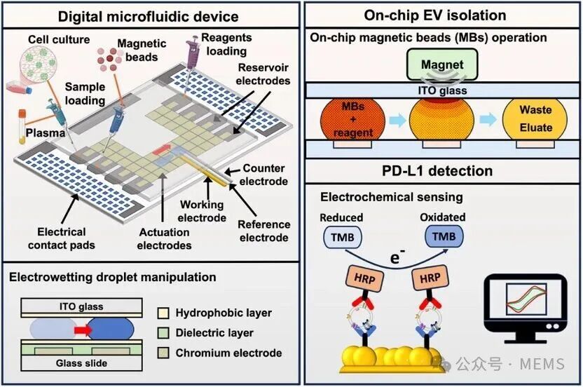
Digital microfluidic device based on electrowetting principle
In the proof of concept, researchers selected the immune checkpoint molecule PD-L1 as the target for EV binding, as its circulation in the blood is associated with resistance to PD1/PD-L1 blockade, a cutting-edge therapy in the field of immunotherapy. The developed digital microfluidic device can automatically extract EVs from 20 µL of cell culture and human plasma samples within 25 minutes and detect PD-L1+ EVs on-chip using electrochemical sensors, with a detection limit of 1 x 10⁴ EVs/mL. This study demonstrates the feasibility of simplifying the extraction of EVs from various biological fluids and detecting specific EV subpopulations, which can be applied in biological research and clinical applications.
Digital microfluidic device platform
The digital microfluidic device consists of two glass substrates with a gap between them for droplet manipulation. The bottom substrate is patterned with driving electrodes, storage electrodes, and side electrical contact pads, while the top substrate is coated with indium tin oxide (ITO) as the grounding electrode, allowing for visual observation of the liquid from the top. The digital microfluidic device can be programmed to control the opening/closing of electrodes to process droplets in a timed manner. Samples and reagents (e.g., culture media, plasma, magnetic microspheres) can be dispensed onto different storage electrodes and then split into smaller droplets based on the electrowetting principle, processed on the driving electrodes. In this study, researchers achieved multiple functions within a single device, including functionalization of magnetic microspheres, capturing EVs from biological fluids, sample washing, and elution of EVs. Subsequently, researchers inserted gold wire-based electrochemical sensors into the device to detect PD-L1+ EVs.
On-chip TIM-4 magnetic microsphere preparation
In this study, researchers utilized immunomagnetic microspheres for the extraction of EVs on-chip. First, researchers functionalized TIM-4 onto immunomagnetic microspheres within the digital microfluidic device. In brief, 10 µL of streptavidin-coated magnetic microspheres were dispensed from the storage electrode on the chip, and the microspheres were retained using a magnet while the supernatant was separated and removed. Then, a solution containing biotinylated TIM-4 was added and suspended in a washing buffer, mixed with the magnetic microspheres, and TIM-4 was fixed onto the microspheres through biotin-streptavidin coupling. Multiple 20 µL washing buffer droplets were then used to remove unbound molecules in the same manner. The microspheres were resuspended in a 20 µL washing buffer droplet before starting the EV separation process. It is noteworthy that many microfluidic processes use pre-functionalized microspheres prepared outside the chip, while this study combines the microsphere functionalization process with EV extraction, demonstrating the feasibility of on-demand functionalization of different biological recognition agents onto microspheres for specific detection.

On-chip extraction of extracellular vesicles
Based on surface-modified working electrodes, researchers constructed an electrochemical immunosensor to detect the PD-L1 expression levels of EVs extracted on-chip.
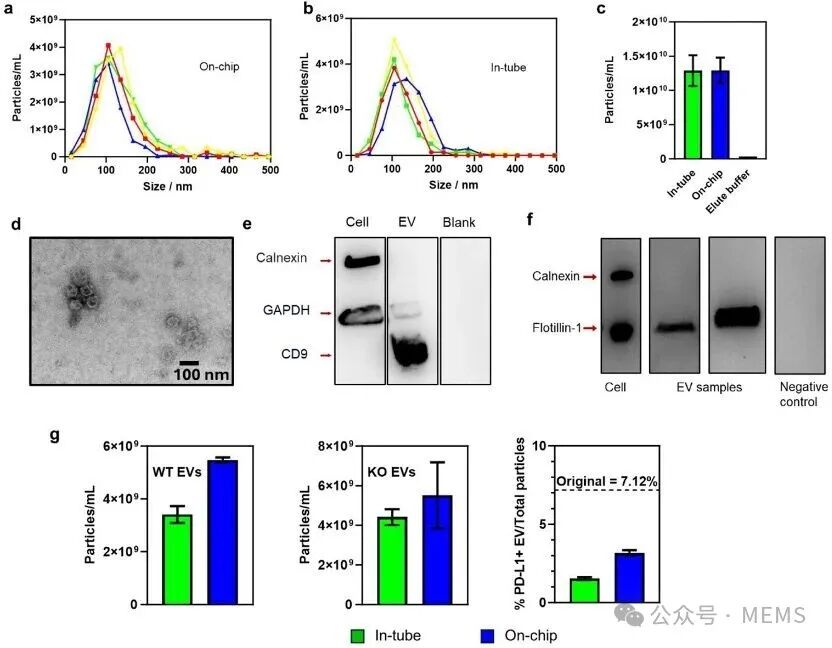
Characterization of extracellular vesicles
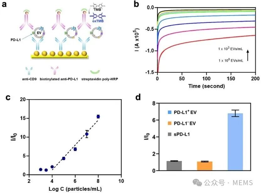
Detection of PD-L1+ extracellular vesicles using the designed electrochemical sensor
Conclusion
A key contribution of this work is the development and integration of a miniaturized electrochemical sensor into a digital microfluidic device to simplify and automate the extraction and detection of EVs on-chip. This proof-of-concept platform can perform magnetic microsphere functionalization and EV extraction from 20 µL of cell culture media and clinical plasma samples within 25 minutes, and the electrochemical sensor can detect as low as 10⁴ PD-L1+ EVs/mL within 5 minutes. Over time, these sensors have demonstrated the required specificity and stability. Compared to standard EV analysis, this portable system significantly reduces the demand for specific infrastructure, eliminating multiple time-consuming and labor-intensive processes, bringing hope for biomarker analysis based on EVs in biological research and clinical applications. Ultimately, the researchers envision that this platform could be transformed into a routine benchtop tool, making biomarker analysis based on EVs possible in life science laboratories and clinical settings.
Paper link:
https://doi.org/10.1002/smll.202504335
Further reading:
“Biosensor Technologies and Market for Instant Diagnostic Applications – 2022 Edition”
“Analysis of Apple’s Invention Patents and Industry Layout in Non-invasive Blood Glucose Monitoring”
“Patent Landscape Analysis of Raman Spectroscopy-based Blood Glucose Monitoring – 2024 Edition”
“Abbott FreeStyle Libre Continuous Glucose Monitoring Sensor Product Analysis”
“Diabetes Management Technologies and Market – 2025 Edition”
“Analysis of Apple’s Invention Patents and Industry Layout in Blood Pressure Monitoring”