Hello everyone, today I would like to introduce a paper on an engineered supramolecular fluorescent chemosensor for multiscale visualization of glutamate dynamics in living systems.
Glutamate (Glu) is the main excitatory neurotransmitter in the central nervous system and plays a crucial role in maintaining normal neural activity. Once the level of Glu in the central nervous system exceeds the normal range, it can trigger pathological conditions such as oxidative stress, depression, Alzheimer’s disease, and Parkinson’s disease. Therefore, achieving high-sensitivity specific recognition and accurate quantification of Glu, and tracking its dynamic changes in brain regions to establish related molecular networks, is crucial for understanding the potential mechanisms of physiological and pathological processes in the brain. However, Glu has a structure very similar to that of 19 other amino acids (especially aspartate), and its dynamic release process occurs on a millisecond timescale, which poses a significant challenge for developing probes that can achieve specific recognition and rapid response to Glu in the brain.
This paper designs and constructs the first fluorescent chemosensor for real-time imaging and quantitative analysis of neurotransmitters within organelles, using Glu as a model for study. The method is based on the indicator displacement detection (IDA) strategy based on host-guest interactions (Figure 1). Pillar[5]arene is a class of macrocyclic host molecules with unique rigidity and intrinsic symmetrical structures that can exhibit excellent binding capabilities with various guest molecules. The previously reported bifunctional fluorescent pillar[5]arene supramolecular probe (CN-DFP5) can recognize seven neurotransmitters, providing important inspiration for subsequent research. In this study, a pillar[5]arene (Tymp) with primary amine substituents was designed as the host molecule, and a series of guest molecules (Gn) with different spectral properties and binding sites were synthesized. These guests can complex with the electron-rich cavity of Tymp, forming a series of host-guest systems. Among the ten designed molecules, TympG2 exhibited the best binding energy and affinity, showing significantly higher specificity for Glu compared to other neurotransmitters and amino acids. This chemosensor achieved rapid ratio quantification of Glu (response time approximately 145 milliseconds) and can perform real-time imaging and quantitative analysis in multiple organelles such as the plasma membrane, cytoplasm, mitochondria, and lysosomes. Hypoxia stimulation significantly induced an increase in Glu levels in the plasma membrane, cytoplasm, and mitochondria, with the concentration in mitochondria increasing by 5.6 times within 8.5 seconds. Furthermore, two-photon imaging of brain tissue slices revealed that different brain regions exhibited regional dependence in their sensitivity to hypoxic Glu levels: SB1F, CA1, and LD subregions responded significantly stronger than CPU. Consistent with these results, in vivo fluorescence imaging of juvenile zebrafish also confirmed a significant increase in Glu induced by hypoxia. Furthermore, a high-density fluorescent fiber array was constructed to apply the developed chemosensor for synchronous imaging and biosensing of Glu in 24 brain regions. The results showed that the increase in Glu under hypoxic conditions was mainly concentrated in the cortex, hippocampus, and thalamus. More notably, the functional network mapping of these regions indicated that the correlation between adjacent brain regions in hypoxic mice, especially within the cortex-hippocampus-thalamus axis, was significantly weakened compared to the normoxic control group. This reduction in correlation suggests that hypoxia may lead to disruption of neural connectivity and function.
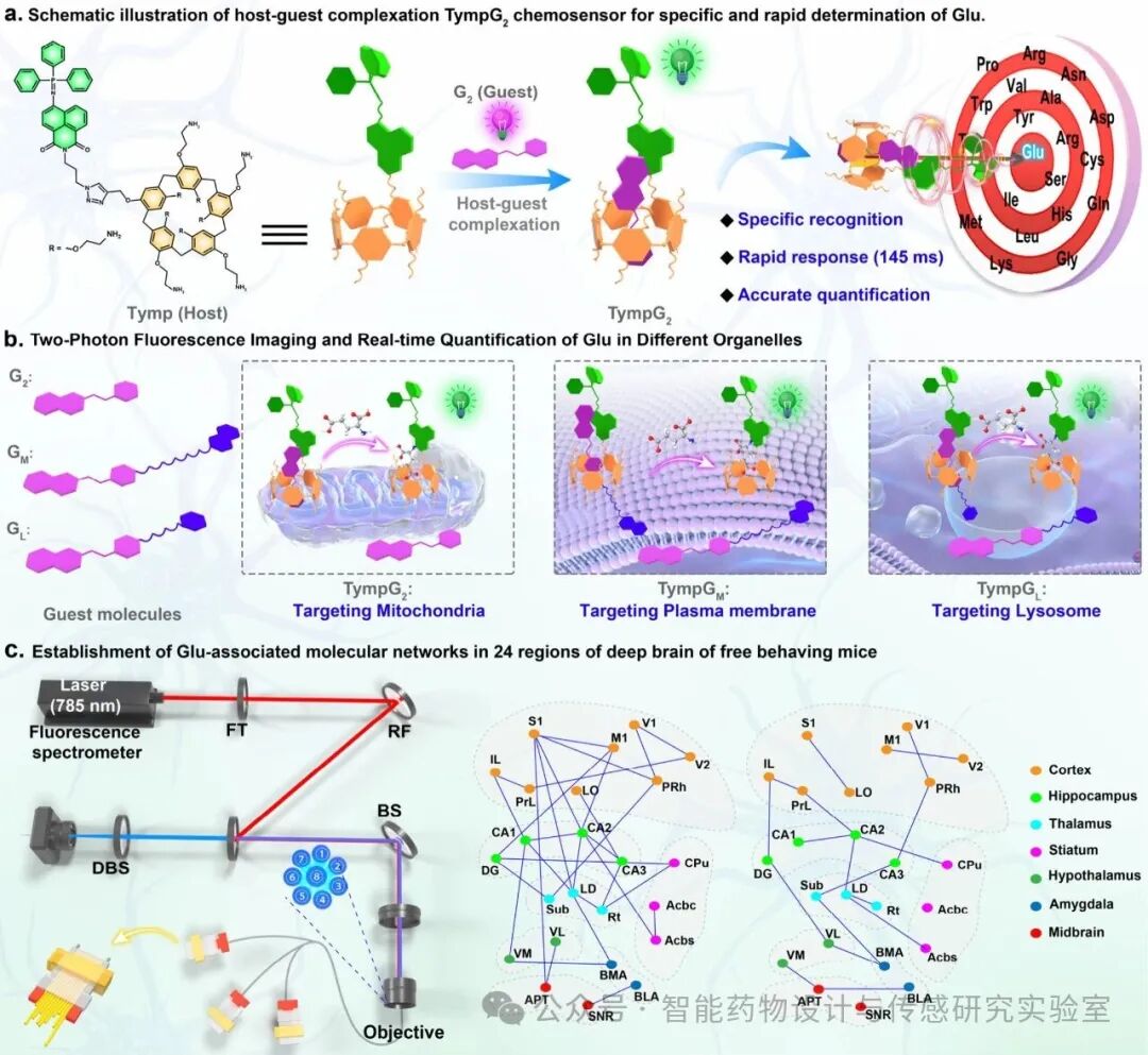
Figure 1. Host-guest type TympG2 chemosensor for in vitro and in vivo visualization and quantitative detection of glutamate.
This paper developed a two-photon ratio-type supramolecular chemosensor for rapid response quantitative recognition of Glu based on the fluorescence resonance energy transfer (FRET) strategy. As shown in Figure 2a, a functionalized pillar[5]arene (Tymp) with an electron-rich cavity was synthesized as the host molecule, with primary amine groups modified at both ends to act as hydrogen bond donors. Meanwhile, a naphthalimide substituted with triphenylphosphine was introduced on one of the branches of Tymp as the fluorescent group. A total of ten different guest molecules were synthesized, including pyridinium salt-derived dyes (G1–5) and heteroaromatic ring-derived coumarin dyes (G6–10). The terminal pyridinium or heteroaromatic ring groups of these guest molecules can form complexes with the electron-rich cavity of Tymp, resulting in a series of host-guest systems. The fluorescent guest molecules are divided into two categories: pyridinium salts (G1–G5) — acting as fluorescent acceptors, regulating the electrostatic interactions with Tymp by changing the electronegativity of the dye substituents (G1 > G2 > G3 > G4 > G5).
Heteroaromatic ring-derived coumarins (G6–G10) — acting as fluorescent donors, regulating the binding sites with Tymp by changing the size of the heteroaromatic rings (G6 > G7 > G8 > G9 > G10). This molecular design allows for precise regulation of host-guest interactions, optimizing the selectivity of the chemosensor in recognizing Glu against other structurally similar neurotransmitters and amino acids.
To evaluate the binding affinity of Tymp with guest molecules (G1–10), fluorescence titration experiments were conducted. As shown in Figure 2b, the host molecule Tymp exhibited a significant fluorescence emission peak at 525 nm (F525). The guest molecules G2–5 acted as fluorescent acceptors, emitting at 635 nm (F635); G1 acted as a fluorescent acceptor emitting at 700 nm (F700); while G6–10 acted as fluorescent donors, emitting at 460 nm (F460). Upon the addition of G1–5, the fluorescence intensity of F525 significantly decreased: among them, G1 induced a gradual increase in F700 fluorescence intensity, while G2–5 enhanced the emission of F635. Conversely, the addition of G6–10 led to a decrease in F460, while F525 increased). The binding ratios of Tymp with the ten guest molecules indicate a 1:1 stoichiometric ratio (Figure 2c, 2d).
The selectivity of the constructed host-guest chemosensor for Glu was evaluated. The results showed that TympG1 had the least response to Glu, while upon the addition of Glu, the fluorescence intensity of TympG2–5 at F525 and that of TympG6–10 at F460 underwent significant changes. Therefore, among all the developed host-guest systems, the TympG2 chemosensor exhibited the highest selectivity for specific recognition of Glu (Figure 2e).
To reveal the recognition mechanism of TympG2 for Glu, density functional theory (DFT) calculations were performed. The optimized structures of Tymp and its guest molecules showed that the methylpyridinium or imidazolium groups of the guest molecules (G1–5) are located at the center of the cavity of Tymp, while the coumarin fluorescent group is distributed at the upper edge of Tymp. As shown in Figure 2f, the negatively charged Tymp electron-rich cavity attracts positively charged guest molecules through electrostatic interactions. In contrast, the heteroaromatic ring-derived guest molecules (G6–10) form host-guest complexes with Tymp through hydrogen bonding.
Subsequently, the designed chemosensor’s kinetic response to Glu was evaluated using a rapid mixing stopped-flow technique (mixing time less than approximately 8 ms). As shown in Figure 2g, upon the addition of Glu, the fluorescence intensity of TympGn (n = 1–5) at F525 or that of TympGn (n = 6–10) at F460 immediately increased and reached a plateau at different times. The order of response times was: TympG10 (~55 ms) ≈ TympG9 (~55 ms) < TympG8 (~65 ms) < TympG7 (~70 ms) < TympG6 (~85 ms) < TympG5 (~90 ms) < TympG4 (~105 ms) < TympG3 (~120 ms) < TympG2 (~145 ms). Notably, TympG1 showed almost no response to Glu. The reaction rate constants (kobs) of the nine host-guest chemosensors were also determined and summarized in Figure 2h, ranging from 1.30 × 10⁻² ms⁻¹ to 2.44 × 10⁻² ms⁻¹, showing a trend consistent with the response times. Further comparison revealed that the stronger the host-guest binding affinity, the slower the reaction rate. This result is consistent with the DFT calculations, further demonstrating the role of host-guest interactions in regulating kinetics. Although TympG2 has a slightly slower response speed to Glu, it exhibits the highest selectivity for recognizing Glu, making it the optimal supramolecular fluorescent chemosensor for subsequent in vivo imaging and sensing studies.
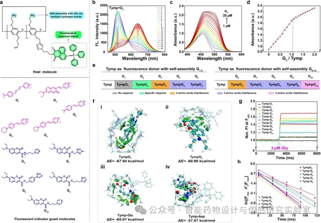
Figure 2 Design and synthesis of the host-guest type TympGn chemosensor for selective and rapid response to glutamate.
After establishing the optimized chemosensor platform, the photophysical properties of TympG2 in the detection of Glu were characterized. As shown in Figure 3a, as the concentration of Glu increased, the intensity of the F635 channel (collection range 590–700 nm) rapidly decreased, while the intensity of the F525 channel (collection range 490–570 nm) significantly increased. The ratio of F525/F635 showed a good linear correlation with Glu concentrations in the range of 0.1–14.0 μM (R² = 0.997). The detection limit was determined to be 32.8 ± 1.5 nM (n = 10, S.D., Figure 3b). In summary, these results fully demonstrate that TympG2 possesses good sensitivity, stability, and long-term applicability for long-term use in living systems. To explore the recognition mechanism of TympG2 for Glu, high-resolution mass spectrometry (HR-MS) and nuclear magnetic resonance (NMR) spectroscopy were utilized to study the host-guest binding and its stoichiometry. Based on the above results, it is believed that the branched amine groups of Tymp can interact with the carboxyl groups of Glu through hydrogen bonding, thereby facilitating the replacement of Glu in the TympG2 complex by G2. Due to the lower binding energy of Glu, this process can be smoothly achieved.
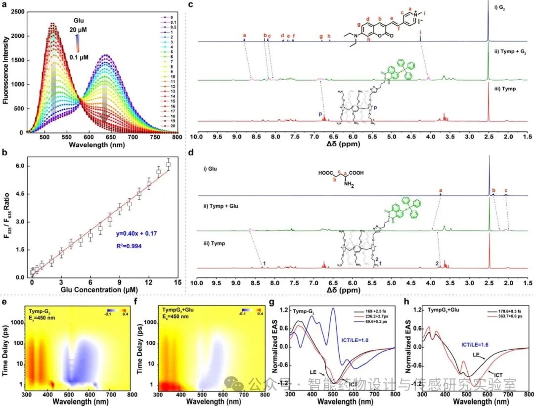
Figure 3 Fluorescence titration experiments and mechanism evaluation of the TympG2 chemical sensor for glutamate.
To achieve multi-organelle imaging, three fluorescent guest molecules with different organelle-targeting functions were designed and synthesized (Figure 4a). Among them, the positively charged pyridinium group in the G2 molecule endows the chemosensor with good water solubility and mitochondrial targeting ability; the terminal morpholine group in the GM molecule enhances the targeting effect towards lysosomes; while the long-chain naphthalene ring introduced at the terminal of the GL molecule anchors to the cell membrane by embedding into the phospholipid bilayer.
In the experiments, neurons were co-cultured with TympG2, TympGM, TympGL, and TympGC along with commercial targeting probes (MitoLite, Cell-Tracker CMDiR, Lyso-Tracker Blue, and Cell-Tracker Blue) for co-localization imaging. The results showed (Figure 4b), that the fluorescence signals of TympG2, TympGM, TympGL, and TympGC in the F635 channel highly overlapped with those of the commercial probes, with Pearson correlation coefficients of 0.96, 0.97, 0.94, and 0.95.
This paper further utilized the constructed chemosensor to study the real-time response dynamics of Glu in the plasma membrane, cytoplasm, mitochondria, and lysosomes under external stimulation. First, baseline levels were quantified: the concentrations of Glu in the plasma membrane, cytoplasm, mitochondria, and lysosomes were 0.20 ± 0.04 μM, 0.54 ± 0.03 μM, 1.16 ± 0.08 μM, and 86.2 ± 4.5 nM (Figure 4c). A large number of experimental and clinical evidence has shown that the dysregulation of Glu homeostasis is closely related to the pathological progression of ischemic/hypoxic injury. Subsequently, the responses of TympG2, TympGM, TympGL, and TympGC under hypoxic stimulation were detected. The results showed that as the oxygen (O₂) concentration decreased, the concentrations of Glu in the plasma membrane, cytoplasm, and mitochondria significantly increased, while the concentration of Glu in lysosomes remained almost unchanged. When the O₂ concentration dropped to 1%, the concentrations of Glu in the plasma membrane, cytoplasm, and mitochondria increased to 1.17 ± 0.03 μM, 1.55 ± 0.05 μM, and 5.96 ± 0.18 μM. In further evaluating the dynamic process under 1% O₂ stimulation, this paper found significant differences in Glu concentrations among the three organelles (Figure 4d–j).
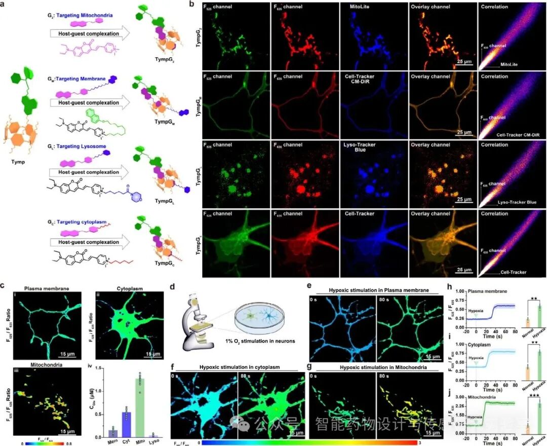
Figure 4 Two-photon fluorescence imaging and real-time quantitative analysis of glutamate in different organelles of neurons.
Thanks to the two-photon performance of the TympG2 probe, this paper further applied it to imaging studies in brain tissues and zebrafish. Two-photon fluorescence images showed significantly deeper penetration depth (over 660 micrometers, excitation wavelength of 780 nanometers), while single-photon images showed shallower penetration depth (less than 360 micrometers, excitation wavelength of 488 nanometers). Considering the functions of the hippocampus, cerebral cortex, and thalamus are closely related to glutamate, this paper further quantified the glutamate concentrations in these brain regions through two-photon imaging. As shown in Figures 5a-f, the normal glutamate concentrations in the four brain regions were 1.28 ± 0.05 μM (S1BF), 1.62 ± 0.08 μM (CA1), 0.55 ± 0.03 μM (LD), and 0.33 ± 0.01 μM (CPU). Interestingly, under hypoxic stimulation, the glutamate levels in the SB1F, CA1, and LD regions significantly increased to 5.98 ± 0.18 μM, 3.66 ± 0.12 μM, and 2.97 ± 0.
To achieve monitoring of Glu in freely moving individuals, this paper developed a multi-channel optical fiber fluorescence spectroscopy platform (Figure 5k). In the experiment, 24 optical fibers were implanted into three anterior regions of the unilateral hemisphere of the brains of three mice, distributed at different locations to reduce randomness in data collection and extend the fiber array to a larger range of brain networks (Figure 5l). The optical field distribution volume is approximately 5.92 × 10⁻³ mm³, extending about 200 μm from the conical fiber end face. The selected brain regions belong to different neurobiological networks, specifically including: medial prefrontal cortex (IL), prefrontal cortex (PrL), lateral orbital frontal cortex (LO), primary motor cortex (M1), primary somatosensory cortex (S1), piriform cortex (PRh), visual cortex (V1), secondary visual cortex (V2), dentate gyrus (DG), hippocampus CA1 region, CA2 region, CA3 region, subthalamic nucleus (Sub), lateral dorsal thalamic nucleus (LD), thalamic reticular nucleus (Rt), caudate putamen (CPU), nucleus accumbens core (Acbc), nucleus accumbens shell (Acbs), ventromedial thalamic nucleus (VM), ventrolateral thalamic nucleus (VL), medial basolateral amygdala (BMA), lateral basolateral amygdala (BLA), anterior pretectal nucleus (APT), and substantia nigra reticular part (SNR). Through three-dimensional (3D) reconstruction, the positions of the fiber endpoints and the labeled neurons in front were analyzed, verifying the precise localization of the constructed fiber array in the target brain regions (Figure 5m–o).
Glutamate, as the main excitatory neurotransmitter, mediates critical neurophysiological responses under hypoxic stimulation. Utilizing the developed multi-channel optical fiber fluorescence spectroscopy platform, this paper detected the changes in Glu levels after injecting the TympG2 probe into 24 specific brain regions of mice undergoing hypoxic stimulation. The results showed (Figure 5p), that in the cortex, hippocampus, and thalamus regions, the Glu levels significantly increased under hypoxic stimulation, while no significant changes were observed in the remaining 18 brain regions. This indicates that hypoxia primarily induces an increase in Glu concentrations in the cortex, hippocampus, and thalamus regions. Subsequently, this paper compared the distribution of Glu activity in 24 brain regions between normal and hypoxic mice (1% O₂ stimulation). The results showed that Glu activity exhibited regional heterogeneity, with the hippocampus and striatum regions showing the most significant activity. In hypoxic mice, the activity of Glu in the cortex (IL, PrL, LO, M1, S1, PRh, V1, V2), hippocampus (CA1, CA2, CA3, DG) and thalamus (LD, Sub, Rt) regions increased by approximately 3.65 times, 2.17 times, and 3.22 times, respectively, while no significant changes were observed in other brain regions. Furthermore, this paper independently analyzed the cross-regional functional networks related to Glu in normal and hypoxic mice. Based on the calculation of Pearson correlation coefficients (r), the results showed that in the brains of normal mice, there were 18 positive correlations and 14 negative correlations (Figure 5q); while in hypoxic mice, the positive and negative correlations decreased to 10 and 8 (Figure 5r). These results indicate that hypoxia impairs the cross-regional correlation of Glu levels in the brain, especially between cortical and non-cortical regions (such as cortex-hippocampus, cortex-lateral dorsal thalamic nucleus).
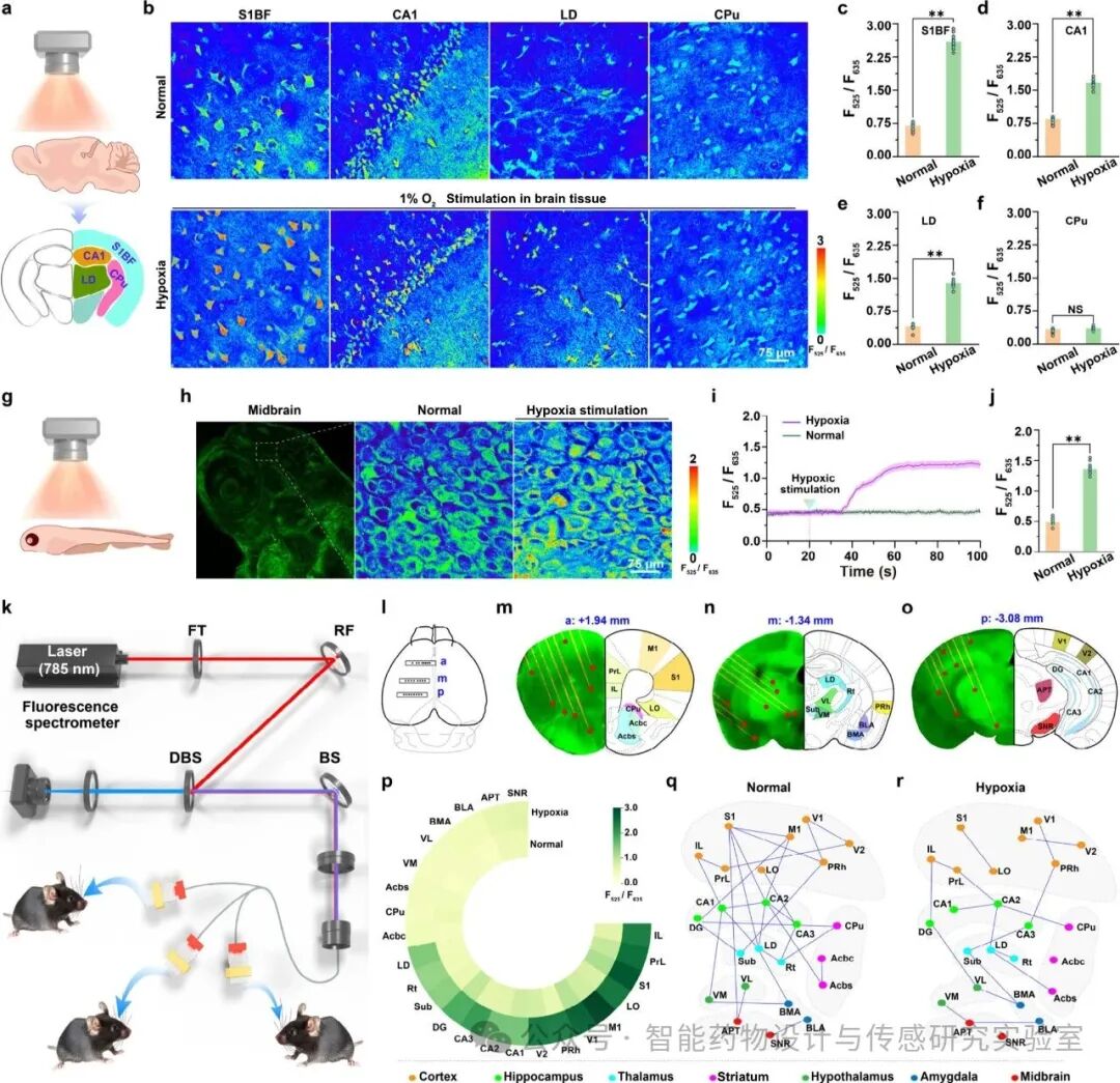
Figure 5 Real-time quantitative analysis of glutamate (Glu) in brain tissues, zebrafish, and freely moving mice using two-photon fluorescence imaging technology.
In this study, based on host-guest interactions and using the indicator displacement method (IDA) strategy, a series of Glu sensors (TympGn) were designed and validated. Among them, the optimized TympG2 can monitor Glu activity in vivo and in vitro, possessing high temporal resolution, high affinity, high sensitivity, and cell-type specificity. With its two-photon performance, TympG2 can track the dynamics of Glu in brain slices and zebrafish under hypoxic stimulation. More importantly, this study first constructed a Glu-related molecular network in 24 regions of the deep brain of freely moving mice. The TympG2 sensor demonstrated highly specific recognition of Glu, effectively distinguishing it from other neurotransmitters and amino acids, and achieving high precision in ratio quantification with a response time of approximately 145 ms. Compared to existing gene-encoded indicators, TympG2 shows significant advantages: its F525/F635 signal-to-noise ratio reaches 6.09, which is 1.35 times higher than that of iGlusnFr. In addition, the dual-channel signal-to-noise ratio and built-in calibration mechanism of TympG2 effectively avoid interference from the environment, light, and probe concentration, thus achieving precise quantification of Glu in vivo. Superior to reported gene-encoded indicators, the developed chemosensor can not only image and detect Glu in the plasma membrane but also extend to the cytoplasm and mitochondria. Furthermore, this paper successfully applied this probe to achieve real-time monitoring and concentration quantification of Glu dynamics in the plasma membrane, cytoplasm, and mitochondria under hypoxic stimulation for the first time. Meanwhile, this sensor was also successfully applied to the dynamic detection of Glu in brain tissues and zebrafish, and to analyze the Glu molecular network in the deep brain regions of freely moving mice. The study found that under hypoxic conditions, the correlation between adjacent brain regions significantly decreased, especially between the cortex, hippocampus, and thalamus, with more pronounced differences compared to normal mice.
In summary, the TympG2 sensor can achieve dynamic spatiotemporal precision measurement of Glu in various model organisms and is suitable for research under complex behavioral contexts. This study provides a new approach for real-time monitoring of Glu concentrations, distributions, and dynamic changes from organelles to the whole brain, thereby contributing to a deeper understanding of brain physiological and pathological processes related to Glu. At the same time, this research also provides new methodological references for designing and synthesizing chemical sensors capable of specific and highly sensitive detection of neurotransmitters, amino acids, and proteins in organelles, tissues, and the brains of freely moving animals.
References: Mei Y, Sun J, Liu Z, Zhao Y, Zhang Q, Tian Y. An Engineered Supramolecular Fluorescent Chemosensor for Multiscale Visualization of Glutamate Dynamics in Living Systems. J Am Chem Soc. Published online September 3, 2025. doi:10.1021/jacs.5c11915
Research Group Introduction: Professor Tian Yang, Dean of the School of Chemistry and Molecular Engineering at East China Normal University. He is dedicated to the research of in vivo brain and intracellular chemical signaling molecular bioimaging and in situ sensing, making systematic innovative research contributions in the qualitative and quantitative analysis of brain neurochemical substances, rapid and sensitive imaging of freely moving animal brains, etc. He has received funding from the National Outstanding Youth Fund and has been selected for the National Hundred-Thousand-Ten Thousand Talents Project, the Ministry of Education New Century Talent Program, Shanghai Excellent Academic Leaders, Shanghai Leading Talents, and the Baosteel Excellent Teacher Award.In 2015, he received the Female Analytical Chemist Award from the Chinese Chemical Society; In 2018, he received the First Prize of Shanghai Natural Science. To date, he has published over 170 papers, including Nat. Commun., Sci. Adv., JACS, Angew. Chem., Adv. Mater. , Sci. Adv., with over ten thousand citations. He currently serves as the associate editor of Chem. Commun. journal.