Expert Guide
—— Department of Dermatology, Peking University People’s Hospital
Professor Zhang Jianzhong
Case Summary
The patient is a male adolescent with skin features including cerebriform connective tissue nevi, epidermal nevi, venous and capillary malformations, lipomas, and a mutation in AKT1 causing c.49G > A, resulting in 17 amino acids being changed from glutamic acid to lysine, clinically diagnosed as deformation syndrome. He underwent acne assessment at the age of 15 (Figure 1), where acne began to develop from the face and gradually spread to the upper left part of the back. Previous treatments included topical retinoids, benzoyl peroxide, clindamycin, tazarotene, and oral antibiotics, but with little effect.
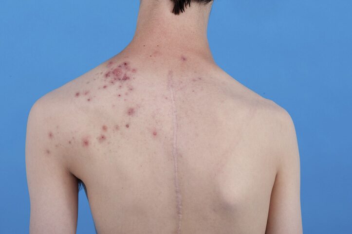
Figure 1. At age 15, acne primarily occurred in the upper left part of the back
Physical Examination Information
At the age of 17, the physical examination showed: scars on the face, chest, and upper back, scattered comedones and erythematous papules. In addition, there were large inflammatory papules, pustules, and nodules aggregated along the Blaschko lines on the upper left part of the back and left side of the neck (Figure 2).
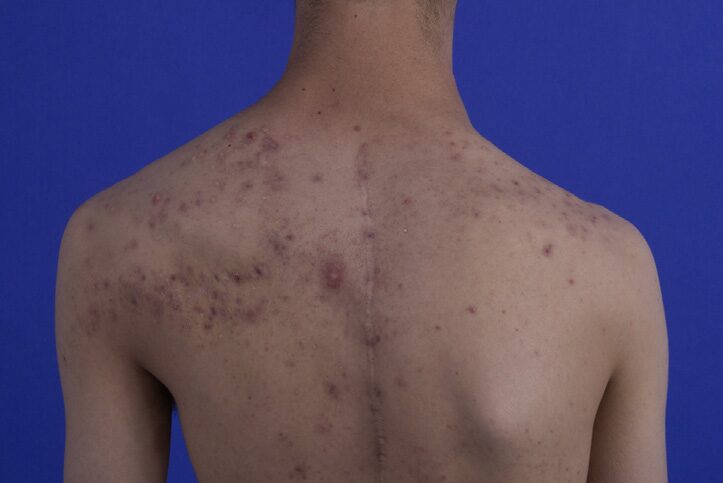
Figure 2. At age 17, acne on the back, more densely distributed on the left side
Clinical photos from ages 11 to 13 revealed no significant skin changes in the area prior to the onset of acne. The patient reported that the hair on the left side grew faster than on the right side. There was left-sided lumbar scoliosis, overgrowth of the left maxilla, capillary malformations on the left side of the trunk, and varicose veins in the legs, with the left leg being more prominent. Cerebriform connective tissue nevi were observed on both feet, and an epidermal nevus was present on the left heel (Figure 3).

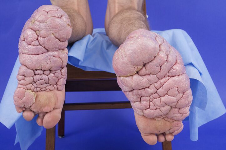
Figure 3. At age 17, cerebriform connective tissue nevi appeared on the soles of the feet
At the age of 19, the patient’s physical examination showed: persistent acne on the back (Figure 4), with slight improvement after oral doxycycline, but significant scarring was present. The patient subsequently underwent photodynamic therapy, but the linear comedones and disseminated lesions persisted.
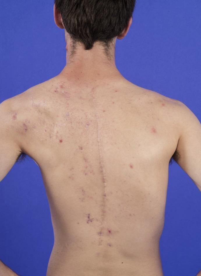
Figure 4. At age 19, acne on the back, with significant scarring in the upper left part
Case Discussion
Several mosaic diseases in dermatology include narrow Blaschko lines, broad Blaschko lines, checkerboard patterns, leaf-like patterns, and patchy patterns without midline separation. The patient’s acne lesions included typical distribution on the face and back, with more severe lesions in the upper left part of the back, corresponding to several narrow Blaschko lines. These areas did not show acne or other skin changes at birth or in early childhood. The authors hypothesize that the severe acne along Blaschko lines in this patient represents a mosaic phenomenon of deformation syndrome.
The most prominent Blaschko-like acne lesions in this patient occurred in the upper left part of the back. Several mosaic manifestations of deformation syndrome were also predominantly left-sided, including epidermal nevi, vascular malformations, maxillary overgrowth, and facial hypertrichosis. Blaschko pigmentation was significantly distributed on the left side, indicating that the skin areas with severe acne may consist of cells influenced by activated AKT1 variants. In deformation syndrome, the type of skin damage seems to partially depend on the types of cells affected by AKT1 variants. In epidermal nevi, AKT1 variants are primarily present in keratinocytes, while in cerebriform connective tissue nevi, fibroblasts are affected. It is possible that the hair follicles in the Blaschko-like acne regions are rich in cells influenced by AKT1 variants.
In this patient, the acne distributed along Blaschko lines that appeared during puberty occurred in silent areas of clinical skin mosaic manifestations. Unilateral Blaschko acne lesions began in puberty, covering areas of hypopigmentation, which have been reported to be caused by pathogenic variants of FGFR2, believed to represent mosaic Apert syndrome.
It has been reported that a 13-year-old male adolescent had Blaschko-like acne superimposed on epidermal nevi. This patient did not have deformation syndrome, but interestingly, AKT1 variants were detected in similarly affected individuals. In the patient described in this article, more severe Blaschko acne manifestations began in adolescence, possibly due to the emergence of factors leading to acne, which may not be related to AKT1 variants.
Acne is caused by an increase in sebaceous secretion, keratinization changes, inflammation, and colonization of Propionibacterium acnes in hair follicles due to androgens. Interestingly, in this patient with deformation syndrome, there was no increased hair growth in the acne lesion area on the back. Hypertrichosis is also androgen-mediated, which has been shown to distribute primarily along Blaschko lines in Proteus syndrome. The patient indeed had hypertrichosis and acne on the left side of the face, but not on the back.
Acne is ineffective against various conventional topical or systemic medications. The authors believe that AKT inhibitors such as miransertib may have a role in reducing cerebriform connective tissue nevi and could be beneficial for Blaschko-like acne in Proteus syndrome, but this remains to be studied.
Conclusion
Localized acne distributed along Blaschko lines may represent a rare mosaic phenotype of Proteus syndrome. The Blaschko-like acne in Proteus syndrome may be due to the mosaic phenomenon affecting hair follicles and sebaceous glands. Since many acne patients receive treatment during puberty, the damage may not be obvious. So far, no better therapies have been found.
Expert Introduction: Professor Zhang Jianzhong

Professor Zhang Jianzhong, Director of the Department of Dermatology, Peking University People’s Hospital, 13th Chairman of the Dermatology and Venereology Branch of the Chinese Medical Association, Chairman of the Dermatology and Venereology Branch of the Chinese Rehabilitation Medical Association, Council Member of the Asian Dermatology Association, Council Member of the International Society for Atopic Dermatitis, Vice President of the Chinese Association of Dermatologists, Vice President of the Dermatology and Venereology Branch of the Chinese Medical Association, Vice President of the Plastic Surgery and Regenerative Medicine Branch of the Chinese Association of Plastic Surgery, Chief Expert of the Atopic Dermatitis (Eczema) Research Center of the Dermatology and Venereology Branch of the Chinese Medical Association, and Head of the Hair Research Group.
He serves as Associate Editor for journals such as the Chinese Journal of Dermatology, Clinical Dermatology, and is on the editorial board of journals including the Journal of the American Academy of Dermatology, Chinese Medical Journal, and SKINmed. He has conducted in-depth research on atopic dermatitis, psoriasis, and hair diseases. He has organized the formulation of various guidelines for the diagnosis and treatment of skin diseases in China, served as the chief editor of the first long-term education textbook “Dermatology and Venereology” planned by the National Health Commission, led the development of a new Class 1 drug, benvetomide, and has led dozens of clinical trials for skin disease drugs, published over 500 papers, and edited and contributed to more than 50 works. He has trained more than 50 graduate students and has received numerous awards including the Chinese Medical Association Award, the International Society of Dermatology Outstanding Contribution Award, and the National Science and Technology Progress Second Prize, and was awarded the title of “National Famous Doctor” in 2018.
Original Literature:
Cartron A M, Pithadia D J, Buser A, et al. Acne Following Blaschko’s Lines in Proteus Syndrome[J]. JAAD Case Reports, 2020.
References:
[1]Biesecker LG, Sapp JC. Proteus syndrome. Gene Rev. 2019. In:Adam MP, Ardinger HH, Pagon RA, et al., eds. Gene Reviews? [Internet]. Seattle, WA: University of Washington, Seattle;1993-2020. Available at: https://www.ncbi.nlm.nih.gov/books/NBK99495/. Accessed September 13, 2020.
[2]Nguyen D, Turner JT, Olsen C, Biesecker LG, Darling TN. Cutaneous manifestations of Proteus syndrome: correlations with general clinical severity. Arch Dermatol. 2004;140(8):947-953.
[3]Happle R, Steijlen PM, Theile U, et al. Patchy dermal hypoplasia as a characteristic feature of Proteus syndrome. Arch Dermatol. 1997;133(1):77-80.
[4]Pithadia DJ, Roman JW, Sapp JC, Biesecker LG, Darling TN. Hypertrichotic patches as a mosaic manifestation of Proteus syndrome. J Am Acad Dermatol. https://doi.org/10.1016/j.jaad.2020.01.078.
[5]Sapp JC, Buser A, Burton-Akright J, Keppler-Noreuil KM, Biesecker LG. A dyadic genotypeephenotype approach to diagnostic criteria for Proteus syndrome. Am J Med Genet C Semin Med Genet. 2019;181(4):565-570.
[6]Happle R. Mosaicism in human skin. Understanding the patterns and mechanisms. Arch Dermatol. 1993;129(11):1460-1470.
[7]Lindhurst MJ, Wang J-A, Bloomhardt HM, et al. AKT1 gene mutation levels are correlated with the type of dermatologic lesions in patients with Proteus syndrome. J Invest Dermatol. 2014;134(2):543-546.
[8]Kiritsi D, Lorente AI, Happle R, Bernabeu Wittel J, Has C. Blaschko line acne on pre-existent hypomelanosis reflecting a mosaic FGFR2 mutation. Br J Dermatol. 2015;172(4):1125-1127.
[9]Hivnor CM, Yan AC, Honig PJ. Acne arising in an epidermal nevus. Pediatr Dermatol. 2007;24(5):534-535.
[10]Keppler-Noreuil KM, Sapp JC, Lindhurst MJ, et al. Pharmaco-dynamic study of miransertib in individuals with Proteus syndrome. Am J Hum Genet. 2019;104(3):484-491.
This Issue’s Learning Interaction
Cover and article images source: Original literature
Translation:mocovi, Lee
Editor: Lay
Note: This article is an original article of the platform and cannot be reproduced without authorization. Reminder: The content of this article is for academic exchange only and is intended for reading by medical professionals.
Recommended Reading

Empowering Dermatologists’ Value to Shine
Long-term collection of professional submissions in the field of dermatology
Submission/Reprint inquiries contact