A 60-year-old female with cognitive developmental delay presented with seizures that began in childhood and skin abnormalities affecting the left side of her face and eyes. Physical examination revealed alopecia, a lipomatous nevus (naevus psiloliparus), subcutaneous fat accumulation, and exophthalmos (Figure 1). Brain MRI showed intracranial lipomatosis and left-sided hemimegalencephaly (Figure 2), confirming the diagnosis of encephalocraniocutaneous lipomatosis (ECCL).
ECCL, also known as Haberland or Fishman syndrome, is a rare congenital neurocutaneous disorder characterized by ectodermal dysplasia with unilateral involvement of the skin, eyes, and brain. Neuroimaging features include intracranial lipomatosis, meningeal enhancement, polymicrogyria, and lateral ventricular enlargement. Differential diagnoses include sebaceous naevus syndrome, Proteus syndrome, oculocerebrocutaneous syndrome, Sturge-Weber syndrome, and hemimegalencephaly.
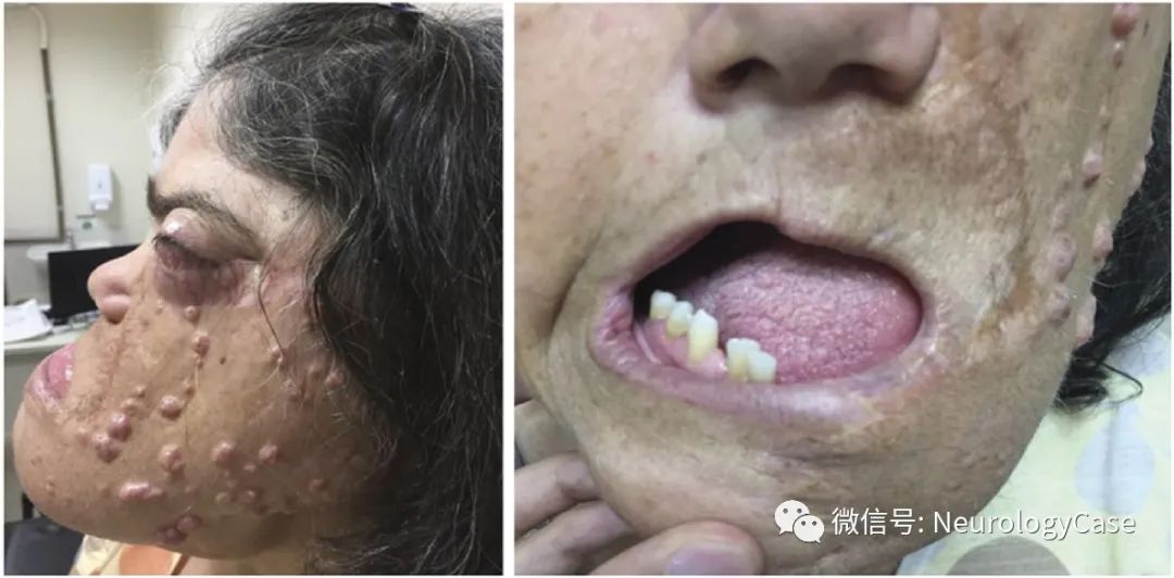
(Figure 1: Patients with this syndrome present with skin tumors, lipomatous nevi, large and protruding soft tissue masses [lipomas] on the scalp, alopecia, exophthalmos, and mandibular tumors leading to facial asymmetry.)
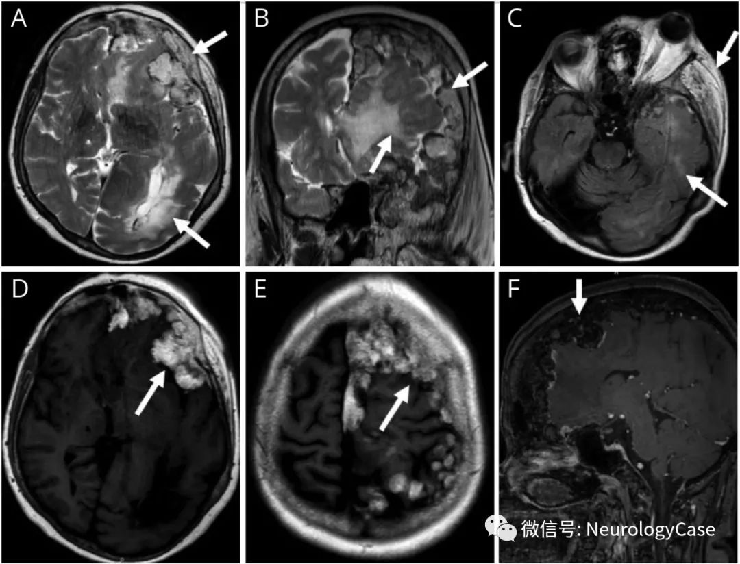
(Figure 2: Axial and coronal T2WI [A-B], axial T2/FLAIR [C], T1WI [D, E], and fat-suppressed sagittal T1WI show a large lipomatous mass in the left temporal region [A, C]; the image also shows infiltration extending into the temporal muscle and retro-orbital fat; intracranial lipomatosis in the left frontal-parietal region [B, D, E] extends to the convex surface; loss of fat suppression signal indicates its lipomatous nature; [F] left hemisphere enlargement and white matter signal changes characteristic of hemimegalencephaly [A-C].)
[References]
de Moraes MPM, Ferreira de Abrantes F, Tonholo Silva TY, Pedroso JL, Marussi VHR, Meneses A, Barsottini OGP. Clinical and Neuroimaging Features of Encephalocraniocutaneous Lipomatosis. Neurology. 2021 Oct 19;97(16):785-786.