Blood Routine
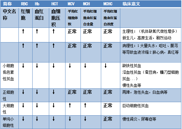
 Blood routine is abbreviated as blood Rt. Rt stands for Routine Test. The history of blood routine testing is relatively short; in 1953, W.H. Coulter invented the world’s first automatic blood cell counter using the principle of electrical impedance. In 1962, the first blood cell counter in China was successfully developed in Shanghai, and in the 1980s, white blood cell classification was developed.The blood picture = blood routine = blood cell analysis, checking a blood picture at the hospital means checking a blood routine. In colloquial terms, a high blood picture specifically refers to a high white blood cell count. (Differences in terminology across different eras and specialties)Why can blood routine be a routine check?Because its significance is broad and substantial. Blood circulates throughout the body, and most diseases leave traces in the blood.By observing the white blood cells (WBC) responsible for fighting and maintaining stability, one can determine if there is an invasion by external enemies and the severity of the threat. Observing red blood cells (RBC) can reveal nutritional status, while observing platelets (PLT) can indicate issues with blood coagulation and hematopoiesis.The main reasons are as follows:1. To assess whether there is inflammation and its severity (most diseases in the body are associated with inflammation, which is a defensive reaction of the body to stimuli). It can also determine if there is an allergic reaction through eosinophils and basophils.2. To determine if there is an infection and its type (bacterial or viral?)3. To assess nutritional status (by evaluating red blood cells and hemoglobin to determine the type and degree of anemia, as well as the cause of blood loss).4. To evaluate bone marrow hematopoiesis (by assessing white blood cells, red blood cells, and platelets to determine if hematopoiesis is healthy, and to preliminarily assess the body’s coagulation status through platelets).1. Red Blood Cells
 MCV, MCH, MCHC reference range
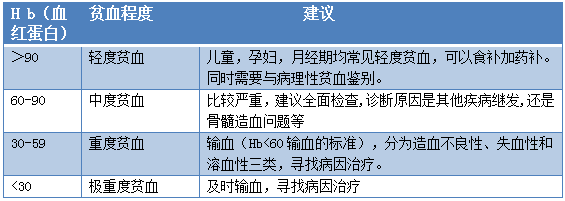
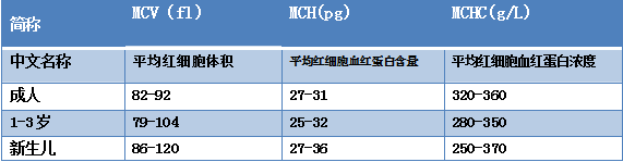 MCV, MCH, and MCHC are calculated from (RBC Hb HCT data).MCV = RBC (HCT) (Understanding: Assume there is one liter of whole blood. HCT is the volume of red blood cells in one liter of whole blood, and RBC is the number of red blood cells in one liter, so the ratio of the two is the volume of a red blood cell. The unit is fL, which is 10^-15L). MCH = RBC (Hb) (Understanding: Assume there is one liter of whole blood. Hb is the weight of hemoglobin in one liter of whole blood, and RBC is the number of red blood cells in one liter, so the ratio is the weight of a red blood cell. The unit is pg, which is 10^-12g). MCHC = HCT (Hb) (Understanding: Assume there is one liter of whole blood. Hb is the weight of hemoglobin in one liter of whole blood, and HCT is the volume of red blood cells in one liter, so the ratio is the weight of red blood cells in one liter. The unit is g/L).Thus, MCHC is also equal to MCV (MCH) (RBC (Hb) divided by RBC (HCT)) (Understanding: the weight of a red blood cell compared to its volume, i.e., the weight of red blood cells in a unit volume). In macrocytic anemia, the volume of red blood cells increases (MCV), and the weight of the cells also increases, so MCHC remains normal.Grading of Anemia Severity:
 Reference Ranges for Red Blood Cell Parameters: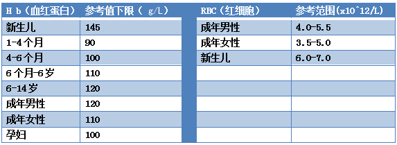
Data reference from the 8th edition of Internal Medicine, People’s Health Publishing House
2nd edition of Clinical Laboratory Basics, People’s Health Publishing House
It is also noted that there is another standard for anemia: for children aged 6-11 years, hemoglobin concentration below 115 g/L; for children aged 12-14 years, hemoglobin concentration below 120 g/L; for newborns, 170-200 g/L.2. White Blood CellsCBC:Complete Blood Count (CBC), also known as blood routine, blood picture, blood cell analysis, etc.In CBC mode, white blood cells are not classified but only the total count of white blood cells is measured.White Blood Cell Tripartite Group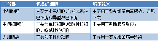 Common questions: Why is the largest cell group neutrophils instead of the largest monocytes?Answer: Because the blood cell analyzer counts dehydrated white blood cells, and neutrophils are the largest when dehydrated.White Blood Cell Five Classification
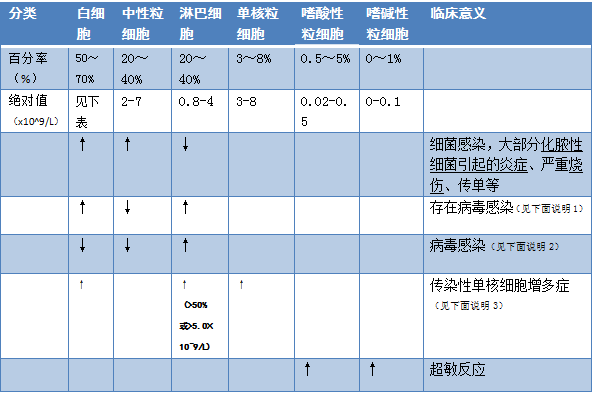 Explanation 1:Generally, in viral infections, white blood cells decrease. Why do lymphocytes increase while white blood cells increase in viral infections?Most viral infections present with normal or decreased white blood cell counts, but some viral infections can cause white blood cell counts to increase, such as EB virus infections (causing infectious mononucleosis), and viral encephalitis, which can both lead to elevated white blood cell counts. In cases of viral infections combined with bacterial infections, white blood cell counts may also increase.Explanation 2:What are the reasons for low white blood cells and high lymphocytes?Viral infections (measles, chickenpox, mumps, viral hepatitis, hemorrhagic fever, and also seen in whooping cough, tuberculosis, brucellosis, syphilis, etc.), severe infections, certain blood diseases (aplastic anemia, non-leukemic leukemia, granulocyte deficiency, benzene poisoning… that suppress hematopoiesis), factors damaging blood cells (typhoid, paratyphoid, black fever, malaria, X-ray and radium exposure, tumor chemotherapy) [Viral invasion of white blood cells → hence viral infections also belong to factors damaging blood cells], transplant rejection reactions.Explanation 3: A simple increase in monocytes accompanied by cold symptoms is most commonly due to viral infections, but it may also result from a relative increase in monocytes due to a decrease in neutrophils and lymphocytes.Reference Range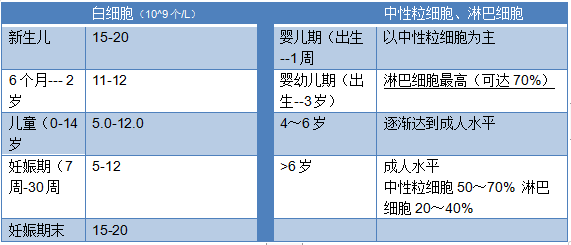
(Rule: The upper limit of white blood cells for newborns is 20. For those under 14 years, the upper limit is 12. It may vary depending on the specific instrument used.)
3. PlateletsIncreased ↑:Reactive thrombocytosis (acute suppurative infections, trauma, non-infectious inflammation… various stress responses).Thrombocytosis, chronic myeloid leukemia, polycythemia… Increased due to testing factors: small red blood cells being misidentified as platelets by the instrument, leading to elevated platelet counts (common in microcytic hypochromic anemia, such as iron deficiency anemia).Decreased ↓:Excessive destruction or consumption of platelets → primary thrombocytopenic purpura, splenomegaly, SLE, DIC.Hematopoietic dysfunction → acute leukemia, aplastic anemia.Note: Colds can cause low platelet counts.Possible Causes:1. Sudden thrombocytopenic purpura. Generally caused by viral infections leading to decreased platelets. If the platelet count is not too low, observation can be done before taking further action; once the viral infection resolves, platelets may return to normal. Most of these patients are children under 10 years old, with about 80% of them spontaneously resolving within 6 months.2. Drug-induced thrombocytopenia (DITP). Antibiotics (chloramphenicol, penicillin, cephalosporins, fluoroquinolones, sulfonamides, etc.), antipyretic analgesics (ibuprofen, sodium salicylate, butazolidin, acetaminophen, aspirin, etc.), and other medications.
CRP
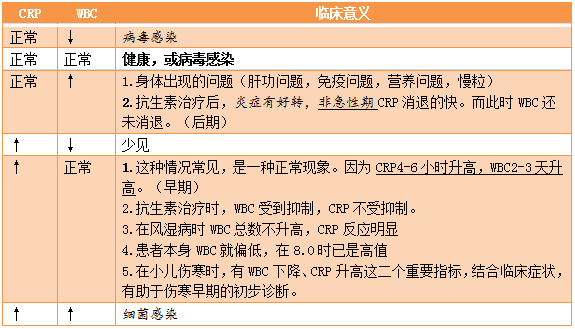
 C-Reactive Protein (CRP) is a typical acute-phase protein and one of the first acute-phase proteins recognized in history, discovered in 1930 by Tillet and Francis. In 1944, Jones used CRP for disease diagnosis. In 2003, the European guidelines for hypertension prevention and treatment (ESH/ESC) officially recommended that hypertensive patients should test hs-CRP levels. In recent years, CRP has been widely used in clinical settings in China.Characteristics:The characteristic of CRP is that it rises significantly during acute phases (by dozens or hundreds of times); however, if a recent illness has improved and is no longer in the acute phase, CRP will quickly drop. CRP concentration begins to rise 6 hours after a bacterial infection, reaching its peak after 24-48 hours. “CRP levels of 10-50 mg/L indicate mild inflammation, while levels ≥100 mg/L suggest a more severe bacterial infection.”Main Clinical Significance:(Reference range: 0-10 mg/L)1. Differential diagnosis of bacterial ↑ and viral infections.2. Various stress responses ↑ (trauma, cardiovascular diseases…) and immune system diseases ↑ (rheumatism, SLE…)Appendix: WBC and CRP Combination Table