To address the critical challenge of “how to provide functional vascular networks for new tissues,” a joint research team from Linköping University and Sahlgrenska University Hospital in Sweden has developed a biphasic particle bioink based on gelatin and hyaluronic acid, named μInk. This material can construct dermal tissue substitutes with ultra-high cell density through 3D bioprinting technology, successfully achieving neovascularization, tissue integration, and scar repair in animal experiments, opening new pathways for the treatment of chronic wounds and severe burns. The research results have been published in the international journal Advanced Healthcare Materials.
Clinical Pain Points: Traditional Therapies Struggle to Rebuild
Functional Dermis
The skin, as the largest organ of the human body, not only serves a protective barrier function but is also closely related to social interaction and self-perception. Globally, severe skin injuries such as burns lead millions of patients to seek medical treatment each year, with the U.S. healthcare system alone spending as much as $96.8 billion annually on wound care.
Current mainstream clinical treatment options, such as autologous skin grafts and cultured epidermal autografts, have many limitations. The former leaves new wounds at the donor site, and the transplanted skin lacks a complete dermal structure; the latter can only repair the epidermal layer and cannot achieve functional regeneration of dermal tissue.Traditional tissue-engineered skin often results in significant scarring due to low cell density, insufficient intercellular signaling, and limited extracellular matrix (ECM) secretion, making it difficult to restore the structure and function of normal skin.
“The complexity of the dermis exceeds imagination; its rich cellular network and ECM architecture have yet to be fully elucidated, which has been a key barrier for tissue-engineered skin for a long time,” said Johan Junker, a professor of plastic surgery at Linköping University and the research leader. Existing biomaterials either lack mechanical stability or have degradation rates that do not match tissue regeneration, failing to provide continuous growth support for cells.
Technical Breakthrough: Biphasic Structure Creates a “Cell-Friendly” Bioink
μInk
The innovation of μInk lies in its unique biphasic particle structure design, which perfectly balances biocompatibility, mechanical stability, and printability.
The research team used porous gelatin microcarriers (PGMs) as the core, first culturing and expanding human dermal fibroblasts (HDFs) in a bioreactor, allowing the cells to fully attach and grow on the surface and inside the microcarriers. These porous microcarriers have an average diameter of about 256μm, forming interconnected pores of approximately 20μm inside, providing ample space for cell proliferation and nutrient exchange. Subsequently, the cell-loaded gelatin microcarriers were mixed with hyaluronic acid (HA)-based hydrogels and crosslinked using copper-free click chemistry to form a biphasic particle bioink with both shear-thinning properties and rapid recovery capabilities.
“This design gives the bioink three major advantages: ultra-high cell density ensures a cellular basis for tissue regeneration, the biphasic structure provides physical support similar to natural dermis, and the bioorthogonal crosslinking technology avoids the toxic effects of traditional chemical crosslinking on cells,” explained Dr. Daniel Aili, a biomaterials expert involved in the research. Rheological tests showed that μInk has a storage modulus of about 1400Pa, which highly matches the mechanical properties of natural reticular dermis, and can quickly recover elasticity after printing, allowing for direct 3D printing of over 10 layers of self-supporting structures without the need for additional support baths or secondary crosslinking treatments.
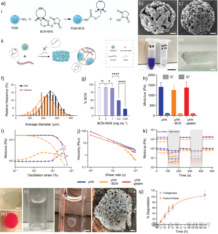
Figure 1. Preparation, synthesis, and characterization of μInk
Animal Experiment Validation: Achieving Functional Tissue Integration in 28 Days
To validate the in vivo repair effects of μInk, the research team implanted 3D printed cell-loaded μInk constructs subcutaneously in an immunodeficient mouse model and conducted a 28-day follow-up observation.
The experimental results showed that the transplanted fibroblasts maintained over 90% survival rate, continued to proliferate, and maintained a dermis-specific phenotype. By 14 days post-surgery, new blood vessels began to form within the construct; by 28 days, it had achieved close integration with the host tissue and successfully secreted key ECM components such as collagen I, III, and laminin, forming a tissue architecture similar to natural dermis.Immunofluorescence staining confirmed that the transplanted cells not only continued to proliferate but also recruited host cells to participate in tissue remodeling, significantly enhancing vascularization levels.
Compared to the acellular control group, the degradation rate of the fibroblast-loaded μInk constructs better meets the needs of tissue regeneration—the gelatin microcarriers can be degraded by collagenase, and the hyaluronic acid matrix can be broken down by hyaluronidase, gradually freeing up space for new tissue.More importantly, the construct did not trigger significant inflammatory responses or fibrous capsule formation, achieving the treatment goal of “regeneration rather than repair,” effectively avoiding excessive scar formation.
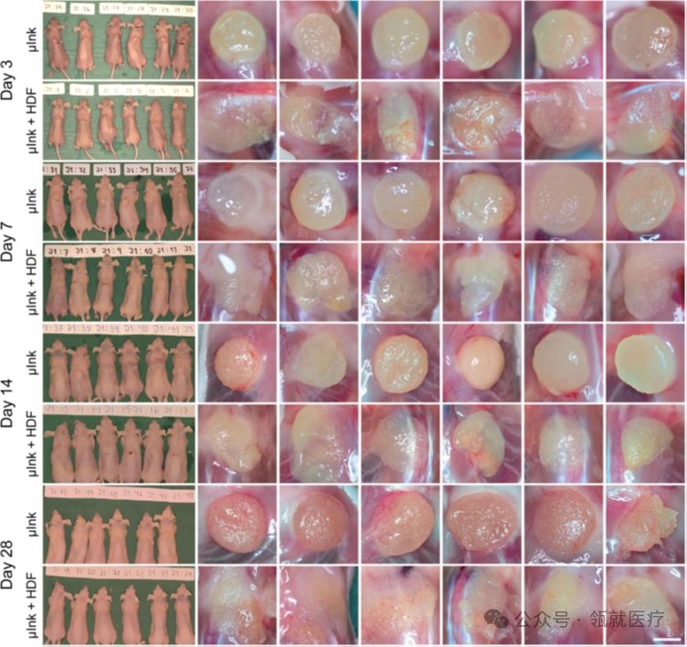
Figure 2. Macroscopic evaluation results of μInk and μInk+HDF containing human dermal fibroblasts
Conclusion
This study organically combines cell expansion and bioprinting technology to address core issues such as insufficient cell density, poor mechanical properties, and difficult tissue integration in traditional tissue-engineered skin. μInk is not only suitable for dermal regeneration, but its modular design concept can also be extended to other tissue repair fields.
“With the improvement of technology, we expect to bring this 3D printed dermal tissue to clinical use in the next 3-5 years, providing patients with burns and chronic ulcers with repair solutions that result in less scarring and more complete functionality,” added Lars Kölby, a professor of plastic surgery at Sahlgrenska University Hospital. This research has received significant funding from the European Research Council, the Swedish Research Council, and other institutions, providing strong support for technology transfer.
Literature Link:
https://doi.org/10.1002/adhm.202501430
Scan to follow us for more information
Zhihu: Lingjiu Medical

Xiaohongshu: Lingjiu Technology

For product and customization services, please contact WeChat: Captain_WangCharly;MeiQingClaire
Shanghai Lingjiu Medical Technology Co., Ltd.
Shanghai LinGel Medical Technology Co., Ltd.
Rooted in Innovative Soil | Achieving Rapid Recovery
Phone|021-62213281, 17316396996
Email| [email protected]
Website| www.lingeltech.com
Address|9th Floor, Building 1, No. 58 Yuanmei Road, Minhang District, Shanghai