
BioLegend is a company focused on biomedical research, offering a wide range of products, including but not limited to the following categories: flow cytometry-related products, stem cell research products, ELISA kits, Western blotting-related products, totalseq-related products, and many immunofluorescence-related products.
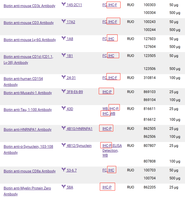
Immunocytochemistry (ICC) is characterized by imaging cultured primary cells or cell lines. Immunohistochemistry (IHC) refers to detecting antibodies in tissue sections. Both ICC and IHC applications can utilize colorimetric or fluorescent detection methods. In our product technical data sheets, we use IHC-P to indicate that the antibody is useful in formalin-fixed paraffin-embedded (FFPE) sections, and IHC-F to indicate that the antibody is useful in tissues fixed and frozen prior to sectioning. If only the IHC name is provided, please check literature references to confirm the designation.

Newly Validated IHC Antibodies
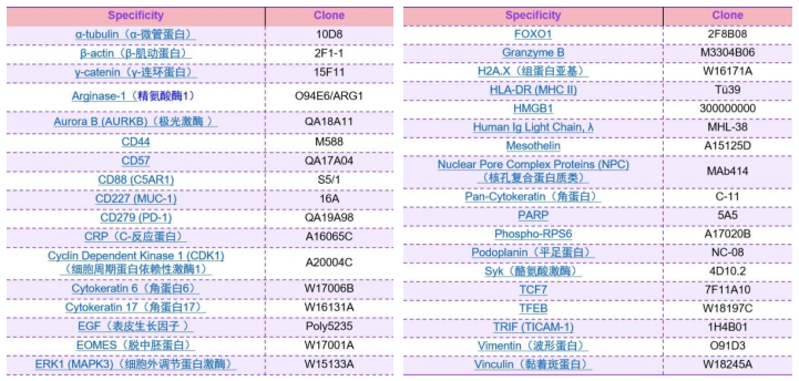
BioLegend offers a wide and diverse range of antibodies, boasting one of the largest clone libraries in the industry. With twenty years of experience in antibody production, we provide high-quality antibodies that can be used for various applications, including western blotting, flow cytometry, and microscopy. In microscopy, immunohistochemistry can drive discoveries in research fields such as oncology, immunology, and neurology. With this in mind, BioLegend is continuously working to add more application areas to our existing products. Below are the antibodies for the latest validated immunohistochemistry applications.
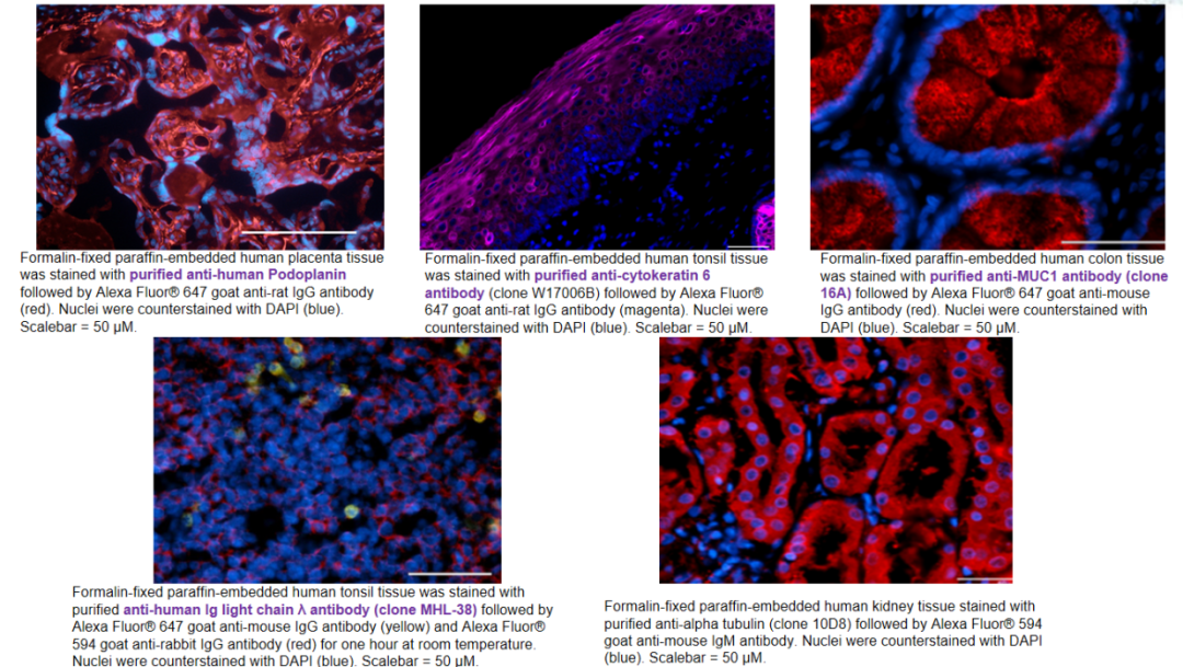
Example Results:
Fluorophores Suitable for Confocal Microscopy
Fluorescent detection allows for the visualization of multiple markers simultaneously, most commonly through the use of optimal lasers to excite fluorophore groups. Fluorescent detection also brings opportunities for advanced imaging applications such as live-cell imaging, multiphoton imaging, super-resolution microscopy, FLIM, and FRET. While sensitivity may be limited by fluorescent microscopy at certain wavelengths, particularly with reagents emitting in the 350-450 nm range, improved signal-to-noise ratios can be achieved using different enzymes and immunoamplification techniques, higher sensitivity instruments, and near-infrared emitting fluorophores.
BioLegend provides a vast array of fluorophore antibody conjugates for your microscopy research. The table below displays the optimal excitation light, emission light, and luminescence color of the fluorophores.
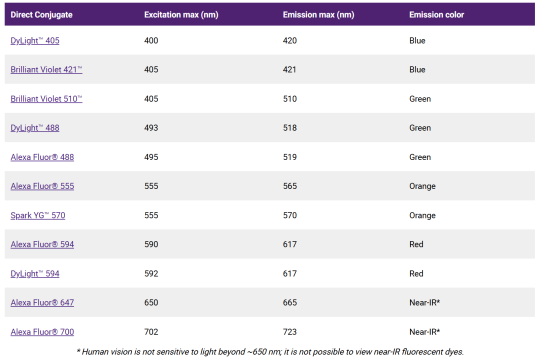
Primarily three series of fluorophores: Alexa Fluor® Dyes, Brilliant Violet™ Fluorophores, and Spark YG™ 570. Each series of fluorophores has unique characteristics.
✦
•
(1) Alexa Fluor® Dyes Alexa Fluor® dyes are very commonly used fluorophores in immunofluorescence applications. Alexa Fluor®488, Alexa Fluor®594, Alexa Fluor®647 are the most commonly used fluorophores, in addition to Alexa Fluor®555.
✦
•
(2) Brilliant Violet™ Fluorophores Brilliant Violet 421™(BV421™) is typically used in the “blue” channel usually occupied by DAPI or Alexa Fluor®405. Directly conjugated antibodies can visualize antigens, enhancing the capability of using multicolor microscopy. Brilliant Violet 510™(BV510™) is also excited at 405 nm but emits at 510 nm. When the instrument filter configuration is optimized, BV421™ and BV510™ can be used simultaneously. This series of fluorophores has undergone detailed stability testing.
For more details, see the link https://www.biolegend.com/en-us/microscopy/ihc-icc-applications
✦
•
(3) Spark YG™ 570 Spark YG™570 is an ideal choice for building multicolor microscopy schemes and is a reliable addition to any multicolor microscopy scheme. The maximum excitation is at 555 nm, and the maximum emission is at 570 nm, which can be shared with common filters for Alexa Fluor®555, Cy3, or TRITC in laser confocal microscopy.
Purified and Biotinylated Primary and Secondary Antibodies
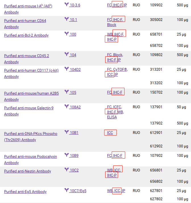
Purified antibodies are commonly used reagents for monocolor staining or when amplification of secondary antibodies is needed. Biotinylated antibodies are also effective tools for signal amplification or multicolor applications. BioLegend offers hundreds of purified/biotinylated antibody products and dozens of biotinylated antibodies that have been quality tested for ICC or IHC.
Purified Antibodies:
https://www.biolegend.com/en-us/search-results?PageSize=25&Applications=ICC&Applications=IHC&Applications=IHC-F&Applications=IHC-P&Format=PURE
Biotinylated Antibodies:
https://www.biolegend.com/en-us/search-results?PageSize=25&Applications=ICC&Applications=IHC&Applications=IHC-F&Applications=IHC-P&Format=BIOTIN
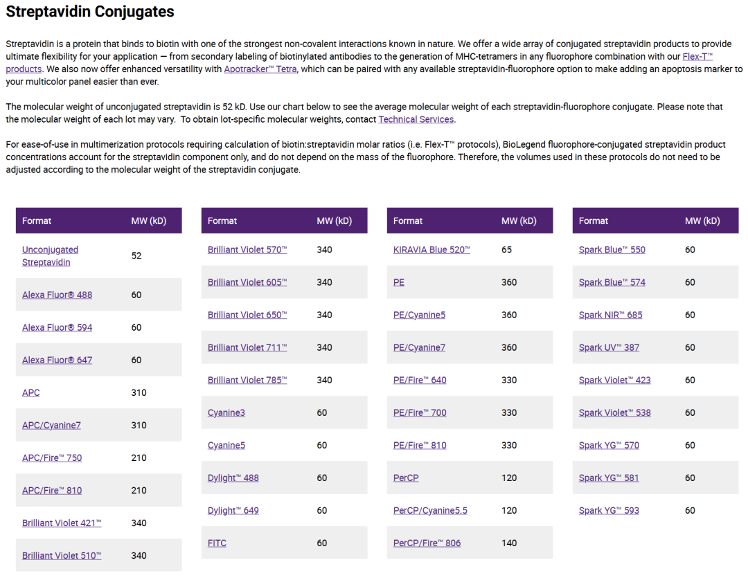
Secondary Reagents:
https://www.biolegend.com/en-us/antibodies-reagents/secondaries
Chemical Probes
In addition to primary antibodies, many dyes or probes can be used for specific staining of subcellular structures. These can be used for various fluorescent microscopy applications, including staining of nuclei, organelles, or cytoplasmic structures, such as actin and microtubules.
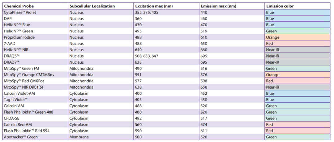
The figure shows some non-antibody chemical probes, their subcellular localization and emission profiles.
Colorimetric Detection Reagents

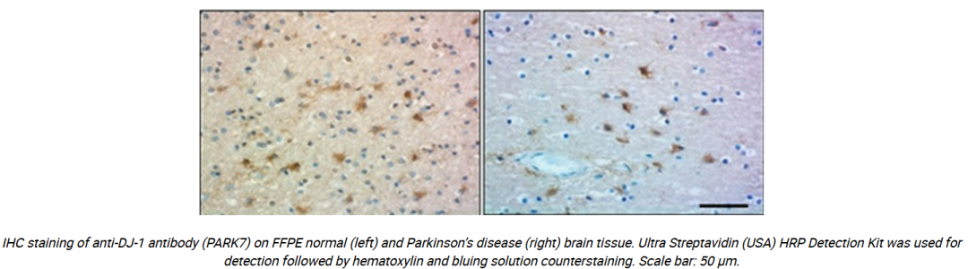
In addition to fluorescent-related reagents, colorimetric reagents such as DAB, AEC, or BCIP/NBT are also available. These are enzyme-catalyzed substrates. Antibodies or streptavidins commonly used for colorimetric immunoreactions covalently bind to HRP or ALP, with substrates like DAB, AEC, or BCIP/NBT. These enzymes catalyze their substrates, leaving a layer of color where antibodies are attached to cells or tissues.
We provide a variety of ancillary reagents for immunohistochemistry, including: Ultra Streptavidin (USA) HRP Detection Kit (Multi-Species, DAB) (biolegend.com) and ACUITY Advanced Biotin Free Polymer Detection System DAB Kit (biolegend.com).
Official website: www.bio-city.net
Email: [email protected]

Toll-free hotline: 400 819 7199
Shanghai Dakewei: 021-33887795
Beijing Dakewei: 010-64828744/54/84/94
Guangzhou Office: 020-87381715/87350260