
Subhajit Pal, Erika E. Salzman, Dominic Ramirez, Hannah Chen, Cynthia A. Perez, Katelyn Dale, Sujoy K. Ghosh, Liwei Lin, Phillip B. Messersmith, et al. 🎉🎉🎉 They conducted research on the issues of poor water solubility and high hydrophobicity of poly α-lipoic acid (αLA) when used as a medical adhesive building block, which requires high temperatures, organic solvents, or complex preparation processes. They successfully developed αLA-based powders and low-viscosity liquid superglues; these superglues can quickly polymerize and bond upon contact with wet tissues, can incorporate and release small molecule regenerative drugs without altering physical and bonding properties, and possess good cell and tissue compatibility, biodegradability, high conductivity due to inherent ionic properties, and sensitivity to deformation, making them suitable for developing tissue adhesive strain sensors. The related results “Versatile Solid-State Medical Superglue Precursors of α‑Lipoic Acid” were published in the Journal of the American Chemical Society!! 🎉🎉🎉 Corresponding authors are Subhajit Pal and Phillip B. Messersmith.

Currently, synthesizing medical adhesives that possess strong bonding strength, rapid action, good adhesion to wet tissues, high biocompatibility, biodegradability, and tissue-like flexibility is quite challenging. Different surgical procedures and tissue structures require adhesives to have different forms to ensure optimal performance, and controllable loading and release of active drugs in biodegradable tissue adhesives are of great value in chronic wound care and regenerative medicine. Cyanoacrylate-based medical superglues, while having high strength and rapid curing characteristics due to polymerization triggered by moisture and nucleophilic functional groups in tissues, are mainly limited to external wound closure due to cytotoxicity, rigidity, and non-biodegradability. Solid forms (such as powders and films) of superglues are attractive for mechanical bonding or sealing upon contact with tissues but have not been reported. Although α-lipoic acid (αLA) polymers have attracted attention due to their good biocompatibility, biodegradability, adhesion, and flexibility, their spontaneous depolymerization issues hinder practical applications. Previous studies have attempted to improve this through copolymerization, chain-end capping agents, or the addition of other substances, but still face problems such as weak bonding strength, slow curing, rapid degradation, and complex preparation processes. Moreover, previously developed αLA polymerization methods required organic solvents to prepare monomer precursor solutions, limiting in vivo applications.
In this context, the authors developed a method for in situ copolymerization of αLA and its derivatives under physiological conditions, successfully preparing αLA-based powders (αLA-PS) and aqueous liquid superglue (αLA-LS) precursors; they explored the effects of different component ratios on the performance of the powders and liquid superglues, such as studying their thermal and mechanical properties through differential scanning calorimetry and rheological analysis, and testing their bonding strength to wet bovine pericardium through lap shear experiments; they also conducted in vitro degradation experiments, cell co-culture experiments, and subcutaneous implantation experiments in mice to evaluate biocompatibility and degradability, and investigated the feasibility and drug release of the liquid superglue (αLA-LS2) loaded with small molecule regenerative drugs (NaDPCA). Additionally, they tested the conductivity of αLA-LS1 and its potential as a strain sensor.

(1) Experimental drugs include α-lipoic acid (αLA), sodium lipoate (NaLA), activated esters of αLA (S1, S2), ethanol, dimethyl sulfoxide (DMSO), isotonic saline, 1×PBS (phosphate-buffered saline), glutathione (GSH), N-bromosuccinimide, 4-prolylhydroxylase inhibitor 1,4-dihydropyrrole-4-keto-3-carboxylate sodium salt (NaDPCA), and mouse embryonic fibroblasts (NIH 3T3).
(2) Preparation of powder superglue (αLA-PS): Mix αLA, NaLA, S1/S2 in different molar ratios (e.g., αLA-PS1 is 67:27:6, αLA-PS2 is 49:45:6) to form a uniform powder mixture; then study its transitions under different conditions, such as open storage at room temperature (22°C), open storage at 37°C, and treatment at 37°C with 90% relative humidity for 5 minutes, observing the transition from powder to gel. Preparation of liquid superglue (αLA-LS): Dissolve αLA, NaLA, and S1 in a molar ratio of 66:27:7 in ethanol containing different amounts of water, and evaporate the ethanol to obtain a low-viscosity injectable liquid; during the preparation of αLA-LS2, 2wt% of NaDPCA was also added. Performance testing steps: Analyze the melting point and thermal properties of the materials through differential scanning calorimetry; evaluate the mechanical properties of the gel and liquid precursors through rheological analysis, such as storage modulus, loss modulus, viscosity, and shear rate relationships; test the bonding strength of the adhesive to wet bovine pericardium through lap shear experiments, applying the powder or liquid precursor to the wet bovine pericardium, forming a lap joint, and incubating in isotonic saline at 37°C, measuring shear strength at 2 minutes and 24 hours; in vitro degradation experiments: place the polymerized αLA-LS1 in isotonic saline containing 0.1mM GSH and monitor degradation over 15 days; cell compatibility experiments: co-culture NIH 3T3 cells with in situ polymerized αLA-PS1/αLA-LS1 for 24 hours and assess cell viability; in vivo biocompatibility experiments: subcutaneously implant αLA-LS1 in the back of mice, observe behavioral changes and inflammation at the injection site, and perform histological analysis of the implanted skin and other organs after 3 days; drug release experiments: monitor the release of drugs from αLA-LS2 and the biological activity of drugs in co-cultured cells; conductivity testing: measure the current-voltage characteristics of αLA-LS1 after incubation in isotonic saline for 30 minutes and 4 hours, test its ability to light up an LED, and monitor resistance changes during stretching and bending.
(3) Testing methods include: differential scanning calorimetry (DSC) to determine the melting point (Tm) of materials and analyze changes in the crystalline phase of monomers, such as analyzing the thermal behavior of pure αLA, NaLA, and different powder mixtures; rheological analysis through frequency scanning, shear rate scanning, etc., to determine parameters such as storage modulus, loss modulus, and viscosity, evaluating the elasticity of the gel, the shear-thinning characteristics of the liquid precursor, and the relationship between polymerization degree and water content; lap shear strength testing to evaluate the adhesive performance of the adhesive to wet bovine pericardium by measuring shear strength at different incubation times, analyzing changes in bonding strength and failure modes; in vitro degradation testing by monitoring the degradation of materials in isotonic saline containing GSH over time to assess biodegradability; cell viability testing using co-culture methods to compare cell viability of cells in contact with the adhesive and control group cells, determining cell compatibility; histological analysis through hematoxylin-eosin (H&E) staining to observe the structure of tissues and organs at the implantation site in mice, assessing in vivo biocompatibility; dynamic light scattering (DLS) analysis to detect the particle size and polydispersity index of NaDPCA forming nanoparticles in solution; current-voltage (I-V) characteristic testing to determine the conductivity of materials by lighting up an LED and monitoring resistance changes during stretching and bending, assessing its potential as a strain sensor.
Q:What is the role of each component?🟢️α-lipoic acid (αLA): It is the core building block of the adhesive, and its polymer has good biocompatibility, biodegradability, adhesion, and flexibility, providing the basic structure and biological performance for the adhesive; at the same time, the conjugate acid-base pair formed by αLA and NaLA endows the mixture with hygroscopicity, facilitating water-induced polymerization.🟢️Sodium lipoate (NaLA): Forms a low eutectic system with αLA, lowering the melting temperature of the mixture, allowing polymerization to occur at lower temperatures (such as physiological temperature); its content affects the non-covalent interactions between the adhesive and tissue surfaces, thereby altering bonding strength.🟢️Activated esters of αLA (S1, S2): As chain-end stabilizers, they can inhibit the spontaneous depolymerization of αLA polymers, ensuring the stability of the adhesive, which is crucial for achieving optimal bonding performance; in the absence of these, the adhesive’s adhesion to tissues would significantly decrease; among them, S1 is more suitable for the preparation of liquid superglue (αLA-LS) due to its lower electrophilicity and higher stability.🟢️Ethanol: Used as a solvent in the preparation of liquid superglue, dissolving components such as αLA, NaLA, and S1, and after evaporation, a low-viscosity injectable liquid is obtained; only a small amount of ethanol remains in the final product, which does not affect performance.🟢️Glutathione (GSH): Used in in vitro degradation experiments to simulate the in vivo environment, as poly αLA can be biologically degraded through GSH-mediated pathways, monitoring the degradation process of materials through solutions containing GSH.🟢️N-bromosuccinimide: Used to stain the adhesive, assisting in observing the failure modes of the adhesive in lap shear experiments, determining whether the failure is cohesive or interfacial.🟢️4-prolylhydroxylase inhibitor 1,4-dihydropyrrole-4-keto-3-carboxylate sodium salt (NaDPCA): As a small molecule regenerative drug, it is used to evaluate the drug loading and release capabilities of the adhesive, can stably exist in the adhesive and be released slowly, and can exert biological activity in cells (such as inhibiting 4-prolylhydroxylase, stabilizing HIF-1α) without affecting the physical and bonding properties of the adhesive.🟢️Mouse embryonic fibroblasts (NIH 3T3): Used in in vitro cell compatibility experiments, co-culturing with the adhesive to detect cell viability and assess the adhesive’s toxicity to cells.

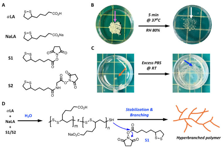 ▲ Figure 1A: Shows the chemical structures of the monomers (αLA, NaLA) and stabilizers (S1, S2); Figure 1B: Presents photos of αLA-PS1 powder before and after treatment at 37°C and 90% relative humidity for 5 minutes, with the left purple arrow indicating the powder before treatment and the right white arrow indicating the state after treatment; Figure 1C: Is a video screenshot showing the state of αLA-LS1 before and after contact with excess isotonic saline at room temperature, with the left orange arrow indicating the liquid before contact and the right blue arrow indicating the state after contact; Figure 1D: Illustrates the overall reaction process of copolymerization and chain-end stabilization.
Figure 1: Mainly presents the monomer structure and polymerization-related information of α-lipoic acid-based superglues. Part A clarifies the chemical structures of αLA, NaLA (sodium lipoate), and stabilizers S1, S2, which is fundamental to understanding the material composition; Part B visually reflects the changes in αLA-PS1 powder after treatment at 37°C and 90% relative humidity for 5 minutes, intuitively demonstrating the transition from powder to subsequent states; Part C shows a video screenshot demonstrating the differences in αLA-LS1 before and after contact with excess isotonic saline at room temperature, reflecting the rapid changes of the liquid precursor upon contact with water; Part D clearly presents the process of copolymerization and chain-end stabilization, explaining how to inhibit depolymerization through chain-end stabilization, providing theoretical support for material preparation, focusing on the core composition and key reaction processes of the material, laying the foundation for subsequent performance studies.
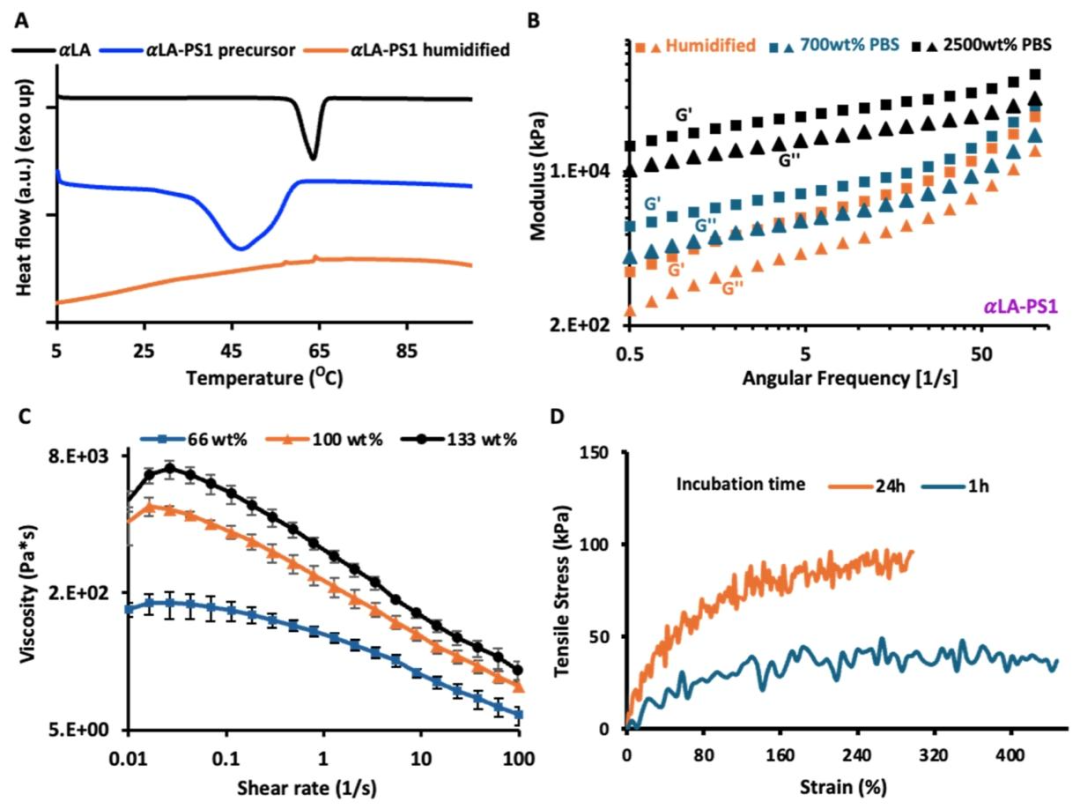 ▲ Figure 2A: Is the differential scanning calorimetry (DSC) curve (first heating cycle), showing the thermal behavior of pure αLA, αLA-PS1 precursor powder, and αLA-PS1 after treatment at 37°C and 90% relative humidity for 5 minutes; Figure 2B: Is the rheological analysis graph, presenting the frequency scanning results of αLA-PS1 after humidification and polymerization with isotonic PBS at 700wt% and 2500wt% for 10 minutes at a constant strain of 0.4% and 37°C; Figure 2C: Is the viscosity versus water content relationship graph of αLA-LS1 at room temperature, with all viscosity tests completed within 3 days after preparation; Figure 2D: Is the stress-strain curve of αLA-LS1 after incubation in excess isotonic saline at 37°C for 1 hour and 24 hours.
Figure 2: Focuses on the thermal and mechanical properties of αLA-PS and αLA-LS. Part A of the DSC curve shows that the melting point of pure αLA is about 65°C, while the melting point of αLA-PS1 precursor powder drops to 45°C, and after treatment at 37°C and 90% relative humidity for 5 minutes, the crystalline phase of the monomer completely disappears, indicating a change in the crystallinity of the treated material; Part B of the rheological analysis shows that after humidification, the storage modulus of αLA-PS1 is higher than the loss modulus, and significantly increases with the amount of PBS, such as the modulus at 2500wt% PBS being much higher than at 700wt% PBS, indicating that water content promotes polymerization, resulting in stronger elastic solid characteristics; Part C of the viscosity graph shows that the viscosity of αLA-LS1 increases with water content at room temperature, and remains stable within 3 days after preparation, reflecting its shear-thinning characteristics and storage stability; Part D of the stress-strain curve indicates that αLA-LS1 exhibits high flexibility after incubation for 1 hour and 24 hours, with tensile strength increasing and fracture strain decreasing after 24 hours, indicating that the polymer network continues to mature over time, optimizing mechanical properties.
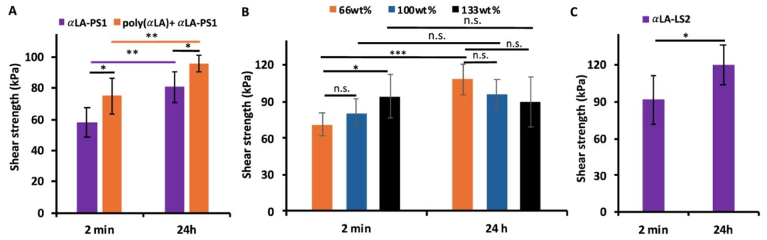 ▲ Figure 3A: Is a comparison graph of lap shear strength, comparing the shear strength of αLA-PS1 and the coated αLA-PS1 poly αLA film after incubation in 37°C 1×PBS for 2 minutes and 24 hours to wet bovine pericardium; Figure 3B: Shows the comparison of lap shear strength of αLA-LS1 with different water contents after incubation in 37°C 1×PBS for 2 minutes and 24 hours; Figure 3C: Presents the lap shear strength of αLA-LS2 containing 2wt% NaDPCA and 100wt% water after incubation in 37°C 1×PBS for 2 minutes and 24 hours, with sample sizes (N=5) and significant differences determined by Student’s t-test (n.s. indicates p>0.05, * indicates p≤0.05, ** indicates p≤0.01, *** indicates p≤0.001).
Figure 3: Focuses on the lap shear strength of αLA polymers to wet bovine pericardium, with N=5 and significance analyzed by Student’s t-test. Part A data shows that the shear strength of αLA-PS1 after 2 minutes is 58±9kPa, increasing to 80±9kPa after 24 hours; the coated αLA-PS1 poly αLA film has a shear strength of 74±11kPa after 2 minutes and reaches 96±5kPa after 24 hours, with both groups showing significantly higher strength at 24 hours compared to 2 minutes, indicating that the maturation of the polymer network enhances bonding strength, and the coating strategy also improves the bonding performance of the poly αLA film; Part B shows that for αLA-LS1 with different water contents, the strength of the 66wt% water group is 71±10kPa after 2 minutes, the 100wt% water group is 80±12kPa, and the 133wt% water group is 94±18kPa, showing linear growth, while after 24 hours, an inverse trend is observed, with the 66wt% water group at 108±13kPa, the 100wt% water group at 96±12kPa, and the 133wt% water group at 89±20kPa, indicating that water promotes polymerization to enhance cohesive strength in the short term, while in the long term, interfacial adhesion is more affected by water; Part C shows that αLA-LS2 containing 2wt% NaDPCA has a strength of 91±19kPa after 2 minutes and 119±12kPa after 24 hours, with no significant difference compared to related groups without drugs (n.s., p>0.05), confirming that drug addition does not affect bonding performance.
 ▲ Figure 4A: Is the in vitro degradation curve of αLA-LS1, showing the degradation of the material over time; Figure 4B: Is the viability graph of NIH 3T3 mouse fibroblasts after 24 hours of contact with αLA-LS1, comparing different concentrations of αLA-LS1 (5μL/mL, 2μL/mL) with the control group (growth medium); Figure 4C: Is the histological image of the implanted skin stained with hematoxylin-eosin (H&E); Figure 4D: Is the histological image of healthy control skin, with a scale bar of 100μm, and no acute inflammation or structural abnormalities observed.
Figure 4: Evaluates the biological performance of αLA-PS and αLA-LS. Part A of the in vitro degradation curve shows that αLA-LS1 gradually degrades in isotonic saline containing 0.1mM GSH over 15 days, with a smooth degradation process, reflecting good biodegradability; Part B of the cell viability experiment shows that NIH 3T3 cells in contact with 5μL/mL and 2μL/mL αLA-LS1 for 24 hours show no significant difference in viability compared to the control group (growth medium), both close to 100%, proving that the material has no significant cytotoxicity; Part C histological images of implanted skin compared to Part D healthy control skin show no acute inflammatory cell infiltration, tissue necrosis, or other structural abnormalities, and histological analysis of other organs (such as heart, liver, etc.) also shows no adverse reactions, indicating that the material has good biocompatibility in vivo, with no significant toxic side effects.
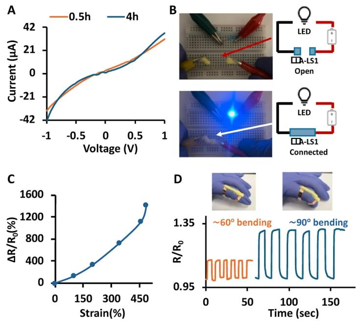 ▲ Figure 5A: Is the current-voltage characteristic curve of αLA-LS1 without applied strain after incubation for 0.5 hours and 4 hours; Figure 5B: Demonstrates the conductivity of αLA-LS1, with the upper image showing the state of the circuit before contact with the material and the lower image showing the state after contact with the adhesive when the LED lights up; Figure 5C: Presents the relative change in resistance of αLA-LS1 under stretching strains from 0% to 485%; Figure 5D: Is the relative change in resistance over time of the adhesive device attached to the finger during bending (at 60° and 90° bending angles), including illustrations of the device attached to the finger.
Figure 5: Investigates the conductivity of αLA-LS1. Part A of the current-voltage characteristic curve shows that after incubation for 0.5 hours and 4 hours, the material’s conductivity is 1.74±0.16mS/cm and 1.65±0.08mS/cm, respectively, with the curve being nonlinear, indicating that the conductivity is ionic and that incubation time has little effect on conductivity; Part B of the experiment shows that αLA-LS1 can light up an LED when connected to a circuit with a 9V battery, while it does not light up when not in contact, intuitively proving that the material has sufficient conductivity; Part C of the relative change in resistance graph shows that as the stretching strain increases from 0% to 485%, the relative change in resistance gradually increases, indicating that at a strain of 485%, the relative change in resistance is much higher than at lower strains, demonstrating that the material is sensitive to stretching deformation; Part D of the time-resistance change curve shows that during finger bending at 60° and 90°, the relative change in resistance synchronously responds, with the change being greater at 90° bending than at 60°, proving that the material can be used as a strain sensor for human-computer interaction, such as monitoring finger movements.

Q: Why does the material prepared in this paper have excellent performance??The materials prepared have excellent performance because:🟢️First, due to its unique component design and polymerization mechanism: αLA as the core module, its polymer itself has good biocompatibility, biodegradability, and flexibility, laying the foundation for the material’s basic performance; the low eutectic system formed by αLA and NaLA lowers the melting temperature to near physiological temperature, combined with the “hydroeutectic” effect (combining hygroscopicity and low eutectic characteristics), allowing the powder and liquid precursors to quickly polymerize upon contact with wet tissues or water without requiring high temperatures or complex conditions, while chain-end stabilizers S1/S2 effectively inhibit the spontaneous depolymerization of αLA polymers, ensuring material stability and bonding strength.🟢️Secondly, the material has excellent and controllable mechanical properties: By adjusting the ratio of αLA to NaLA, water content, etc., the material’s viscosity, elasticity, tensile strength, and other mechanical parameters can be optimized, such as the liquid precursor having shear-thinning characteristics that facilitate injection, and forming a highly flexible solid after polymerization, with the polymer network maturing over time to further enhance mechanical properties, meeting the demands of different medical scenarios for morphology and mechanics.🟢️Furthermore, the material possesses good biological safety and functionality: In vitro cell co-culture and in vivo implantation experiments confirm that it has no significant cytotoxicity or inflammatory response, can biodegrade to avoid in vivo residues; it can stably load and slowly release small molecule regenerative drugs (such as NaDPCA) without affecting core performance, aiding in wound healing and regeneration;🟢️At the same time, due to the inherent ionic properties brought by the conjugate acid-base pair of αLA and NaLA, the material has high conductivity and sensitivity to deformation, making it suitable as a strain sensor in the field of bioelectronics, achieving multifunctional integration.
In summary, the innovation of this paper mainly lies in the following aspects: First, it developed two forms of medical superglue precursors based on α-lipoic acid (αLA), filling the research gap of solid medical superglues, and both forms of precursors can quickly polymerize and bond with wet tissues or water under physiological conditions without requiring high temperatures, organic solvents, or complex preparation processes, solving the problem of traditional αLA-based adhesives needing organic solvents and limited applications, while elucidating the mechanism of rapid polymerization through the “hydroeutectic” effect (combining hygroscopicity and low eutectic characteristics). Second, it achieved efficient integration of multifunctionality in materials, confirming for the first time that αLA-based adhesives can stably load and slowly release small molecule regenerative drugs without altering core performance, while the inherent ionic properties endow the material with high conductivity and sensitivity to deformation, successfully applying it to tissue adhesive strain sensors, expanding the application scenarios of medical adhesives in drug delivery and bioelectronics. Third, it optimized the material performance control strategy, allowing precise control of thermal properties, mechanical properties, bonding strength, and degradation rate by adjusting parameters such as the ratio of αLA to NaLA, the addition of chain-end stabilizers (S1/S2), and water content, providing customized solutions for different medical needs.
The DOI is: 10.1021/jacs.4c18448