Original: Customized 3D-Printed Titanium Mesh Developed for an Aesthetic Zone to Regenerate a Complex Bone Defect Resulting after a Deficient Odontectomy: A Case Report
Published in: Medicina 2022, 58, 1192
A 27-year-old female patient reported that her aesthetics and function were affected ten years after a complex tooth extraction. She was dissatisfied with the temporary removable prosthesis, which was uncomfortable and impacted her social interactions. After detailed examination and CBCT evaluation, it was found that the patient was missing teeth 13 and 12, with severe alveolar bone defects.
After discussing with the patient, it was decided to use a customized 3D-printed titanium mesh for bone augmentation, postponing the implant placement.
The surgical procedure is shown in Figure 3. First, connective tissue was harvested from the palatal side for soft tissue augmentation to improve gingival thickness. After 10 weeks, bone augmentation was performed by mixing allogenic bone with bovine bone in a 1:1 ratio, secured with the titanium mesh. Six months later, the titanium mesh was removed to assess the bone augmentation results. Two months later, two implants were placed. Four months later, a second-stage surgery was performed to place the healing abutments.

Figure 1: Keratinized gingival graft was performed before bone augmentation. a, Buccal view. b, Occlusal view.
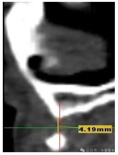
Figure 2: Initial CBCT: The defect area measures approximately 4.19mm x 4.2mm.

Figure 3: Surgical procedure steps.
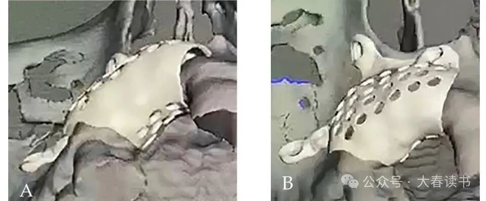
Figure 4: Digital design: a, Occlusal view. b, Buccal-palatal view.
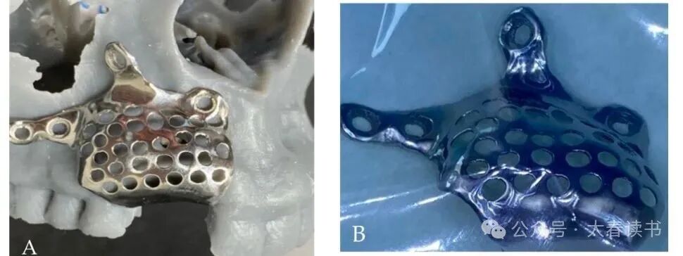
Figure 5: Customized titanium mesh: a, Titanium mesh positioned on the model. b, Preoperative disinfection.
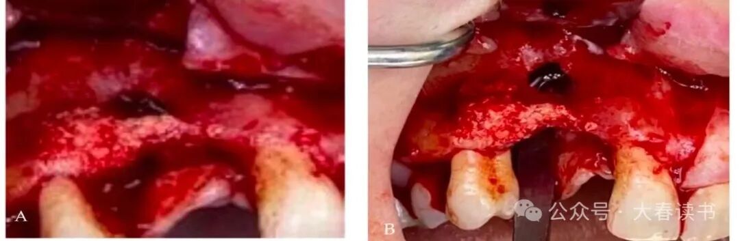
Figure 6: Intraoral view: a, Insufficient alveolar bone width, defect reaching the apex of adjacent teeth. b, Flap exposing the defect area.

Figure 7: Titanium mesh (0.5mm thick) secured with screws.
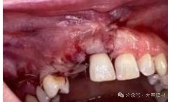
Figure 8: Suturing to close the wound.
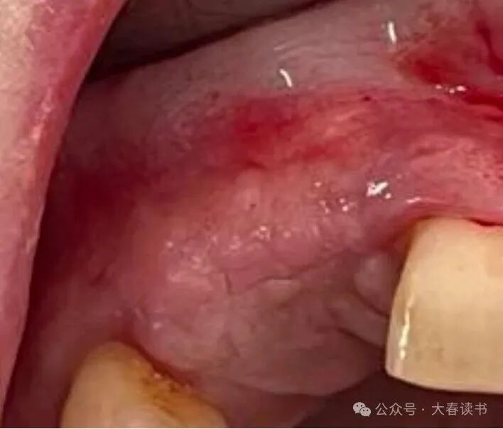
Figure 9: Intraoral view of soft tissue six months postoperatively.
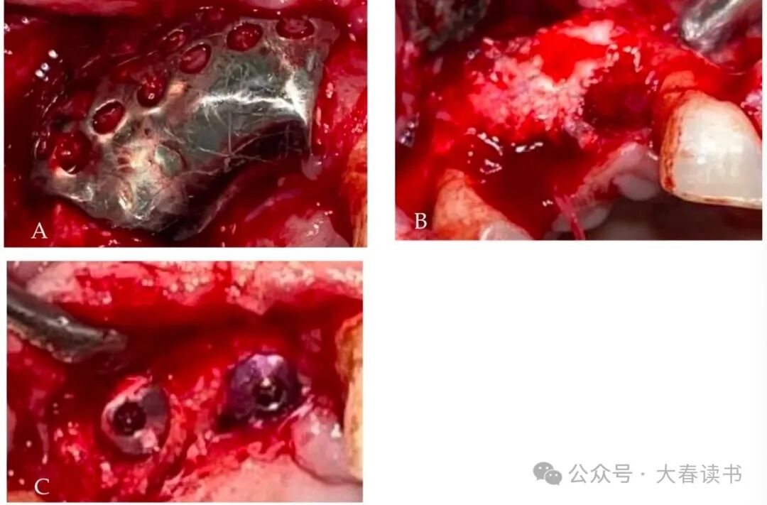
Figure 10: Intraoral examination: a, Titanium mesh exposed after flap reflection. b, Titanium mesh removal. c, Adequate alveolar bone width and height after bone augmentation, two months later, two implants were placed.
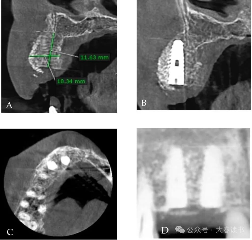
Figure 11: CBCT image after implant placement.
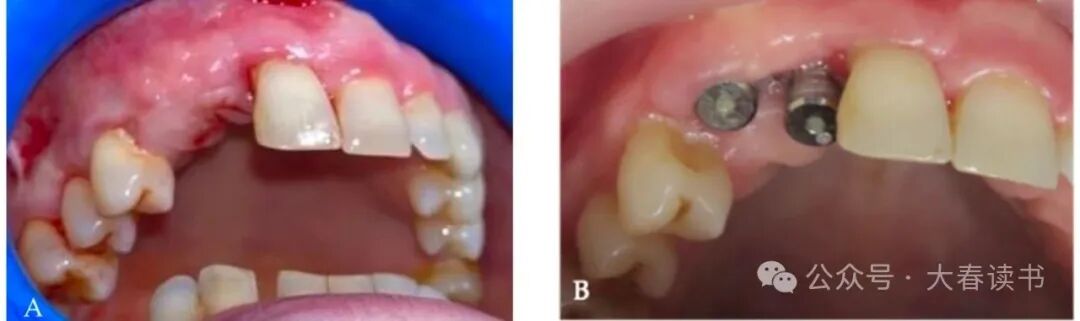
Figure 12: Intraoral view: a, Gingival status before placing the healing abutment. b, Placement of the healing abutment.
END
All literature shared in this public account belongs to the original publication and may not be used for commercial purposes; it is for educational use only. Please respect intellectual property rights. Feel free to share directly in your social circles. If there is any infringement, please contact for removal.