 Introduction
All cells, both eukaryotic and prokaryotic, secrete extracellular vesicles, and exosomes are a type of extracellular vesicle, ranging in size from 40 – 160 nm (average 100 nm). Depending on their cellular origin, they contain various components, including DNA, RNA, lipids, metabolites, and cytoplasmic and cell surface proteins. Their physiological purpose is not yet clear, but it is speculated that their role may be to remove excess and/or unnecessary components from the cell to maintain intracellular homeostasis.Recent studies have shown that the accumulation of specific cellular components in exosomes that are functional, targeted, and driving plays an important role in regulating intercellular communication.
Exosomes are associated with immune responses, viral pathogenicity, pregnancy, cardiovascular diseases, central nervous system-related diseases, and cancer progression. Different exosome payloads can be designed for therapeutic purposes, including siRNA, antisense oligonucleotides, chemotherapeutic agents, and immunomodulators, and delivered to intended targets.The lipid and protein composition of exosomes may affect their pharmacokinetic properties, and their natural components may play a role in enhancing the efficacy of biopharmaceuticals and reducing adverse reactions. Besides their therapeutic potential, exosomes also have potential in aiding disease diagnosis.
Exosomes have been confirmed to exist in all biological fluids, and their complex components can be easily obtained through sampling biological fluids (liquid biopsy). They have potential utility in diagnosing and determining the prognosis of cancer and other diseases. Disease progression and response to treatment can also be determined through multi-component analysis of exosomes.
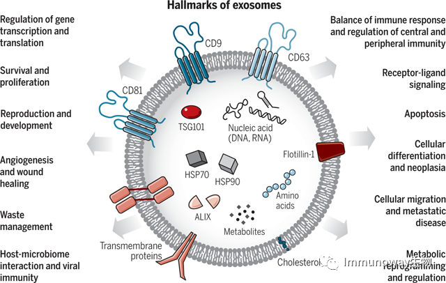
Exosomes: The Transport System Between Human Cells, with Various Functions
Exosomes are the transport system between human cells, with various functions. They are extracellular vesicles produced by all cells, carrying nucleic acids, proteins, lipids, and metabolites. They mediate both close-range and long-range intercellular communication in health and disease, influencing various aspects of cell biology.
Biogenesis and Heterogeneity of Exosomes
The process of exosome formation is summarized as follows: the cytoplasmic membrane invaginates, encapsulating some extracellular components and membrane proteins, forming early endosomes (ESEs). These endosomes can exchange materials with other organelles or fuse between different endosomes to form late endosomes, further forming intracellular multivesicular bodies (MVBs), which contain many intraluminal vesicles (ILVs) that may be released as exosomes. After the cell forms multivesicular bodies, they may be degraded by fusing with autophagosomes or lysosomes, or they may fuse with the plasma membrane, releasing their contents, including intraluminal vesicles, which are the exosomes that are ultimately formed.
Ras-related GTPase Rab, Sytenin-1, TSG101,ALIX, syndecan-1, ESCRT proteins, phospholipids, tetraspanins, ceramide, sphingomyelinase, and SNARE complex proteins are involved in the origin and biogenesis of exosomes.
Exosomal heterogeneity reflects their size, inclusions, impact on target cell functions, and cellular origins. Different exosomes have different effects on target cells, and such functional heterogeneity may lead one exosome to induce cell survival, while another induces apoptosis, and others induce immune modulation, targeting different types of target cells. Heterogeneity can also be based on the organs and tissues from which exosomes originate, including whether they come from cancer cells, granting them unique characteristics such as tropism for specific organs and uptake by specific cell types. The combination of all these features may enhance the complexity and heterogeneity of exosomes.
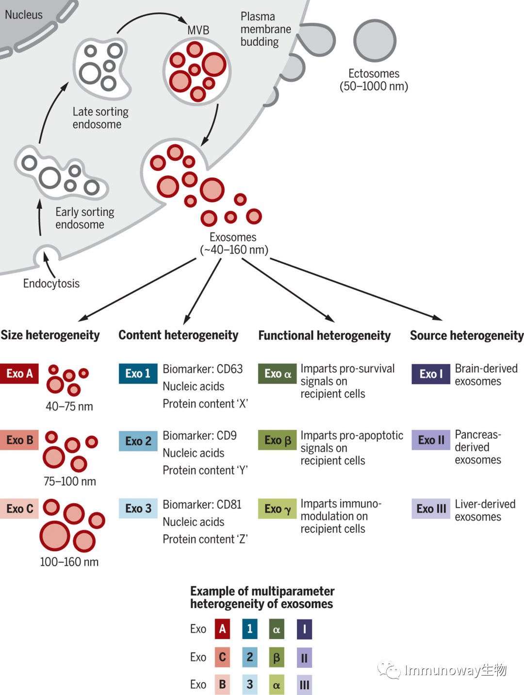
Fig. 1. Characteristics and Heterogeneity of Extracellular Vesicles and Exosomes
Extracellular vesicles are divided into two main categories: exosomes and ectosomes. Ectosomes are released by budding from the plasma membrane, ranging in size from 50 nm to 1 μm. Exosomes originate from the endosomal pathway, formed through the creation of ESEs and LSEs, and ultimately forming MVBs containing ILVs. When MVBs fuse with the plasma membrane, exosomes are released (ranging in size from 40-160nm). Exosomes can be a highly heterogeneous population with unique abilities to induce complex biological responses. Exosomal heterogeneity can be defined based on their size, composition, function, and effects on target and source cells. Different combinations of these characteristics lead to the complex heterogeneity of exosomes.
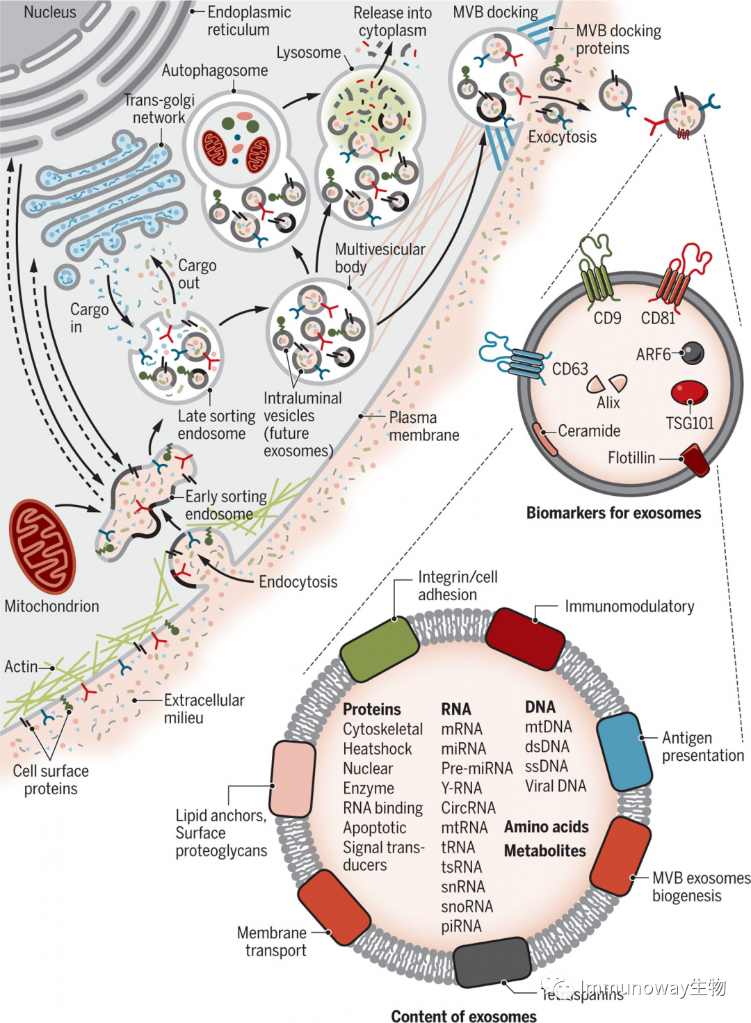
Fig. 2 Biogenesis and Identification of Exosomes
Liquid and extracellular components, such as proteins, lipids, metabolites, small molecules, and ions, can enter the cell along with cell surface proteins through endocytosis and plasma membrane invagination. The resulting membrane buds present an outward-to-inward orientation of the plasma membrane on the luminal side of the cell. This budding process leads to the formation of ESEs or may fuse with ESEs composed of the endoplasmic reticulum (ER), trans-Golgi network (TGN), and mitochondria. ESEs produce LSEs.The second invagination of LSEs leads to the generation of ILVs, which may undergo further modifications to form future exosomes, with cytoplasmic components entering the newly formed ILVs. Depending on the volume of invagination, this process can produce ILVs of different sizes and compositions. LSEs further form MVBs, which can fuse with autophagosomes and ultimately degrade in lysosomes. The degradation products can be recycled by the cell. MVBs can also directly fuse with lysosomes for degradation. MVBs can also be transported to the plasma membrane via the cell’s cytoskeleton and microtubule network, docking on the luminal side of the plasma membrane with the help of MVB docking proteins. As exocytosis occurs, the release of exosomes has a lipid bilayer orientation similar to that of the plasma membrane. Some proteins are involved in exosome biogenesis, including Rab GTPases, ESCRT proteins, and exosome marker proteins (CD9, CD81, CD63, flotillin, TSG101, ceramide, and Alix). Exosomes can contain different types of cell surface proteins, intracellular proteins, RNA, DNA, amino acids, and metabolites.
Intercellular Communication of Exosomes
The questions surrounding the functions of exosomes mainly focus on understanding their compositional makeup and the phenotypic and molecular changes they induce in target cells. Exosomes mediate intercellular communication through various mechanisms. For example, KRAS mutant human pancreatic cancer cells promote exosome uptake through pinocytosis. Human melanoma cells uptake exosomal material by fusing with the plasma membrane, while neurosecretory PC12 cells (derived from rat adrenal medullary tumors) are more reliant on clathrin-mediated endocytosis to uptake exosomes.
Mouse studies have shown that exosomes can also transmit mRNA information to recipient cells, occurring rarely under acute and chronic infection stimuli.
Exosomal protein components are closely related to the source cells. For example, exosomes derived from EGFR-vIII mutant glioblastoma cells often carry more invasive molecules. Inflammatory factors stimulate neural stem cells to release exosomes carrying IFNγ and its receptor, specifically activating the STAT1 pathway in recipient cells.
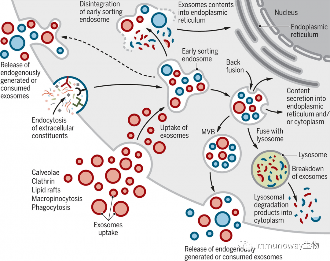
Fig. 3 Cell Journey of Internalized and Endogenous Exosomes.
Exosomes may enter cells directly through different mechanisms (red). Exosomes are formed by cells through endocytosis (blue). Exosomes are continuously produced and absorbed by cells. They are likely a mixture of newly formed and consumed exosomes (red and blue). It is still unclear whether endogenous or consumed exosomes are released together or separately. Absorbed exosomes can be degraded by lysosomes. Exosomes that enter cells may fuse with existing ESEs, subsequently disintegrating and releasing their contents into the cytoplasm. Additionally, nuclear endosomes can also fuse with the plasma membrane and release exosomes extracellularly.
Exosomes and Reproductive Development
Human reproduction, pregnancy, and embryonic development require precise, intricate, and dynamic intercellular communication. Human semen, amniotic fluid, blood, and breast milk all contain exosomes. Seminal exosomes are related to sperm maturation. Multiple analyses of exosomal molecules in human semen detected the presence of let-7a, let-7b, miR-148a, miR-375, and miR-99a, which regulate the expression levels of several interleukins, related to immune residence in the reproductive system. Seminal exosomal sources can also inhibit HIV-1 infection. Exosomes may facilitate viral infection by spreading viral components, and viruses may hijack exosome biogenesis pathways for survival. Exosomes act as pseudo-viral envelopes, invoking a “Trojan horse” strategy that facilitates viral entry into cells, thereby enhancing infectivity. While exosomes shed from virus-infected cells can promote infection, they also participate in antiviral immunity.
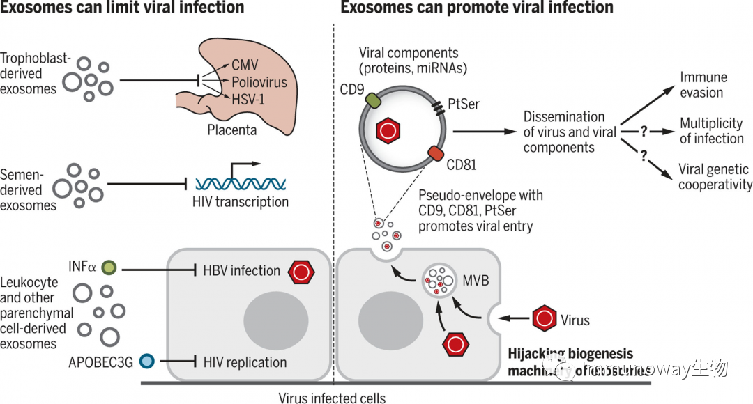
Fig. 4 Exosomes in Viral Infection
Exosomes can limit or promote viral infection. Exosomes releasing interferon α or APOBEC3G can inhibit infection by limiting viral replication or enhancing antiviral immunity. Viruses can also hijack exosome biogenesis mechanisms to facilitate viral spread. Exosomes can serve as pseudo-envelopes, enhancing viral entry into recipient cells through interactions with CD81, CD9, and PtSer, and helping evade antiviral immunity. The co-transport of viral components (proteins and miRNAs) may also enhance infectivity. Exosome-mediated viral transfer may participate in the genetics and diversity of viral infections.
Exosomes may assist in preventing placental infections. The levels of exosomal miRNAs and proteins in the plasma of pregnant women vary continuously. The detection of exosomes in mouse plasma confirms their association with pregnancy and preterm birth.
Exosomal miRNAs derived from breast milk contain immune-related miRNAs, which have also been found to promote the proliferation of porcine intestinal epithelial cells, but it remains unclear how they overcome digestive enzymes and how intestinal epithelial cells preserve nucleic acids and other components. This also suggests that different administration routes may affect tissue uptake of exosomes, impacting treatment strategies.
Exosomes and the Immune System
Studies have shown that low-dose injection of exosomes does not elicit significant immune rejection, and modified exosomes can induce adaptive and innate immune responses, which play significant roles in regulating immune responses and cancer.
The role of exosomes in immune regulation may be due to the transfer and presentation of antigen peptides. Exosomal DNA molecules activate immune effector pathways through the cGAS-STING sensing pathway.
Exosomes carrying MHC-peptides can directly activate the immune system through antigen presentation. Exogenous peptides transferred from exosomes to APCs initiate the activation of naïve T and/or B cells. Mouse dendritic cells can be activated by immunogenic peptides derived from exosomes, indirectly inducing the proliferation of CD4+T cells. It has also been confirmed that in bacteria, exosomes derived from macrophages activate the immune system through antigen presentation.
Exosomes carrying DNA and miRNAs are associated with regulating adaptive and innate immune responses. For example, exosomes carrying bacterial DNA stimulate nearby cells’ cGAS-STING signaling, effectively activating the innate immune response.
Exosomal DNA regulates immune responses and plays a role in cancer progression, with adaptive immune responses triggered by exosomes carrying tumor cell DNA activating dendritic cells, and activation of the cGAS-STING signaling pathway leading to antitumor responses.
Exosomes may also regulate immune responses by affecting gene expression and signaling pathways in recipient cells, primarily through the transfer of miRNAs. Exosomal miRNAs can be exchanged between dendritic cells, suppressing gene expression; this exosome-mediated intercellular communication may influence dendritic cell maturation.
Exosomal regulation of immune responses may also involve the presentation of immunoregulatory molecules such as PD-L1 and FasL. Exosomes derived from melanoma in vivo with PD-L1 inhibit the antitumor function of CD8+T cells, while exosomes from cancer cells obstruct the maturation and migration of dendritic cells through PD-L1. Additionally, PD-L1+ exosomes derived from tumor cells promote T cell exhaustion, facilitating tumor growth.FasL on exosomes from melanoma or prostate cancer induces Fas-dependent T cell apoptosis. In contrast, exosomes derived from mast cells expressing MHC-II, CD86, LFA-1, and ICAM-1 induce the proliferation of B and T cells both in vivo and in vitro. Exosomes derived from cancer cells overexpressing CD40L promote dendritic cell maturation, thereby enhancing T cell proliferation and antitumor activity in vivo.
Exosomes may decrease cytotoxicity to cancer cells in innate immune responses and limit antitumor immune responses in the tumor microenvironment, promoting cancer cell metastasis.
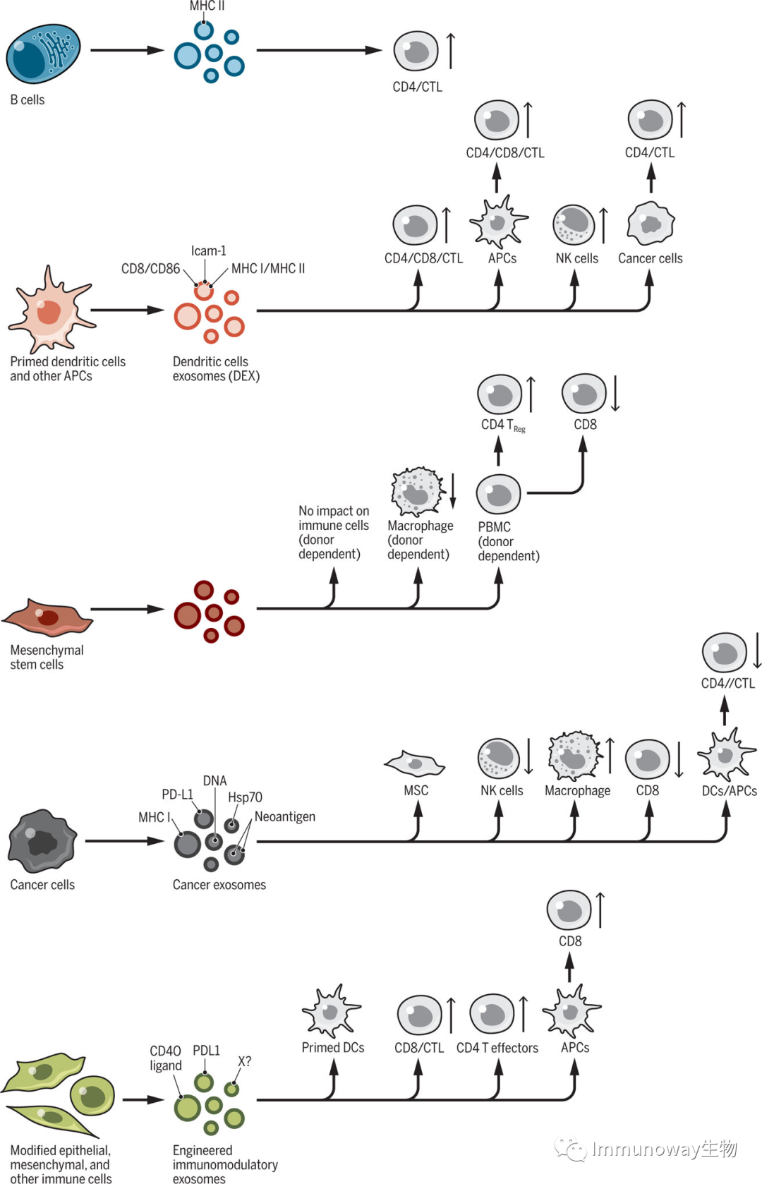
Fig. 5 Regulation of Immune Responses by Exosomes
Exosomes from different cell sources, including immune cells (B cells and dendritic cells), cancer cells, epithelial cells, and mesenchymal cells, release exosomes carrying substances that affect the proliferation and activity of the innate and adaptive immune systems. CD4+ and CD8+T cells [cytotoxic T cells (CTL)] can be directly or indirectly influenced by exosomes, stimulating or inhibiting their proliferation and function.
Exosomes and Metabolic and Cardiovascular Diseases
Exosomes may play roles in metabolic diseases and cardiovascular health, transferring metabolites and facilitating intercellular communication through exosomal miRNAs among mouse and human pancreatic β cells, adipose tissue, skeletal muscle, and liver. Exosomes in the supernatants of mouse and human cell cultures (endothelial cells, cardiac progenitor cells, cardiac fibroblasts, cardiomyocytes) are associated with metabolic diseases, including atherosclerosis, cardiovascular diseases related to diabetes, and metabolic adaptations related to heart failure. For example, exosomes may prevent atherosclerosis, as platelet-derived exosomes reduce the expression of the macrophage scavenger receptor CD36, thereby decreasing the uptake of harmful cholesterol; human smooth muscle cell-derived exosomes may promote thrombosis; and exosomes derived from stem cells protect the cardiovascular system.
Exosomes and Neurodegenerative Diseases
The relationship between exosome biogenesis and the regulation of vesicles secreted by neuronal cells provides new insights into the mechanisms of neurodegenerative diseases. Exosomes can promote or limit the aggregation of unfolded and misfolded proteins in the brain. Exosomes may participate in the clearance of misfolded proteins, thus playing detoxifying and neuroprotective roles, or participate in the propagation and aggregation of misfolded proteins, effectively promoting the “infectious” aggregation of proteins and disease progression. These opposing functions of exosomes may not be mutually exclusive.
Exosomes and Cancer
The research progress of exosomes in cancer is rapid. Exosomes influence tumor formation, growth, and metastasis, paraneoplastic syndromes, and treatment resistance. The role of exosomes in cancer progression may be dynamic and specific to cancer types, genetics, and stages.
Exosomes may induce or promote tumorigenesis. For example, exosomes from breast cancer and prostate cancer cells induce tumors by transferring their miRNAs. The plasticity of cancer cells may also be partially attributed to exosomes, as exosomal miR-200 from metastatic breast cancer cells enhances epithelial-to-mesenchymal transition (EMT) and the metastasis of other weakly metastatic breast cancer cells.
Exosomal effects from cancer cells on stromal cells can be fibroblasts or immune cells. Exosomes carry nucleic acids, signaling proteins, and metabolites that can have tumorigenic effects on stromal cells. For instance, miR-122 derived from breast cancer exosomes inhibits pyruvate kinase and lung glucose uptake, thereby promoting metastasis.
Exosomes from cancer cells induce signaling responses at metastatic sites, effectively remodeling distant microenvironments to enhance metastasis. For instance, TGFβ on the surface of exosomes derived from cancer cells induces fibroblast activation through the expression of αSMA and FGF2. Exosomes from cancer cells play roles in organ-specific metastasis in breast and pancreatic cancers, partly through the expression of exosomal integrins and organ-specific pro-inflammatory responses.
Exosome exchanges from stroma to cancer cells also regulate cancer progression and metastasis. For example, mtDNA in CAF-derived exosomes induces oxidative phosphorylation in breast cancer cells, promoting survival and metabolic dormancy in mice. miR-19a derived from astrocytes transferred to breast cancer cells leads to PTEN inhibition and promotes metastasis. Fibroblast-derived exosomes also stimulate breast cancer cell migration by inducing Wnt-PCP autocrine signaling, among others.
Exosomes also participate in angiogenesis and extracellular matrix remodeling in the tumor microenvironment, which are key steps in tumor growth and metastasis. Exosomal miR-105 from breast cancer cells inhibits the expression of endothelial cell tight junction protein ZO-1, compromising vascular integrity and enhancing vascular permeability, leading to increased metastasis. Hypoxic glioblastoma (GBM) cell-derived exosomes induce endothelial cell pro-angiogenic effects and GBM cell proliferation.
Exosomes shed from cancer cells can promote resistance to various chemotherapeutic drugs and antibodies. For instance, CD20+ exosomes from B-cell lymphomas act as decoys for anti-CD20 binding to B cells, while HER2+ exosomes from breast cancer cells act as decoys for anti-HER2 treatment, limiting their activity against cancer cells. CAF-derived exosomes promote chemotherapy resistance in colorectal cancer by enhancing the growth of cancer stem cells and facilitate the spread of resistance among cancer cell populations. This process may be mediated by the transfer of exosomal miRNAs, among others.
Diagnostic Potential of Exosomes
Exosomes exist in all biological fluids and are secreted by all cells, serving as minimally invasive liquid biopsies to track disease progression. Exosomal biogenesis can capture complex extracellular and intracellular molecules for comprehensive, multi-parameter diagnostic testing. Surface proteins on exosomes also assist in their immune capture and enrichment. The diseases of focus for exosomal diagnostics include cardiovascular diseases, diseases affecting the central nervous system (CNS), and cancer. The applications are rapidly expanding to liver, kidney, and lung diseases.
Small amounts of DNA can be found in exosomes, which is valuable for assessing cancer-related mutations through serum detection. Specific miRNAs or miRNA groups in exosomes may provide diagnostic or prognostic evidence in cancer detection. For instance, elevated circulating exosomal miR-21 is associated with glioblastoma and pancreatic cancer, colorectal cancer, liver cancer, breast cancer, ovarian cancer, and esophageal cancer, while elevated urinary exosomal miR-21 is associated with bladder cancer and prostate cancer.
Advancements have also been made in immune capture, such as capturing circulating tumor exosomes expressing CD147 in colorectal cancer. The potential of combining proteins, lipids, RNA, and miRNA in exosomes for cancer diagnosis and prognostic assessment is currently being considered. The combination of multi-faceted, combinatorial approaches may enhance the specificity and sensitivity of exosome-based diagnostics.
Therapeutic Potential of Exosomes
There is active exploration of using exosomes themselves or as drug carriers to become therapeutic agents. As shown in the figure below:
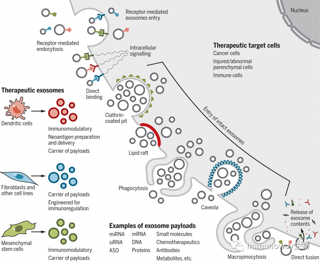
Fig.6.Cell Uptake of Therapeutic Exosomes
Therapeutic exosomes isolated from dendritic cells, fibroblasts, and mesenchymal cells can have specific effects on target cells, including novel antigen presentation, immune modulation, and drug payload delivery. The effects of therapeutic exosomes on target cells may be controlled by different entry or interaction mechanisms. The entry of intact exosomes involves receptor-mediated endocytosis, clathrin-coated pits, lipid rafts, phagocytosis, vesicles, and macropinocytosis. The entry of exosomal contents or signals induced by exosomes may involve ligand-receptor induced intracellular signaling or fusion, depositing exosomal contents into the cytoplasm. Examples of therapeutic payloads are listed. Target cells include cancer cells, damaged parenchymal cells, and immune cells. ASO stands for antisense oligonucleotides.
Unlike liposomes, injected exosomes can effectively enter other cells, providing functional substances in mice with minimal immune clearance. They also exhibit good tolerance. For instance, exosomes derived from mesenchymal cells and epithelial cells do not cause toxicity after repeated injections in mice. Exosomes derived from MSCs used to treat graft-versus-host disease show good tolerance with no significant side effects.
The enrichment of surface ligands on exosomes also facilitates the development of receptor-mediated tissue targeting. The enrichment of modified exosomal ligands can also be used to induce or inhibit signaling in recipient cells or target exosomes to specific cell types. For example, αv integrin-specific RGD (R, arginine; G, glycine; D, aspartic acid) modified peptides (a modified tumor-targeting peptide sequence that serves as an integrin recognition sequence) loaded onto doxorubicin-loaded exosomes from immature dendritic cells have therapeutic effects on breast tumor mice. Other chemotherapeutic compounds have also been loaded into exosomes for cancer therapy and tested in mice, demonstrating antitumor efficacy and low toxicity. For example, macrophage-derived exosomes loaded with paclitaxel can induce responses in lung tumors in mice.
Exosome delivery of miRNA or small interfering RNA has been utilized in the treatment of central nervous system diseases and cancer. Exosomal RNA may be protected from degradation by blood-derived ribonucleases, allowing exosomes to exert their functions distally. Preclinical trials have been conducted for anticancer treatment in breast cancer, gliomas, and pancreatic cancer, as well as exploratory brain-targeted therapies.
Exosomes aim to enhance antitumor immune responses in polarized tumor immune microenvironments. For instance, exosomes extracted from dendritic cells may elicit antitumor effects due to antigen presentation.
Conclusion
These intriguing studies on exosomal biology largely utilize cell culture systems, and further experimental exploration is needed using mouse models and physiologically relevant experimental conditions. Continuous precise understanding of their heterogeneity, composition and functional evolution is required. The physiological levels that exogenous high-dose exosomes can achieve in mice are related to the phenotype of infiltrating cells, including regulating cancer progression, inducing tumors, and tissue regeneration. It is still unclear whether unmanipulated exosomes exert stable regulatory or pathological functions in vivo. Further research is needed through animal model experiments.
Exosomes are produced by cells, yet one cannot help but wonder if they are similar to early primordial particles that contributed to the formation of the first primitive cell. Whether exosomes can grow and divide, and whether they participate in signaling and autonomous biochemical reactions in appropriate environments remains to be determined. The similarities between exosomes and retroviruses also raise the possibility that exosomes may function as primordial particles prior to unicellular organisms.
There are still many unknowns waiting to be explored…
If you are interested in the original text, you can click the “Read Original” button below.
Immunoway Exosome Research Related Product Recommendations:
| Item No. | Name | Item No. | Species | |
| 1 | YN2326 | ANXA5 Polyclonal Antibody | Human, Mouse, Rat | WB, ELISA |
| 2 | YP0041 | Calnexin (phospho Ser583) Polyclonal Antibody | Human, Mouse, Rat | WB, IHC-p, IF/ICC, ELISA |
| 3 | YM0089 | Calnexin Monoclonal Antibody | Human | WB, IF/ICC, ELISA |
| 4 | YT0613 | Calnexin Polyclonal Antibody | Human, Mouse, Rat | WB, IHC-p, IF/ICC, ELISA |
| 5 | YM3424 | Calnexin Polyclonal Antibody | Human, Mouse, Rat | WB, IHC-p, IF(paraffin section) |
| 6 | YT6027 | CD24 Polyclonal Antibody | Human | IHC-p, IF(paraffin section), ELISA |
| 7 | YT0741 | CD24 Polyclonal Antibody | Human, Mouse, Rat | WB, ELISA |
| 8 | YM1258 | CD54(ICAM-1) mouse mAb | Human | WB, ICC |
| 9 | YM1257 | CD54(ICAM-1) mouse mAb | Human | WB |
| 10 | YT5525 | CD63 Polyclonal Antibody | Human | IF/ICC, WB, IHC-p, ELISA |
| 11 | YT0772 | CD63 Polyclonal Antibody | Human | IHC-p, IF(paraffin section), ELISA |
| 12 | YT5394 | CD81 Polyclonal Antibody | Human, Mouse, Rat | IF/ICC, WB, ELISA |
| 13 | YN0174 | CD82 Polyclonal Antibody | Human | WB, ELISA |
| 14 | YT0782 | CD9 Polyclonal Antibody | Human, Mouse, Rat | WB, IHC-p, IF/ICC, ELISA |
| 15 | YM3402 | CYCS Monoclonal Antibody(4B10) | Human, Mouse, Rat, Chicken | WB, IF/ICC, IHC-p |
| 16 | YT5846 | CYCS Polyclonal Antibody | Human, Mouse, Rat | IF/ICC, WB, IHC-p, ELISA |
| 17 | YT1186 | CYCS Polyclonal Antibody | Human, Mouse, Rat, Monkey | WB, IHC-p, IF(paraffin section), ELISA |
| 18 | YT1560 | Endoplasmin Polyclonal Antibody | Human, Mouse | WB, IHC-p, IF/ICC, ELISA |
| 19 | YM0219 | EP-CAM Monoclonal Antibody | Human | WB, IHC-p, IF(paraffin section), ELISA |
| 20 | YM1034 | EP-CAM Monoclonal Antibody | Human, Mouse | WB |
| 21 | YM1242 | EpCAM mouse mAb | Human | WB, IP |
| 22 | YT1572 | EP-CAM Polyclonal Antibody | Human | WB, IF/ICC, ELISA |
| 23 | YT5556 | EP-CAM Polyclonal Antibody | Human, Mouse, Rat | WB, ELISA |
| 24 | YT6292 | Flotillin-1 Polyclonal Antibody | Human, Mouse, Rat | WB, ELISA |
| 25 | YT6291 | GM130 Polyclonal Antibody | Human, Mouse, Rat | WB, ELISA |
| 26 | YM3358 | HSC 70 Polyclonal Antibody | Human, Mouse, Rat | WB, IHC-p, IF(paraffin section) |
| 27 | YT2231 | HSC 70 Polyclonal Antibody | Human, Mouse, Rat | WB, ELISA |
| 28 | YM3482 | HSC70 Monoclonal Antibody(6C7) | Human, Rat, Mouse | WB, IHC-p, IF(paraffin section) |
| 29 | YM3497 | HSC70 Monoclonal Antibody(8G7) | Human, Rat, Mouse | WB, IHC-p, IF(paraffin section) |
| 30 | YM3715 | HSC70 Rabbit Polyclonal Antibody | Human, Mouse, Rat | WB, IHC-p, IF(paraffin section) |
| 31 | YP0137 | ICAM-1 (phospho Tyr512) Polyclonal Antibody | Human | WB, ELISA |
| 32 | YM0351 | ICAM-1 Monoclonal Antibody | Human | WB, ELISA |
| 33 | YM1051 | ICAM-1 Monoclonal Antibody | Human | WB, IF/ICC |
| 34 | YT2269 | ICAM-1 Polyclonal Antibody | Human | WB, IHC-p, IF(paraffin section), ELISA |
| 35 | YT5456 | Rab 5A Polyclonal Antibody | Human, Mouse, Rat | WB, IHC-p, IF(paraffin section), ELISA |
| 36 | YM1100 | Syntenin-1 Monoclonal Antibody | Human, Rat, Dog, Pig | WB |
| 37 | YT4760 | Tsg 101 Polyclonal Antibody | Human, Mouse, Rat, Monkey | WB, IHC-p, IF/ICC, ELISA |
| 38 | YM3359 | HSC 70 Polyclonal Antibody | Human, Mouse, Rat | WB, IHC-p, IF(paraffin section) |
| 39 | YT2232 | HSC 70 Polyclonal Antibody | Human, Mouse, Rat | WB, ELISA |
| 40 | YT0779 | CD83 Polyclonal Antibody | Human | WB, IHC-p, IF(paraffin section), ELISA |
| 41 | YT6283 | Alix Polyclonal Antibody | Human | WB, ELISA |
| 42 | 3YT629 | Thrombospondin 1 Polyclonal Antibody | Human, Mouse, Rat | WB, ELISA |
Exosome-related links:
Exosomes│Understanding Exosomes
Advantages of Immunoway Antibodies

1 The main feature of antibodies is that the immunogen is a polypeptide segment, and the purification process uses peptide-conjugated affinity chromatography columns to purify antibodies, resulting in antibodies with stronger specificity, and better specificity for WB, IF, and IHC.
2 Experimental data is rich, including multi-tissue, multi-species, multi-sample, and multi-application validations. Some pathology-grade immunohistochemistry-specific antibodies are validated using the CRISPR-Cas9 system, with customer feedback and selected data from published articles.
3 We have over 1500 modified antibodies and nearly 200 pathology-grade immunohistochemistry-specific antibodies.
4 There are over 18000 types of antibodies, including more than 15000 polyclonal antibodies and over 2000 monoclonal antibodies.
5 Applications cover routine applications such as WB, IHC, and IF/ICC, IP, Elisa, etc.
6 About 80% are in stock.
7 Reasonable prices and high cost-performance ratio.
Current Activities at Immunoway:
Gifts and Benefits│Great gifts for full orders await your selection!
Product Benefits│Gift packages for labels, internal references!
Other activities will be announced by authorized distributor partners of Immunoway.
Hyperlinks related to WB, IHC, IF:
|
Immunofluorescence Related |
|||
| IF│Application, Experimental Procedures, and Process Analysis | IF│Principles and Color Matching of Secondary Antibody Selection | IF│Common Problems and Solutions, Precautions | mIHC, mIF│Principles, Processes, and Optimization of Different Experimental Methods |
|
Immunohistochemistry Related |
||
|
Summary of Experimental Methods |
Immunohistochemistry (IHC)│Collection of Common Problems and Analyses | |
| Same HRP, Different Color Development Systems | Pathology Grade Secondary Antibodies│PK with International Top Brands | Immunohistochemistry-Specific Antibodies│Exclusive Luxurious Experience |
| When detecting proteins, WB OK, IF, IHC, why not OK? |
|
Western Blot Related |
||
|
Western Blot│Collection of Common Problems and Analyses of Experimental Steps |
Western Blot│RIPA is not a universal lysis buffer | Experimental Methods│What to do if I don’t want to do WB anymore? |
| Western Blot│Principles and Precautions for Internal Reference Selection |
Company Introduction:
ImmunoWay Biotechnology Company (referred to as Immunoway) was established in 2002 and registered its brand in Texas, USA in 2012.
The company invested in the research and development of immunohistochemistry-specific antibodies in 2014, guided by internationally renowned experts in monoclonal antibodies. The immunogens are designed specifically for immunohistochemistry experiments, with screening of monoclonal cell lines, and the verification of antibodies conducted through thousands of positive and negative slice tests, clarifying tissue localization and cellular sub-localization, resulting in strong specificity, high sensitivity, and low background. They are developed and produced in accordance with the registration and filing requirements for in vitro diagnostic reagents. Clinical immunohistochemistry is applied to assist in the diagnosis and differential diagnosis of pathology, providing objective guidance for disease treatment. Currently, the number of antibody types has approached 200, covering the majority of detection indicators required for tumor pathology diagnosis, such as breast cancer, cervical cancer, colorectal cancer, gastric cancer, liver cancer, lymphoma, prostate cancer, thyroid cancer, etc.
The company has been focused on the research and production of antibodies since its establishment, with over 18000+ types of polyclonal antibodies, over 2000+ types of monoclonal antibodies, over 15000+ types of polyclonal antibodies, over 1000+ types of secondary antibodies, over 700+ types of liquid sample detection Elisa kits, and over 3000+ types of cell sample detection Cell-Based Elisa Kits, meeting the needs of a wide range of research users.
Immunoway will continue to develop and produce high-quality antibodies and related products, providing better tools for clinical and research workers, serving the life sciences together.
Welcome to call 400-8787-807
Welcome to visit the website www.immunoway.com
For more exciting content, please long press the QR code▼