
Accurate diagnosis and staging of breast cancer are crucial for guiding clinical treatment strategies and predicting patient survival outcomes. Current clinical methods face limitations in detecting small tumors and micrometastases, such as imaging techniques; although biopsy remains the gold standard, its invasiveness makes it unsuitable for repeated use. Liquid biopsy is a promising solution that offers a non-invasive and convenient sampling method for early diagnosis and monitoring tumor progression during treatment. Key biomarkers targeted in liquid biopsies includeexosomes, circulating tumor cells, and circulating tumor DNA. Higher levels of exosomes are typically observed in advanced cancer stages, positioning them as biomarkers for monitoring tumor progression. However, existing quantitative methods for exosomes, such as nanoparticle tracking analysis (NTA) and flow cytometry, either fail to distinguish subtypes or require complex labeling and separation, which are time-consuming and lack sensitivity. Therefore, there is an urgent need to develop an exosome detection method that can accurately identify, is highly sensitive, and is easy to operate.Based on this, researchers from Sichuan University, Chen Piaopiao, Chen Jie, and Chang Kai from the Army Medical University developed an electrochemical aptamer sensor for rapid and sensitive detection of tumor-derived exosomes. This strategy employs a targeted responsive DNA nanohydrogel that integrates aptamers for specific recognition and uses Pb2+ as a signal output component. The key innovation of this homogeneous method lies in its ability to distinguish free Pb2+ from G-quadruplex-Pb2+ complexes through electrochemical analysis. Epithelial cell adhesion molecule (EpCAM) was targeted as a marker for breast cancer exosomes. The binding of the aptamer to the target triggers the disassembly of the DNA nanohydrogel, exposing a large number of G-quadruplexes that selectively bind Pb2+, thereby amplifying the electrochemical signal for sensitive detection. Additionally, the analytical system completes the process within 45 minutes, with a limit of detection (LOD) of 300 particles/mL. When applied to clinical samples, it accurately distinguishes breast cancer patients (n = 40) from healthy controls (n = 12), with specificity, sensitivity, and accuracy rates of 91.7%, 95%, and 96.2%, respectively. Furthermore, the method achieves an accuracy of up to 86.1% in distinguishing early and late-stage cancers. Overall, this exosome-based liquid biopsy method shows significant potential in improving breast cancer diagnosis and staging, supporting personalized treatment decisions.
Related research is titled “DNA nanohydrogel and Pb2 + -specific recognition enable one-pot electrochemical exosome aptasensor for staging of breast cancer” and will be published in Nano Today on July 30, 2025.
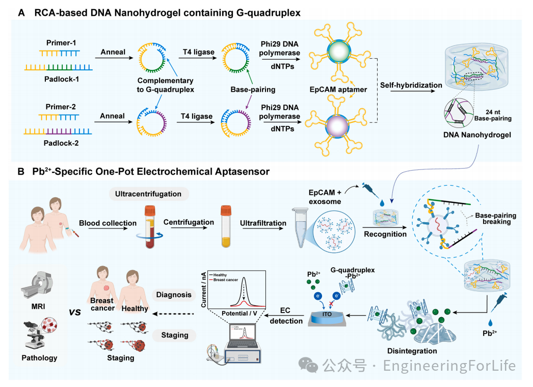
Illustration of electrochemical detection of breast cancer exosomes regulated by target proteins using DNA nanohydrogel without separation
This study developed a homogeneous electrochemical biosensor based on DNA nanohydrogels, integrating G-quadruplex for specific recognition of Pb2+ for detecting breast cancer-derived exosomes, which has potential applications in breast cancer diagnosis and staging (illustration).
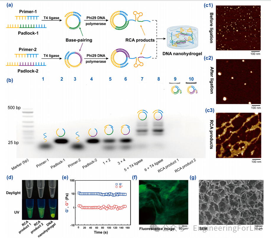
Figure 1 Preparation and characterization of DNA nanohydrogels
Using a reaction center polymer (RCA)-based synthesis strategy, DNA nanohydrogels were successfully prepared (Figure 1a). The effective formation of circular RCA templates and their subsequent products was confirmed by 2.5% agarose gel electrophoresis (Figure 1b). Experimental results showed that the migration speed of the circular RCA template (lanes 7 and 8) was slower than that of the linear single-stranded DNA control group (lanes 1-6), while RCA products 1 and 2 (lanes 9 and 10) were retained in the gel wells, indicating they have larger molecular weights. Meanwhile, atomic force microscopy (AFM) observations showed structural changes in DNA throughout the RCA process (Figure 1c). Mixing RCA products 1 and 2 resulted in a translucent hydrogel (Figure 1d), which exhibited distinct physical properties compared to the initial solution state, confirming successful gelation. Time-sweep experiments further evaluated the mechanical stability of the hydrogel, where G′ consistently maintained its dominance (Figure 1e). Fluorescence imaging after SYBR green I staining showed a uniform distribution of green fluorescence, confirming DNA strands have been successfully integrated into the entire hydrogel network (Figure 1f). High-resolution SEM images further revealed a three-dimensional interconnected fiber network, with fiber diameters ranging from tens to hundreds of nanometers (Figure 1g). This layered porous structure with multi-scale pore structure highlights the significant potential of DNA nanohydrogels in advanced applications for exosome detection and related biomedical fields.
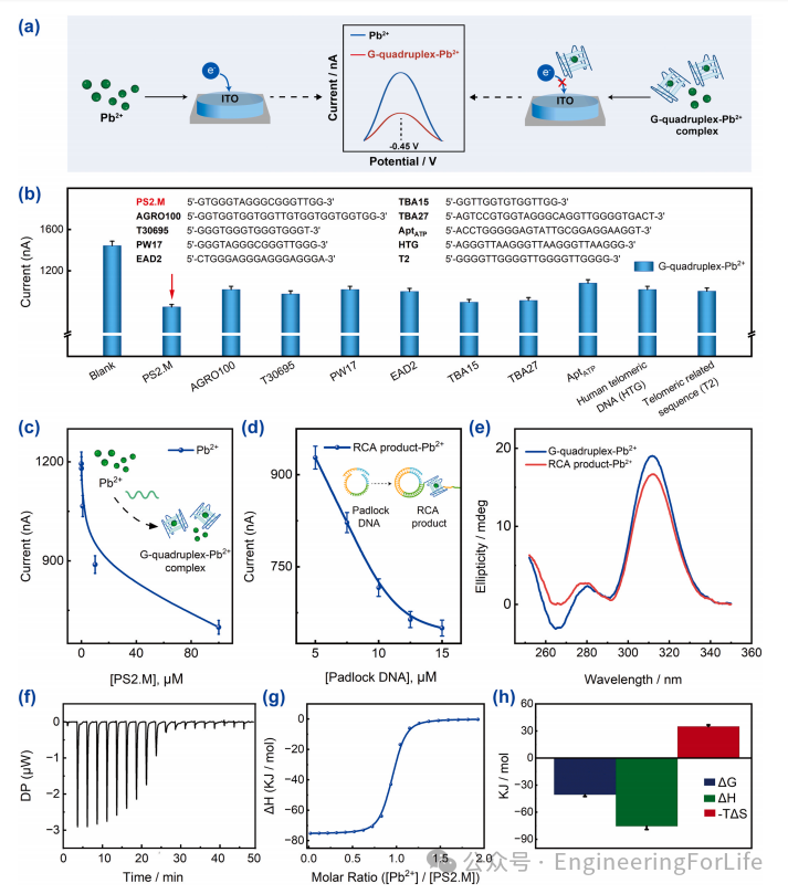
Figure 2 Verification of feasibility for Pb2+ specific recognition
As the concentration of G-quadruplex increases, the electrochemical signal generated by Pb2+ binding gradually decreases, indicating a direct relationship between G-quadruplex concentration and electrochemical signal intensity (Figure 2a). This linear relationship is crucial for ensuring reliable and consistent detection of G-quadruplexes and lays a solid foundation for the design of RCA sequences in subsequent electrochemical biosensing applications (Figure 2b, c). As RCA products accumulate, the electrochemical signal gradually weakens, confirming that RCA products bind to Pb2+ and promote the formation of G-quadruplexes, indicating the significant potential of RCA technology in enhancing electrochemical detection sensitivity (Figure 2d). Circular dichroism (CD) spectral results showed that comparative analysis of artificially synthesized G-quadruplexes exhibited similar spectral characteristics, displaying positive and negative bands at 310 nm and 265 nm, respectively (Figure 2e). The equilibrium dissociation constant (Kd) was determined to be 75.9 ± 22.1 nM through three measurements conducted using isothermal titration calorimetry (ITC), a value comparable to literature data.
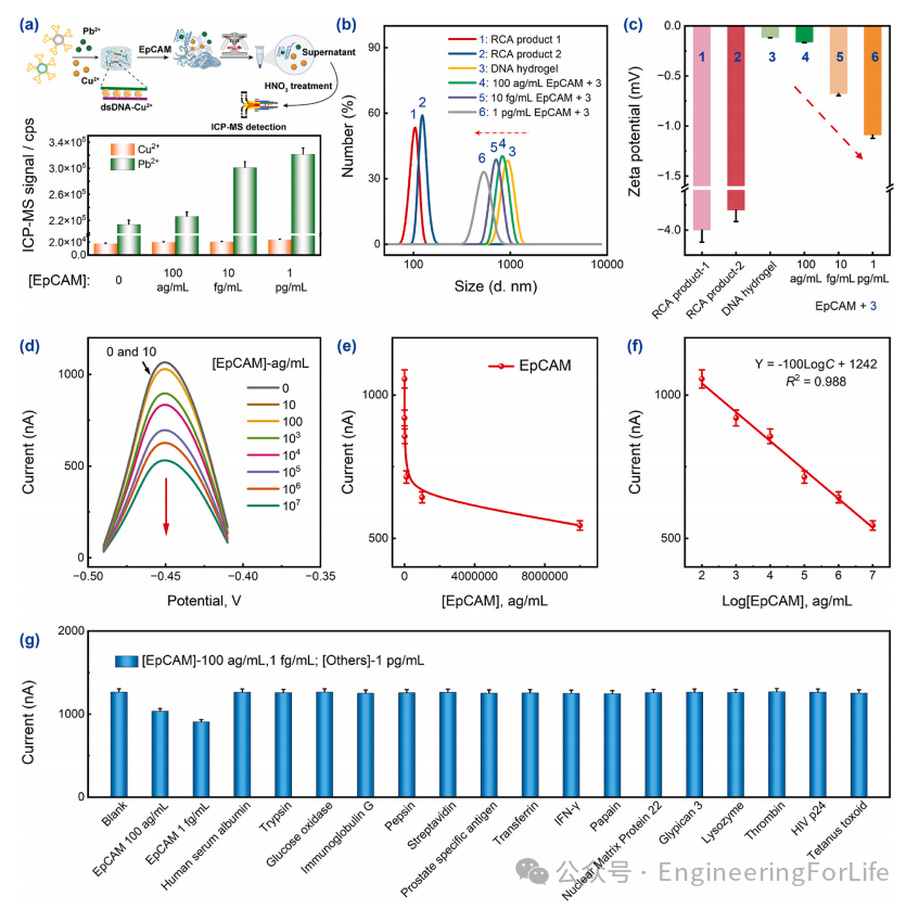
Figure 3 Feasibility and analytical performance of EpCAM analysis
Figure 3a shows the quantitative analysis results of free Cu2+ and Pb2+ concentrations after centrifugation of the system with EpCAM integrated into the DNA nanohydrogel. Dynamic light scattering (DLS) analysis confirmed that when proteins were added at concentrations of 100 ag/mL, 10 fg/mL, and 1 pg/mL, the average sizes of RCA product-1, RCA product-2, and DNA nanohydrogels were 106 nm, 122 nm, 955 nm, 825 nm, 712 nm, and 531 nm, respectively (Figure 3b). Meanwhile, as protein concentration increased, the Zeta potential exhibited a gradual negative shift (Figure 3c). Subsequently, the system’s ability to quantify proteins was studied, revealing a significant correlation between EpCAM concentration and peak current, with higher EpCAM levels resulting in lower Pb2+ current (Figure 3d, e). Similarly, a linear relationship was observed between peak current and the logarithm of EpCAM concentration within the range of 100 ag/mL to 10 pg/mL (Figure 3f). To explore specificity and minimize interference with protein analytical performance, a set of potential interfering proteins from peripheral blood was selected to assess their impact on the electrochemical sensor. The detected proteins included human serum albumin and several cancer biomarkers, such as glypican 3 (Figure 3g). Experimental results indicated that the system exhibited excellent specificity and reliability in complex biological samples, validating its application potential in exosome analysis.
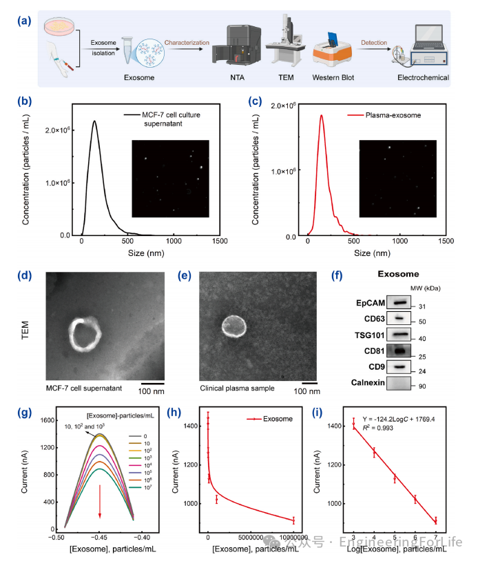
Figure 4 Characterization and analytical performance of exosomes
Exosomes extracted from MCF-7 cell supernatant and clinical plasma samples were characterized, focusing on key parameters such as size, concentration, morphology, and surface protein expression (Figure 4a). Nanoparticle tracking analysis (NTA) technology was employed for quantitative detection of exosome concentration and particle size distribution. The results showed that the concentration of exosomes isolated from MCF-7 cell supernatant was approximately 1.6×10¹⁰ particles/mL (Figure 4b). The concentration of exosomes from clinical plasma samples was slightly lower, at 5.9×10⁹ particles/mL (Figure 4c). TEM images confirmed that exosomes exhibited a typical cup-shaped structure, with a clearly visible bilayer membrane structure (Figure 4d, e). Key exosome markers were successfully detected, such as tetraspanins (CD9, CD63, and CD81), endosomal pathway markers (TSG101), and negative markers (calreticulin), as well as proteins typically associated with cancer cell-derived exosomes, such as EpCAM (Figure 4f). Performance evaluation was subsequently conducted using cell-derived exosomes. The regression analysis established the exosome concentration equation as Y = -124.2LogC + 1769.4, with an R² value of 0.993 (Figure 4g-i).
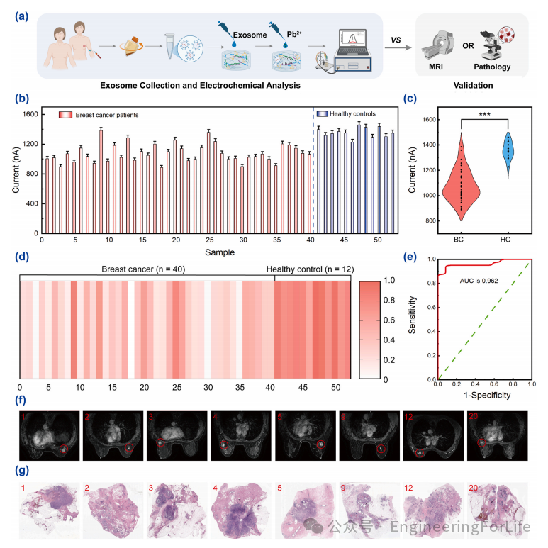
Figure 5 Workflow and results of clinical sample analysis
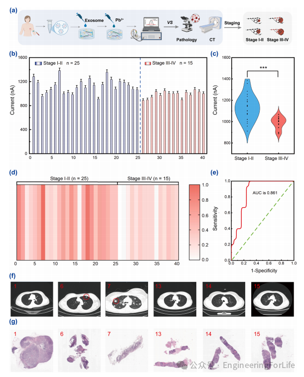
Figure 6 Analysis results of clinical breast cancer patient samples (early vs late)
The complete workflow for sample collection and subsequent analysis is shown in Figure 5a. Electrochemical analysis of clinical samples revealed significant differences in peak currents between healthy controls and breast cancer patients (Figure 5b, c). The expression level of exosomal EpCAM in breast cancer patients was significantly elevated, with a marked decrease in peak current. Additionally, a heatmap visually distinguished breast cancer patients from healthy controls, with lighter colors indicating higher EpCAM concentrations (Figure 5d). To further evaluate the diagnostic performance of the designed system, receiver operating characteristic (ROC) curve analysis was conducted (Figure 5e). The diagnostic specificity of the system reached 91.7% (11/12), with a sensitivity of 95% (38/40). More critically, the area under the curve (AUC) value reached 0.962, further validating the method’s stable diagnostic accuracy and highlighting its significant potential as a reliable tool for clinical breast cancer detection. Furthermore, the exosome detection results were highly consistent with magnetic resonance imaging (MRI) and pathological examination results (Figure 5f, g), further proving the reliability of this method.
Systematic analysis of samples from 40 patients with different disease stages further evaluated the clinical utility of the developed electrochemical detection system in breast cancer staging (Figure 6a). Electrochemical analysis displayed a stage-dependent pattern, with peak currents gradually decreasing as cancer stages progressed (Figure 6b, d). Quantitative comparisons between early (stage I-II) and late (stage III-IV) patients showed significantly lower electrochemical signals in late-stage cases (Figure 6c). ROC curve analysis further validated the diagnostic efficacy of this platform, with an AUC of 0.861 for distinguishing early and late breast cancer, achieving optimal sensitivity of 86.7% (13/15) and specificity of 76% (19/25) (Figure 6e). In this experimental system, comparisons between patient 1 and patient 7, with computed tomography (CT) results and pathological results confirming that patient 1 had no distant organ metastasis while patient 7 had metastasis, indicated a higher degree of breast cancer progression (Figure 6f, g). In summary, these findings emphasize the potential clinical value of a non-invasive electrochemical platform as a reliable auxiliary tool for breast cancer staging and treatment strategy formulation, especially in situations where traditional imaging modalities may be limited.
Conclusion
In summary, this study successfully developed a one-step electrochemical aptamer sensor for detecting tumor-derived exosomes. This method combines aptamer-based recognition with targeted responsive DNA nanohydrogels to induce the formation of G-quadruplex-Pb2+ complexes and utilizes the electrochemical differentiation output signal of Pb2+. The detection can be completed quickly and efficiently within 45 minutes. The method was further applied to the detection of exosomes in clinical samples, showing a high degree of consistency with clinical diagnosis and staging, providing an auxiliary tool for the development of personalized treatment strategies and management. Although EpCAM is a representative marker, it does not comprehensively capture the heterogeneity of breast cancer. Future research should focus on integrating more signaling molecules to develop multi-target systems to address potential limitations associated with subtype variability. Additionally, to further reduce the limitations posed by signal shut-off systems, future work will concentrate on employing background correction methods or exploring new binding ligands, utilizing the multifunctionality of G-quadruplexes to achieve signal activation strategies.
References:
https://doi.org/10.1016/j.nantod.2025.102854
Source: EngineeringForLife
Disclaimer: The views expressed are solely those of the author and are for research purposes. The author has limited expertise, and if there are any scientific inaccuracies, please leave comments below!