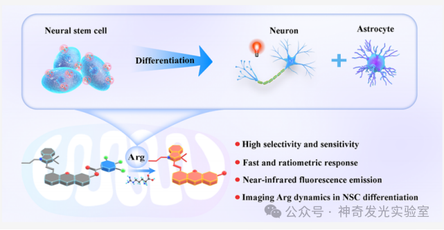
Neural stem cell (NSC) differentiation is one of the core processes in the central nervous system, involving the regulation of various bioactive molecules such as growth factors, reactive oxygen species, and amino acids. Among these, arginine (Arg) plays a crucial role in NSC differentiation. Once the levels of arginine become excessive or uncontrolled, it disrupts the normal differentiation of NSC, impairing their neural repair and regeneration capabilities. This abnormality may trigger various conditions such as neurodegenerative diseases (e.g., Alzheimer’s disease and Parkinson’s disease), inflammation, and cardiovascular diseases. Therefore, developing arginine quantification tools with high selectivity and precision is of great significance, especially to help understand the intrinsic mechanisms of arginine regulation in NSC differentiation.
Traditional mass spectrometry-based methods struggle to achieve real-time monitoring and in situ tracking of arginine dynamics in living cells and the brain. Fluorescence spectroscopy combined with probe technology can provide direct imaging and biosensing information for chemical substances in living systems. Although various fluorescence probes have been reported for studying the role of arginine in biological systems, these probes are often interfered with by arginine analogs such as lysine, histidine, and glutamic acid. This cross-reactivity can affect the accuracy and reliability of arginine detection in complex biological systems.
In this study, a series of phenyl activated ester-derived fluorescence probes Rap1–9 were designed and synthesized, regulating the nucleophilic substitution reaction between phenyl activated esters and arginine’s α-amino group through the electronic effects of substituents. Among them, the near-infrared (NIR) emitting ratiometric fluorescence probe Rap-9, due to the strong electron-withdrawing effect of the trifluoromethyl substituent, exhibited the best performance among the nine designed molecules, achieving high specificity and sensitivity for arginine ratiometric quantification with rapid dynamic changes of 3.6±0.04 seconds. The developed Rap-9 probe was applied to quantitatively track the differentiation process of NSC stimulated by O2·–, discovering that arginine is a key regulatory factor in directing the transition of quiescent neural stem cells (qNSCs) to activated neural stem cells (aNSCs), while also regulating the cell differentiation pathway. Notably, when the arginine concentration was 14.48±0.77μM, the efficiency of NSC differentiation into neurons was highest. Further proteomics analysis indicated that arginine-mediated mTORC1 pathway activation could drive the energy metabolism transition from glycolysis to more efficient oxidative phosphorylation, thereby regulating the transition of qNSCs to neuronal differentiation. These findings significantly advance the understanding of the metabolic regulatory mechanisms of NSC differentiation, providing new insights for neuroscience research and opening new avenues for developing targeted therapeutic strategies.
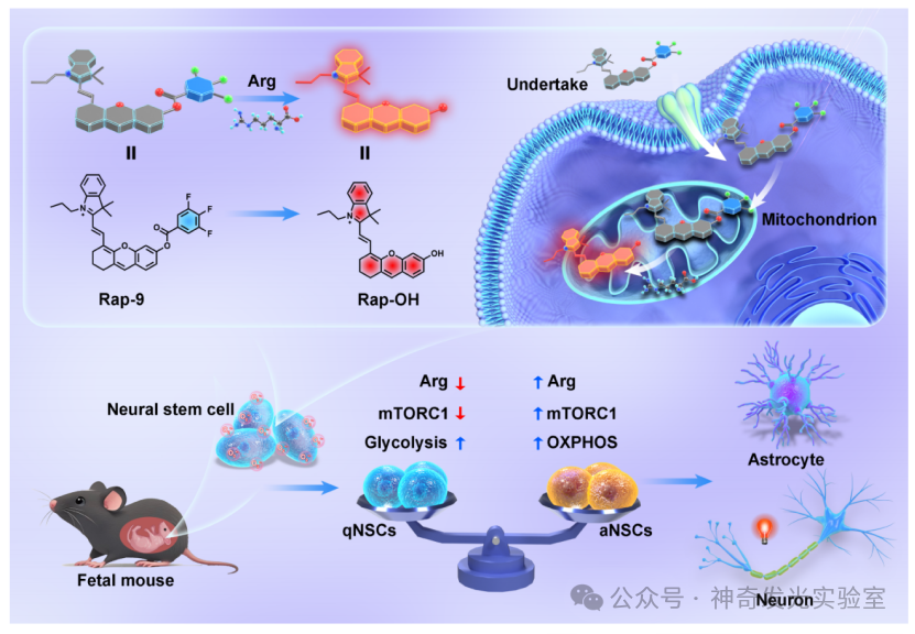
Figure 1: Design Strategy
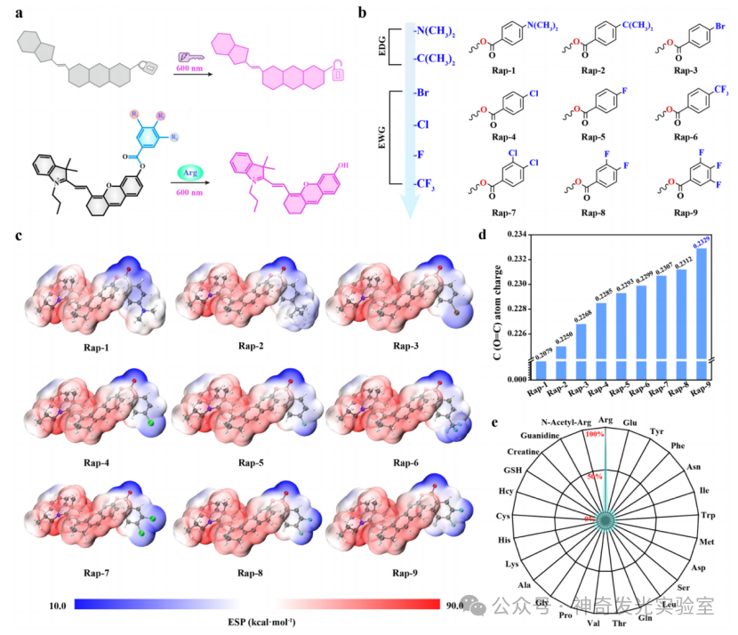
Figure 2: Design strategy and screening process of the probe Rap1−9.
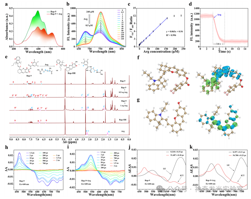
Figure 3: Perception mechanism of Rap-9 in response to Arg
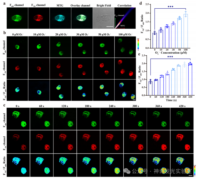
Figure 4: Performance characterization of the probe Rap-9 in NSC.
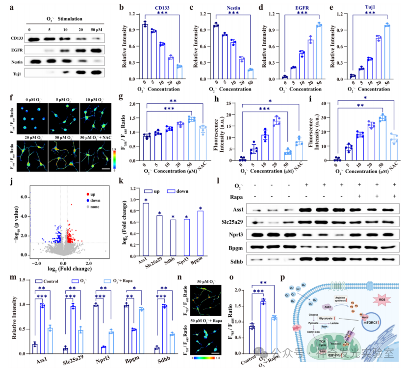
Figure 5: Mechanistic exploration of O2−stimulated NSC differentiation.
[Literature Details] Jin Yan, Yuxiao Mei, Yudan Zhao, and Yang Tian. Lighting Up Arginine Metabolism Dynamics in Neural Differentiation through Ratiometric Fluorescence Imaging. J. Am. Chem. Soc., 2025, https://doi.org/10.1021/jacs.5c05685