
Abstract: Scientists have pioneered a new method to aid in the repair of spinal cord injuries by combining 3D printing, stem cell technology, and lab-cultured tissues.
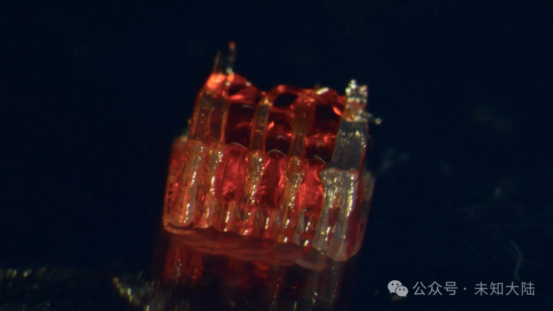
The method involves creating a unique 3D printed framework for lab-cultured organs, known as organoid scaffolds, which feature microscopic channels. Image source: McAlpine Research Group, University of Minnesota.
The research team at the University of Minnesota Twin Cities has demonstrated a groundbreaking process that combines 3D printing, stem cell biology, and lab-cultured tissues for spinal cord injury recovery.
The study was recently published in the peer-reviewed scientific journal Advanced Healthcare Materials.
According to the National Spinal Cord Injury Statistical Center, over 300,000 people in the United States suffer from spinal cord injuries, with no current method to fully reverse the damage and paralysis caused. The main challenges are the death of nerve cells and the inability of nerve fibers to regenerate at the injury site. This new research aims to address this issue.
The method includes creating a unique 3D printed framework for lab-cultured organs, known as organoid scaffolds, which contain microscopic channels. Region-specific spinal cord neural progenitor cells (sNPCs) are then implanted into these channels; these cells are derived from human adult stem cells and have the ability to divide and differentiate into specific types of mature cells.
“We utilize the 3D printed channels on the scaffold to guide the growth of stem cells, ensuring that new nerve fibers grow in the intended manner,” said Guebum Han, a former postdoctoral researcher in mechanical engineering at the University of Minnesota and the first author of the paper, who is now employed at Intel. “This method creates a relay system that can bypass the damaged area when placed in the spinal cord.”
In the study, researchers transplanted these scaffolds into the spinal cords of rats with complete spinal cord transections. These cells successfully differentiated into neurons and extended nerve fibers in both directions—toward the head (rostral) and toward the tail (caudal)—forming new connections with the host’s existing neural circuitry.
Over time, the new nerve cells seamlessly integrated into the host’s spinal cord tissue, leading to significant functional recovery in the rats.
Ann Parr, a professor of neurosurgery at the University of Minnesota, stated, “Regenerative medicine has opened a new era for spinal cord injury research. Our lab is very excited to explore the potential of ‘mini-spinal cords’ for future clinical translation.”
Although this research is still in its early stages, it brings new hope to patients with spinal cord injuries. The team hopes to scale up production and continue developing this technology combination for future clinical applications.
In addition to Han and Parr, the team members include Hyunjun Kim and Michael McAlpine from the Department of Mechanical Engineering at the University of Minnesota; Nicolas S. Lavoie, Nandadevi Patil, and Olivia G. Korenfeld from the Department of Neurosurgery at the University of Minnesota; Manuel Esguerra from the Department of Neuroscience at the University of Minnesota; and Daeha Joung from the Department of Physics at Virginia Commonwealth University.
This work was funded by the National Institutes of Health, the Minnesota Spinal Cord Injury and Traumatic Brain Injury Research Program, and the Spinal Cord Society.
3D Printed Spinal Cord Scaffold (Video)
Journal Reference:
Guebum Han, Nicolas S. Lavoie, Nandadevi Patil, Olivia G. Korenfeld, Hyunjun Kim, Manuel Esguerra, Daeha Joung, Michael C. McAlpine, Ann M. Parr. 3D Printed Scaffolds Promote Spinal Cord Organoid Formation for Spinal Cord Injury Treatment. Advanced Healthcare Materials, 2025; DOI: 10.1002/adhm.202404817