
In recent years, 3D printing technology has played a crucial role in fields such as medical devices, bioengineering tissues, and soft robotics. However, traditional printing processes still rely on users to predefine models through CAD software, and the printers themselves cannot perceive the material composition and structural features in the printing environment. Especially in bioprinting, cells are often randomly distributed, making it difficult to achieve complex structures that fit precisely like vascular networks, thus limiting the construction of functionalized tissues. Despite advancements in embedded sensors and feedback control, the printer’s ability to respond to context remains in its early stages.
Researchers from Utrecht University in the Netherlands led by Riccardo Levatohave proposed a novel printing technology called GRACE (Generative, Adaptive, Context-Aware 3D Printing), which combines three-dimensional imaging, computer vision, and parametric modeling to enable real-time generation of complex geometric structures that adapt to the surrounding environment during volumetric printing.GRACE can print around features ranging from cellular to macroscopic scales without manual intervention, significantly enhancing the functionality and structural accuracy of printed objects. This technology also supports multi-material printing, automatic alignment, and shadow correction, opening new avenues for tissue engineering and regenerative medicine. The related paper titled “Adaptive and context-aware volumetric printing” was published in Nature.
The researchers integrated light-sheet microscopy with the volumetric printing system to achieve rapid three-dimensional imaging and recognition of embedded features within the printed volume. The light-sheet can capture fluorescence signals from multiple angles, and after algorithm processing, point cloud data is generated, which is then used to accurately locate cell clusters, microparticles, or organoids through clustering analysis (e.g., DBSCAN algorithm). Based on this data, parametric modeling software automatically generates complex structures surrounding the features, such as mimicked vascular channel networks, connecting scaffolds, or encapsulating shells, with the entire process taking only a few minutes.
Figure 1 shows a schematic diagram of the experimental GRACE printing system, including light-sheet imaging (green light path), printing light source (purple light path), and image acquisition module. All light paths converge in a printing bottle filled with photosensitive resin, which is aligned in a refractive index matching liquid to minimize optical distortion during imaging and printing.
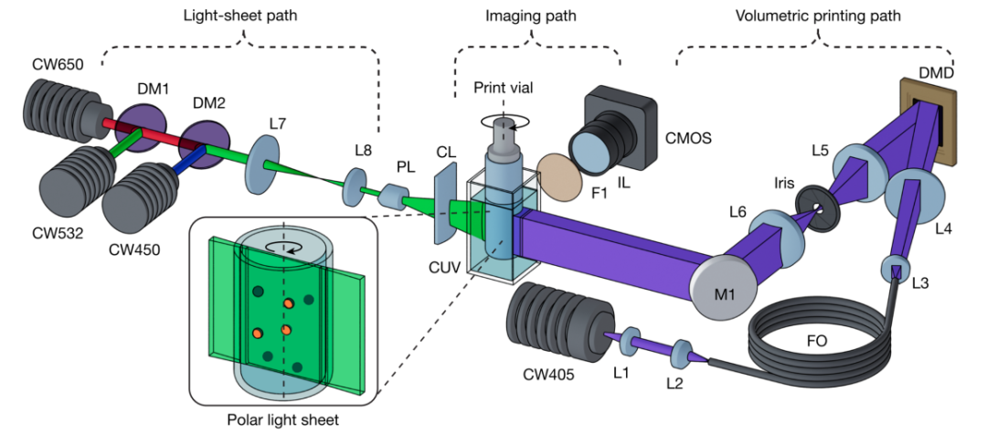
Figure 1: Schematic diagram of the experimental GRACE printing device. The light paths include light-sheet (green) and imaging and printing (purple) paths. The printing light path contains a 405 nm laser source, DMD chip, and relay optical system; the light-sheet path includes three wavelength laser sources, a Powell lens, and a cylindrical lens. The imaging part captures fluorescence signals through a CMOS sensor. The printing bottle is placed in a refractive index matching cubic pool to reduce optical errors.
Figure 2 illustrates the various capabilities of GRACE in generating complex structures. The researchers used fluorescently labeled alginate microspheres to simulate cell clusters or organoids, successfully printing a spherical winding channel network around the microspheres in GelMA hydrogel (Figure 2a), a supporting structure connecting multiple microspheres (Figure 2b), and precise encapsulation of a single feature (Figure 2c). Additionally, the system can distinguish different populations based on microsphere size (Figure 2d) or fluorescence signals (Figure 2e) and generate different geometric forms, such as creating a single channel for smaller microspheres and a complex network for larger or specifically fluorescently labeled microspheres. Figure 2f demonstrates the application of GRACE in automatic alignment sequential printing: through the Iterative Closest Point (ICP) algorithm, the system can accurately align a cartilage model to the surface of a printed femoral head, achieving high repeatability in multi-component constructions.
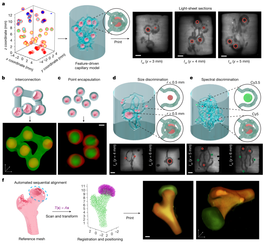
Figure 2: GRACE printing generates adaptive and feature-driven complex geometric structures a, spherical winding channel network generated around alginate microspheres; b, printed supporting structure connecting multiple microspheres; c, encapsulation of a single microsphere; d, channel generation distinguished by size; e, different geometric forms distinguished by fluorescence signals; f, example of automatic alignment sequential printing, aligning the cartilage model to the femoral head.
In response to shadow interference caused by opaque structures during printing, GRACE achieves shadow correction through light-sheet surface mapping and Object Space Model Optimization (OSMO) technology. Figure 3a–h illustrates this process: the light-sheet is used as a profilometer to reconstruct the surface of the occluded structure through reflected signals (Figure 3a), and then the reconstruction results are optimized through OSMO (Figure 3b). The researchers used ten columns as an occlusion model (Figure 3c), and when printing gear structures, the corrected samples showed more uniform crosslinking behavior and clearer details (Figure 3d). In a more complex “caged sphere” model (Figure 3e), the surface error of the printed sphere was significantly reduced after correction, and the sphericity was notably improved (Figure 3f–h), demonstrating that this method is suitable for high-precision printing in complex continuous occlusion scenarios.
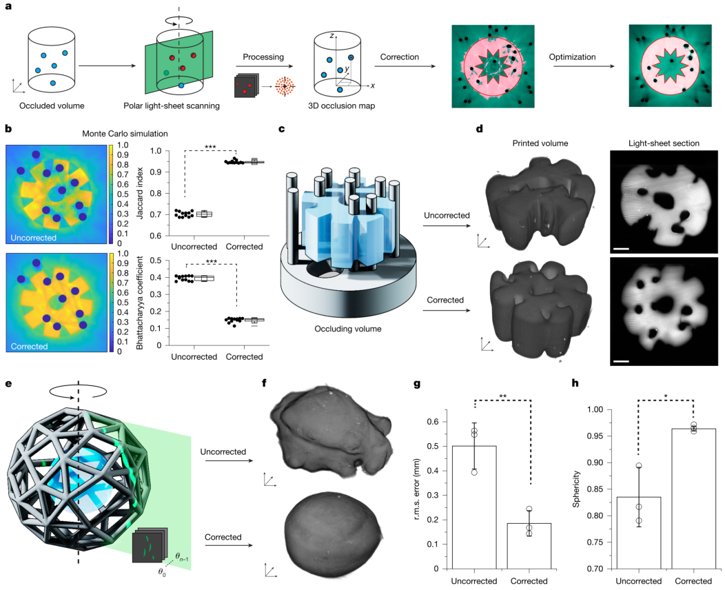
Figure 3: Light-sheet mapping of occluded structures and shadow correction a, scanning, mapping, and correction flowchart; b, comparison of gear reconstruction before and after shadow correction using OSMO; c, occlusion model of columns printed by stereolithography and target gear geometry; d, 3D reconstruction and light-sheet cross-section of corrected and uncorrected prints; e, rendering of the caged sphere model; f, reconstruction of the printed sphere; g, root mean square error of the surface; h, sphericity statistics.
In bioprinting applications, GRACE demonstrates superior performance. Figure 4a–d shows the printing of an adaptive vascular network around insulin-secreting cells (iβ-cells): compared to control groups with random channels or no channels, samples printed with GRACE exhibited higher insulin secretion after dynamic culture, indicating superior material transport capabilities. Figures 4e–f illustrate the bone-cartilage multi-tissue structure printed with automatic alignment: cells remained active and secreted specific matrices after 4 weeks of culture, and histological analysis showed distinct mineralized bone and cartilage regions. Furthermore, combined with Flight printing technology, GRACE can also distinguish different fluorescently labeled cell clusters and encapsulate them in star-shaped or circular fiber structures (Figures 4g–j), demonstrating its compatibility and adaptability to various printing modes.
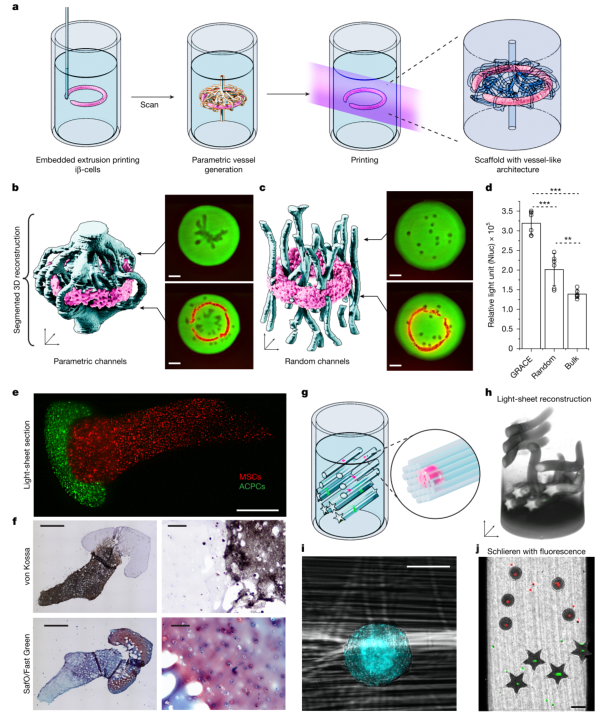
Figure 4: GRACE achieves cell position-driven functional living tissue bioprinting a, experimental process: scanning the annular structure, generating models, and printing scaffolds; b-c, 3D reconstruction and light-sheet cross-section of targeted and random channel structures; d, comparison of insulin release amounts from iβ-cells; e, light-sheet cross-section of the femur-cartilage structure; f, histological slices showing mineralized bone and cartilage regions; g, Flight printing structure model; h, 3D reconstruction after printing; i, fiber structures surrounding the clusters; j, fluorescence overlay images showing precise positioning of clusters in star-shaped/circular structures.
GRACE technology integrates light-sheet imaging, computer vision, and parametric modeling to achieve real-time perception and adaptive manufacturing of features within the printed volume, greatly enhancing the automation level and functional complexity of printing. This workflow is not only applicable to the field of bioprinting but can also be extended to other printing methods such as Xolography and acoustic printing, providing a new platform for soft robotics, multi-material manufacturing, and regenerative medicine. In the future, by integrating a broader range of imaging and printing systems, developing anti-scattering technologies, and merging with self-assembling materials, GRACE is expected to further approach the multi-level complexity of human tissues, achieving precise manufacturing from macroscopic to nanoscale.
 Source: Polymer Science FrontiersDisclaimer: The views expressed are solely those of the author. The author’s expertise is limited, and if there are any scientific inaccuracies, please leave comments below!