Introduction
At present, graphene, as the thinnest, strongest, and most conductive new nanomaterial known, has a promising development prospect recognized by many scientists. As Frank Wilczek, who won the Nobel Prize in Physics in 2004, said: “Graphene may be the only instance from quantum theory to application.” Graphene-based electrochemical biosensors have significant application value in fields such as environmental monitoring, fermentation industry, biomedicine, food analysis, and military. This article reviews the applications of graphene electrochemical biosensors in medical detection over the past five years, providing analytical interpretations to support future applications of graphene biosensors in the biomedical field.
1 Overview of Graphene
1.1 Graphene
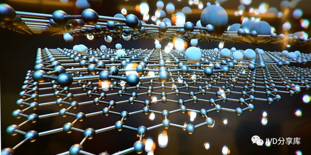
Graphene (Graphene) has a two-dimensional structure and is a crystal formed by sp2 hybridized single-layer carbon atoms, with its basic structural unit being the most stable benzene hexagon in organic materials. Each carbon atom forms three stable C-C bonds through σ bonds, while the remaining π electrons can form delocalized large π bonds with the π electrons of other carbon atoms, thereby enhancing electron mobility and giving graphene excellent conductivity. Chemically, graphene can also be viewed as a network structure formed by removing hydrogen atoms from polycyclic aromatic hydrocarbons. Additionally, whether in a free state or deposited on a substrate, graphene is not completely flat but exhibits its “intrinsic” wrinkles on the surface. The size of these wrinkles affects the electrical and optical properties of graphene. Besides the “intrinsic” wrinkles, graphene also has other “defects” in real three-dimensional space.
1.2 Graphene Oxide
Graphene oxide (GO) is derived from graphite using strong oxidizers, making it an important derivative of graphite, also known as functionalized graphene. The structure of graphene oxide is similar to that of graphene, both having a two-dimensional layered structure with hydrogen bonds and other forces between the layers. However, due to the action of oxidizers, graphene oxide surfaces contain a large number of oxygen-containing active groups. Surface element analysis (XPS), high-resolution solid-state (13C-NMR), and infrared spectroscopy (FT-IR) tests show that these oxygen-containing functional groups mainly include hydroxyl, carboxyl, epoxy groups, and carbonyls. GO has hydroxyl and epoxy functional groups on its basal plane, while many carboxyl and carbonyl groups are found on its edges, resulting in GO exhibiting strong hydrophilicity and good dispersion in water. Moreover, the layers of graphene oxide carry negative charges, promoting electrostatic repulsion, which is a crucial factor for its good dispersibility. The degree of oxidation determines the relative numbers of these two parts, and the presence of certain groups (benzene rings, double bonds, epoxy groups) defines the planar “network” structure of graphene oxide. The carbon atoms in hydroxyl functional groups can undergo angular transformations into tetrahedral structures, resulting in some wrinkles on the surface of GO.
1.3 Reduced Graphene Oxide
During the reduction process, the microstructure and properties of graphene oxide (GO) change, which serves as the basis for determining the degree of reduction of graphene oxide. Reduced graphene oxide (rGO) has increased surface charge carrier concentration and electron mobility, enhancing its reflection of incident light, resulting in rGO films often having a metallic luster. During the reduction of graphene oxide, the brown GO solution transforms into brown or black rGO, which is considered an intrinsic change of the reduction reaction, especially in reactions conducted in solution, attributed to the elimination of oxygen-containing active groups from graphene oxide, leading to increased hydrophobicity of rGO. The formation of CO and CO2 during the reduction process creates “voids” on the basal plane. Graphene oxide itself is fundamentally an insulator and loses the good conductivity inherent in graphene; therefore, another significant purpose of reducing graphene oxide is to restore its intrinsic properties and increase its conductivity. Thus, the improvement in conductivity becomes strong evidence for the reduction of graphene oxide.
2 Applications of Graphene Electrochemical Biosensors in Medical Detection
Graphene’s unique physical structure, along with its chemical and electrical properties, makes it highly suitable for sensor technology. In recent years, graphene and its polymer/metal particle composites have been used to immobilize biomolecules such as antibodies, DNA, and enzymes to create highly sensitive and selective biosensors. Based on the chemical structure of graphene, biomolecules are attached to the surface of graphene primarily through covalent bonds, reactive coupling, and physical adsorption, using straightforward and rapid electrochemical methods to detect and identify various diseases, pathogens, and disease-related biomolecules and viruses.
2.1 Glucose Detection
Glucose plays a crucial role in the metabolic pathway of adenosine triphosphate (ATP) production in the human body. However, abnormal (high) glucose levels in bodily fluids can lead to severe complications such as blindness, heart disease, hypertension, and kidney failure. Therefore, detecting glucose levels in biological fluids is of great significance in the biomedical research for the prevention and control of diabetes. In this regard, glucose oxidase has been used to develop various analytical methods, including quantum dot-based fluorescence, electrochemical, optical, chemiluminescent sensors, and surface plasmon resonance for glucose detection in biological samples. Among these, electrochemical sensors have significant advantages in glucose detection due to their high sensitivity, rapid response, cost-effectiveness, and selectivity.
Yuan et al. used 6-(ferrocenyl)hexanethiol (Fc-C6H12-SH) as an electron transfer medium, sputtering gold nanoparticles onto single-layer graphene (SLG) produced by chemical vapor deposition (CVD) to obtain gold nanoparticle-modified single-layer graphene (Au/SLG). They immobilized glucose oxidase (GOD) on the (Au/SLG) modified glassy carbon electrode to prepare a glucose biosensor (GOD/Fc/Au/SLG/GCE). This process avoids the complex polymer transfer process of graphene, providing good dispersion and clean surfaces for Au NPs, promoting the immobilization of GOD. Due to the clean surface of CVD-generated graphene and low background current, the GOD/Fc/Au/SLG/GCE is suitable for ultra-sensitive detection of low glucose concentrations. This sensor synergistically interacts with the electronic medium, facilitating the electron transfer process and enhancing electrochemical performance, with a detection limit of 0.1 nM (S/N=3) and good selectivity.
Miao et al. employed cyclic voltammetry (CV) to simultaneously deposit graphene-gold nanoparticle hybrid materials on nickel (Ni) surfaces to prepare RGO/Au/Ni electrodes, then covalently immobilized glucose oxidase (GOD) on the RGO/Au/Ni electrode to obtain an enzyme biosensor. The results showed that Au NPs were well distributed on the “wrinkled” reduced graphene oxide sheets, and this biosensor exhibited a glucose sensitivity of up to 32.83 μA·mM.
2.2 Hydrogen Peroxide Detection
Hydrogen peroxide (H2O2) is a very simple compound but is an important substance in many biochemical reactions, as well as an essential medium in the food industry, clinical testing, pharmaceuticals, and environmental analysis. Despite significant advancements in analytical techniques such as spectroscopy, chromatography, and chemiluminescence, electrochemical methods are considered ideal analytical methods due to their high sensitivity, fast response, and low detection limits.
Zhao et al. electrochemically deposited Pt flowers on a glassy carbon electrode (GCE) modified with iron oxide and reduced graphene oxide (Fe3O4/rGO) nanocomposites, obtaining a sensitive amperometric H2O2 biosensor. Due to rapid electron transfer at the Pt/Fe3O4/rGO electrode interface, this biosensor exhibits a rapid and linear amperometric response to H2O2. Moreover, due to the good biocompatibility of the electrode interface, the prepared biosensor also demonstrates good anti-interference capability and long-term stability, making it a reliable and effective tool for H2O2 detection and sensing in complex environments. M. Baghayeri et al. similarly used iron oxide and amino-terminated poly(amidoamine) dendrimer-functionalized magnetic graphene oxide (GO-Fe3O4-PAMAM), modified with palladium nanoparticles (GO-Fe3O4-PAMAM-Pd), for non-enzymatic electrochemical determination of hydrogen peroxide (H2O2). The study found that uniformly dispersed palladium nanoparticles on GO-Fe3O4-PAMAM play a vital role in the electrocatalytic performance of the electrochemical sensor. Under optimal conditions, this sensor exhibited good electrochemical performance for the reduction of H2O2, excellent anti-interference performance, and high sensitivity.
Huang et al. developed a novel non-enzymatic hydrogen peroxide (H2O2) sensor using reduced graphene oxide-tannin-platinum nanocomposites (RGO-PT-Pt). Using ascorbic acid as a reducing agent, they prepared RGO-PT-Pt nanocomposites via reduction, leveraging the high electrocatalytic efficiency of Pt nanoparticles, the high conductivity of reduced graphene oxide, and the large specific surface area. The significant adsorption ability of Pt for metal ions and its ability to prevent agglomeration promote better dispersion of reduced graphene oxide. The RGO-PT-Pt nanocomposite exhibited excellent electrocatalytic activity for the reduction of H2O2 through synergistic effects. Additionally, this sensor demonstrated rapid response time, low detection limits, high stability, and strong selectivity for H2O2 quantification in human serum samples, indicating potential application value in clinical diagnostics.
2.3 Dopamine Detection
Dopamine (DA) belongs to the phenethylamine and catecholamine family and is a widely studied monoaminergic neurotransmitter that plays a chemical signaling role in synaptic communication between cells, significantly impacting cardiovascular regulation, renal system stress responses, and various aspects of brain circuits in organisms. Establishing an extremely precise and ultra-sensitive monitoring method for DA is crucial for clinical diagnostics, pathological analysis, and understanding neurological functions.
Aziz et al. synthesized a Ni Al layered double hydroxide (LDHs) nanosheet with positive charge and a negatively charged single-layer graphene (G) in a periodic stacking manner. By optimizing system parameters, the synthesized Ni Al-LDH/G LBL nanocomposite has been practically applied for sensitive detection of dopamine released from living cells, serving as a diagnostic tool for early-stage Parkinson’s disease (PD). The excellent conductivity of graphene adjacent to the semiconductor LDHs layer, enhanced insertion ability of LDHs, and the large surface area with many active sites resulted in a good synergistic effect in the heterogeneous assembly of Ni Al LDH/G LBL materials, showing strong electrocatalytic capability for DA, especially after modifying the electrode with Nafion, effectively eliminating UA and AA interference with the electrode.
3 Conclusion
Graphene and its derivatives exhibit rich potential in biosensors due to their enormous effective surface area, high electron transfer rates, good conductivity, and enzyme immobilization capabilities. This article reports on electrochemical biosensors based on graphene composites, analyzes the performance results of each sensor, and illustrates their roles in the biomedical field. For instance, antibodies can be used for specific detection of viruses or monitoring certain diseases. On the other hand, enzyme biosensors show good application prospects in detecting glucose in small sample volumes. The miniaturization and commercialization of biosensors for disease diagnostics are urgently needed in sensor technology, as it requires the development of reliable, reproducible sensors with high accuracy, sensitivity, and specificity. However, substantial work is still needed to ensure, guarantee, and verify the biocompatibility and non-toxicity of graphene-based nanomaterials to produce biosensors that can be used in the medical field without posing any health risks over long-term use.
Source: Graphene Alliance