Authors: Enrico Panichi, Marco Tabi, Gaetano Principato, et al. (Veterinary Orthopedic Trauma Center & Department of Veterinary Sciences, University of Messina)
Translator’s Note:
3D printed customized implants have been applied on a small scale in human medicine, accumulating a wealth of cases. The translator has visited all 3D printing companies in the animal treatment industry and found that two companies have done well in both customized solutions and product processes, with many cases that are commendable. In summary, the application of 3D printing in the medical field is bound to become a trend and will serve as a moat for leading hospitals in the region to widen the gap. The original literature is available at the end for old friends to collect.
🐾 Case Summary: 8-Year-Old Golden Retriever Diagnosed with Distal Femoral Chondrosarcoma
An 8-year-old female Golden Retriever was referred to the Veterinary Orthopedic Trauma Center in Arenzano, Italy, due to persistent lameness in the right hind limb, reluctance to exercise, and difficulty standing. Upon palpation, a painful mass approximately 4 cm in size was found at the distal femur, and imaging studies suggested an aggressive bone lesion, ultimately diagnosed as Grade 2 Central Chondrosarcoma (CSA).
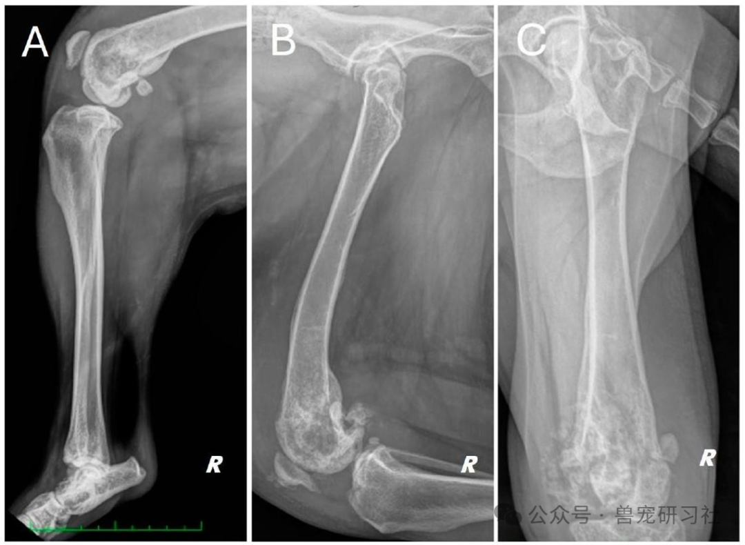 Figure 1: Different radiographic projections of the right hind limb. (A) Medial and lateral projections of the tibia, (B) Medial and lateral projections of the femur, (C) Cranial and caudal projections of the femoral head. Changes in structure and radiographic density are observed near the distal femur and knee joint in all projections.
🩺 Diagnostic Process: From Imaging to Cytology, Precisely Identifying the Tumor
-
X-rays showed “moth-eaten” bone lysis at the distal femoral metaphysis, periosteal reaction, and pathological fractures;
-
CT scans further confirmed the extent of the tumor and soft tissue infiltration, ruling out pulmonary metastasis;
-
Fine needle aspiration cytology indicated spindle cell atypia, consistent with a chondrogenic sarcoma.
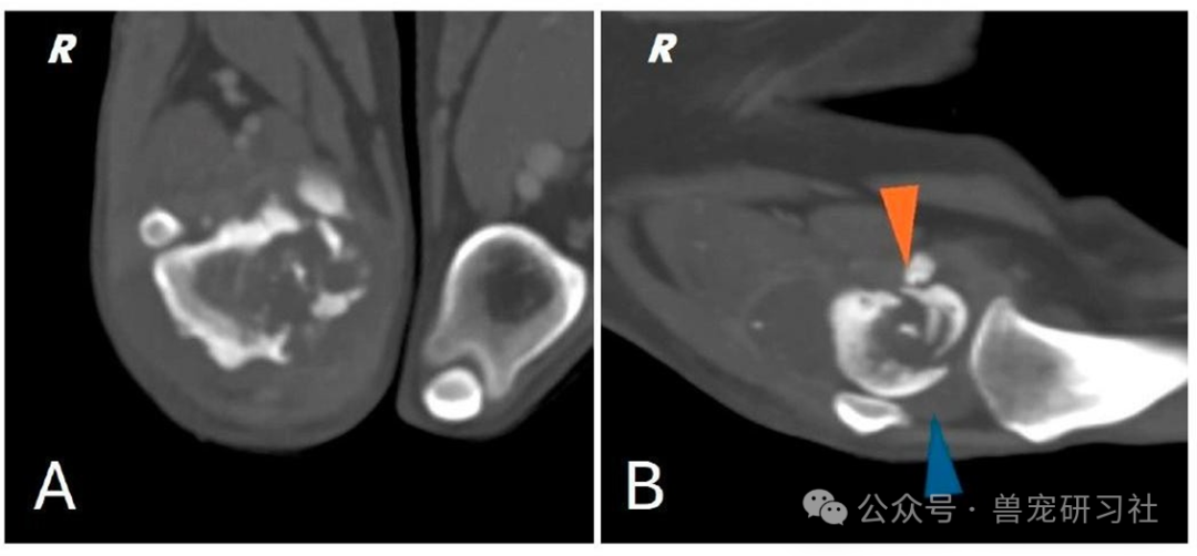 Figure 2: (A) CT scan image of the right distal femur. (B) CT scan image of the knee joint; pathological fracture of the medial femoral condyle (indicated by the orange arrow) with joint effusion (indicated by the blue arrow).
🧠 Innovative Approach: Virtual Surgical Planning + 3D Printed Guides and Prosthetics
To avoid the decline in quality of life associated with amputation, the team decided to perform a limb-sparing surgery, utilizing the following technologies for the first time in canines:
✅ Virtual Surgical Planning (VSP)
-
Reconstructed 3D models of the femur and tibia using CT data;
-
Precisely designed the osteotomy range and prosthetic shape;
-
Simulated the entire surgical process to ensure complete tumor removal.
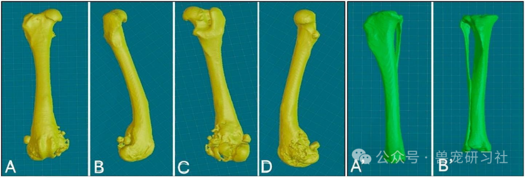 Figure 3: Multi-view representation of the 3D reconstruction of the right femur: (A) Cranial view; (B) Lateral view; (C) Caudal view; (D) Medial view; Multi-view representation of the right tibia: (A”) Caudal view; (B”) Cranial view.
✅ 3D Printed Patient-Specific Guides (PSG)
-
Precisely fitted to the bone surface, ensuring accurate osteotomy angles and positions;
-
Reduced surgical time and intraoperative imaging reliance;
-
Avoided human error, enhancing surgical precision.
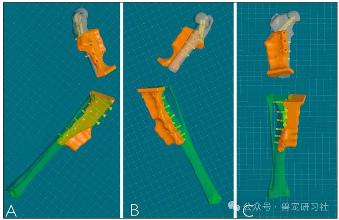 Figure 4: 3D reconstruction of the personalized surgical guides (PSGs): (A) Lateral view; (B) Medial view; (C) Frontal view.
✅ Customized Internal Prosthesis (PSE)
-
Made from Ti-6Al-4V titanium alloy, compliant with FDA standards;
-
Porous structure design to promote bone integration;
-
Optimized screw layout to ensure implant stability;
-
Perfectly matched the shape of the resected bone segment, requiring no intraoperative adjustments.
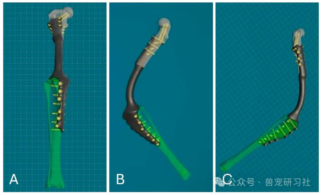 Figure 5: 3D reconstruction of the PSE: Frontal view (A); Medial view (B); Lateral view (C).
🏥 Surgical Process: Precise Osteotomy + Knee Joint Fusion
The surgery utilized a combined approach, sequentially exposing the distal femur and proximal tibia, completing the following steps:
-
Completed osteotomies of the femur and tibia using the 3D printed guides;
-
Implanted the customized prosthesis to reconstruct the bone defect;
-
Performed knee joint fusion, setting the flexion angle;
-
Stabilized the tibial tuberosity, reconstructing the quadriceps mechanism;
-
Started weight-bearing walking 48 hours post-surgery, with good recovery.
📸 Intraoperative and Postoperative Imaging Comparison
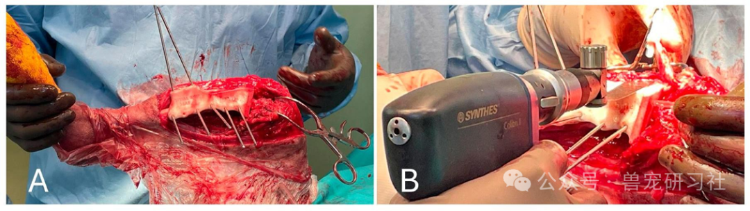
Figure 6: (A) Positioning of the tibial osteotomy guide and preparation for tibial tuberosity osteotomy; (B) Performing the tibial tuberosity osteotomy and exposing the knee joint through medial patellar tendon retraction.
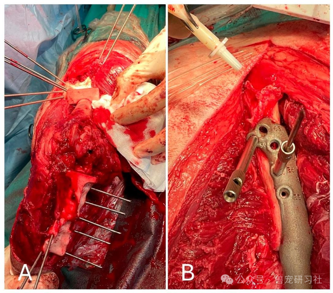 Figure 7: (A) Cranial view of the tibia post-tibial tuberosity osteotomy; (B) Status of the proximal femoral implant.
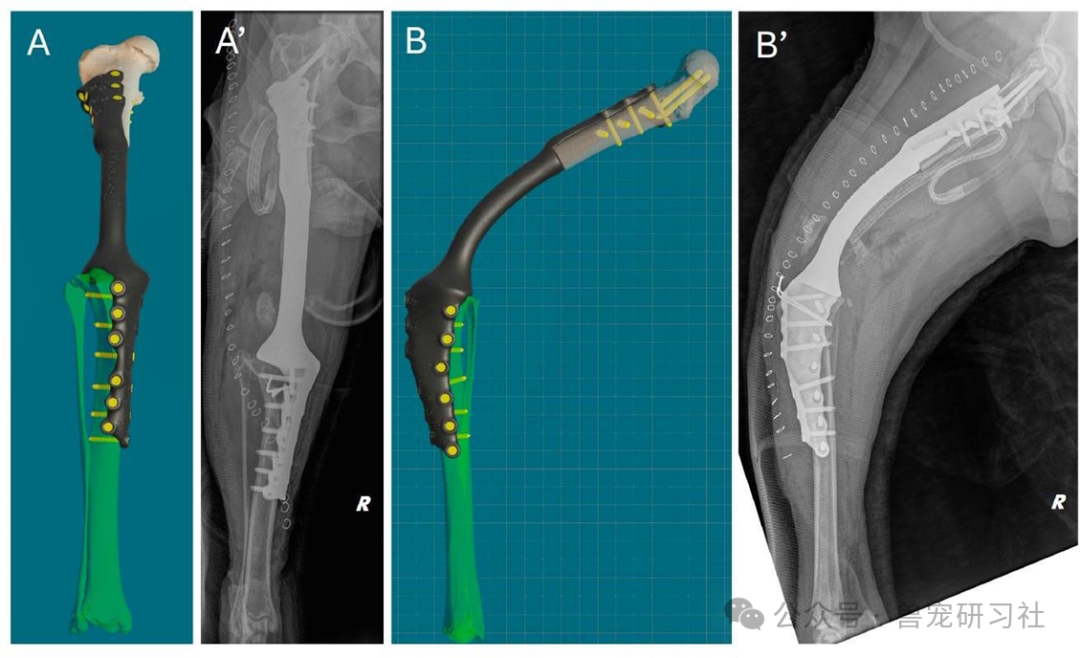 Figure 8: Comparison of virtual surgical planning and postoperative X-rays. (A, A’) Anteroposterior view. (B, B’) Lateral view.
🧪 Histopathological Results: Confirmed Grade 2 Central Chondrosarcoma
-
The tumor cells appeared chondroblast-like, arranged in a hyaline cartilage matrix;
-
Low mitotic activity, no tumor-like osteoid observed;
-
Consistent with a moderately malignant central chondrosarcoma.
📈 Postoperative Recovery and Follow-Up: Weight-Bearing at 2 Days Post-Surgery, 23-Month Survival
-
2 Days Post-Surgery: The affected dog began weight-bearing walking, with lameness grade reduced to II;
-
10 Days Post-Surgery: Sutures removed, stable gait;
-
3, 6, and 12 Months Post-Surgery: No infections, no recurrence, no prosthetic failure;
-
18-Month Telephone Follow-Up: The owner reported significant improvement in the dog’s quality of life, with active behavior, only showing slight fatigue after long walks;
-
Total Survival Time: Approximately 23 months (690 days), significantly higher than some literature reporting only amputation treatment.
💡 Technical Highlights: How 3D Printing Can Change the Game?
| Traditional Limb-Sparing Surgery | 3D Printed Customized Surgery |
|---|---|
| Limited prosthetic sizes, requiring intraoperative shaping | Perfectly matches the bone segment, requiring no adjustments |
| Long surgical time, high infection risk | Shortened surgical time, reduced complications |
| Intraoperative reliance on imaging to determine osteotomy position | Guides provide precise positioning, avoiding errors |
| Slow recovery, poor functional restoration | Early weight-bearing, faster recovery |
📌 Conclusion: Personalized Medicine, the Future is Here
This case successfully applied 3D printed patient-specific guides and prosthetics for the first time in the treatment of chondrosarcoma in canines, achieving not only precise resection and reconstruction but also significantly improving postoperative quality of life. As 3D printing technology continues to mature in the veterinary field, it may provide “tailored” solutions for more complex bone tumor cases in the future.
📎 Postoperative Walking Video
-
Video S1: Gait demonstration at 3-month follow-up