Chondrosarcoma
is one of the common malignant bone tumors,
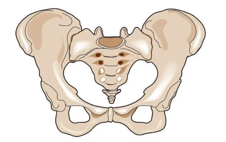
often occurring in the pelvis.
Due to the complex structure of the pelvis,
surgery ishighly traumatic, difficult, and time-consuming,
with many postoperative complications,
which not only affect the recovery of patients,
but can even lead to recurrence,
resulting in impaired limb mobility.
Recently,
Mr. Liu, a chondrosarcoma patient, visited our hospital,
complaining of discomfort in the affected area and pain around the joint,
initially experiencing intermittent pain,
which gradually worsened and limited his movement,
with a hard mass palpable at the base of his thigh,
examination showed that
the tumor had grown to the pubis and ischium of the pelvis,
and had invaded part of the acetabulum.
How to precisely resect the tumor margins,
while preserving Mr. Liu’s limb function,
the doctors from the Bone and Soft Tissue Department decided to proceed as follows~
👇👇👇
The team of Yao Weitao and Du Xinhui from the Bone and Soft Tissue Department of Henan Cancer Hospital utilized artificial intelligence and 3D printing technology to address clinical pain points, creating precise three-dimensional models of pelvic tumors for surgical design, accurately resecting tumor boundaries while maximizing the preservation of the acetabulum, allowing bone tumor patients’ affected limbs to function normally, and reducing the likelihood of recurrence.
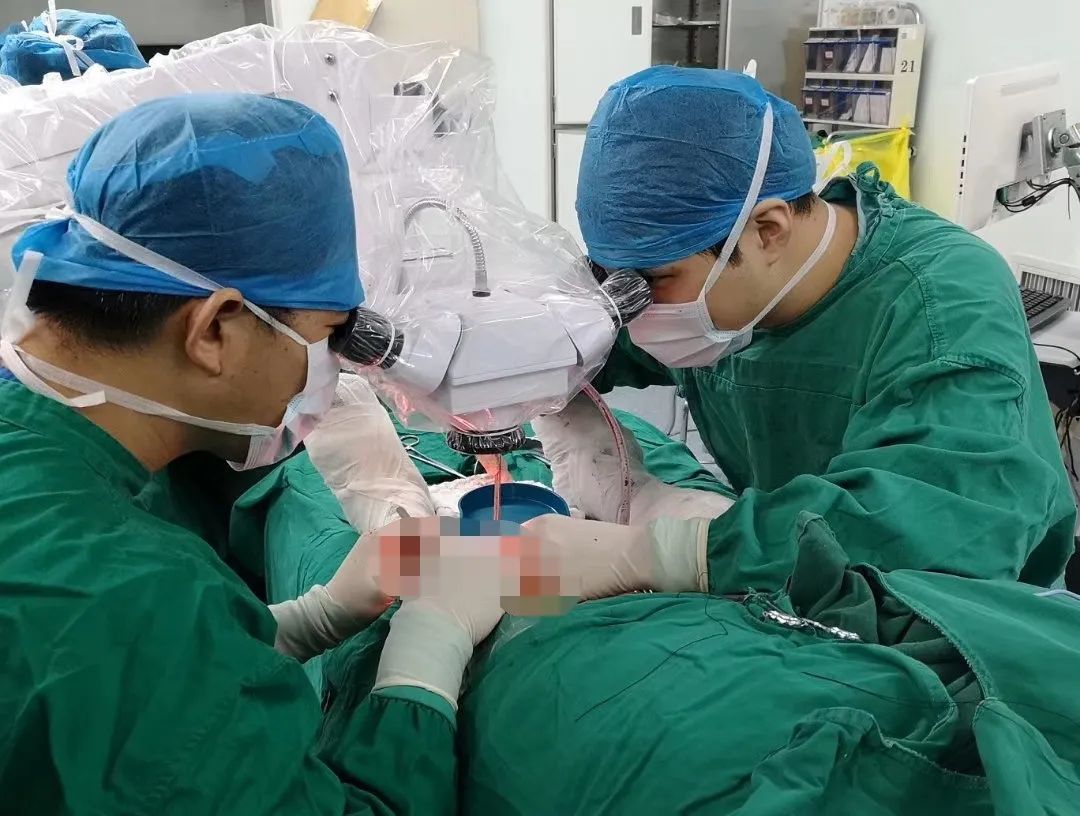
Orthopedic experts performed the surgery with the aid of a microscope
Key to the surgery:
How to preserve the acetabulum?

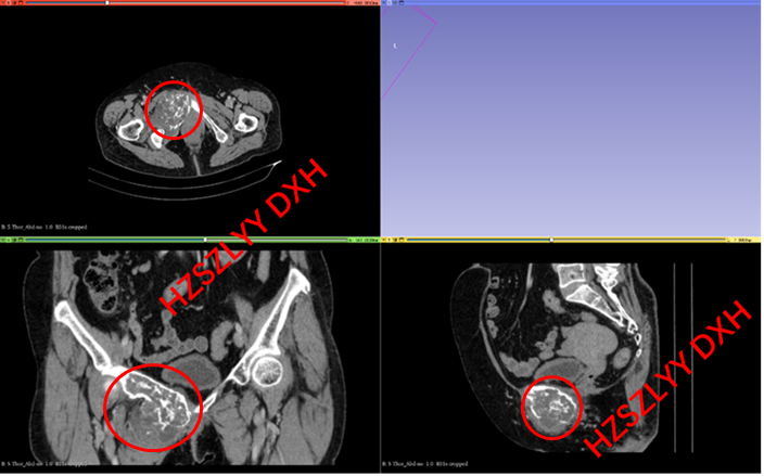
Preoperative CT of the patient, with the red circle indicating the right pubic ramus with expansive bone destruction accompanied by soft tissue mass, showing “popcorn-like” calcification within the mass.
Deputy Chief Physician Du Xinhui explained that preserving the physiological state of the hip joint results in better postoperative function and faster recovery, and hip preservation surgery has less trauma and lower incidence of postoperative complications. However, Mr. Liu’s acetabulum’s medial wall had already been invaded by the tumor. Traditional hip preservation surgical methods involve directly resecting the acetabulum’s inner edge, which often passes through the tumor, causing contamination and turning a wide excision surgery into an intratumoral surgery, increasing the risk of postoperative recurrence and metastasis.Some surgical methods choose to remove the hip joint along with the tumor to achieve a cleaner and safer tumor boundary and reduce postoperative recurrence rates, but this can lead to structural loss of the hip joint, resulting in poor limb function post-surgery, whether left vacant or reconstructed, and an increased incidence of complications.To allow Mr. Liu to treat his tumor while preserving the acetabulum and achieving ideal tumor boundaries, maximizing his normal functional life, the Yao Weitao team utilizeddigital technology to perform precise resection and reconstruction surgery.
Digital Precision
Resection and Reconstruction
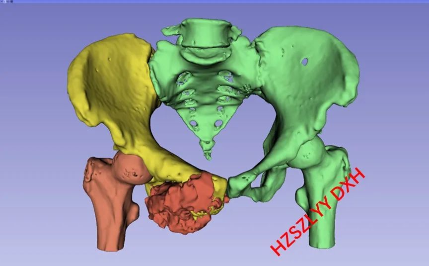
3D modeling and tumor boundary determination assisted by artificial intelligence algorithms
First, the doctors imported the patient’s thin-slice enhanced CT data (<1mm) into image processing software, using artificial intelligence algorithms to segment the images and generate precise 3D models of the pelvis and tumor. This model can display the tumor size at a 1:1 scale and show its relationship with surrounding important structures, such as blood vessels, nerves, and pelvic organs, providing a three-dimensional display of the tumor’s actual extent.
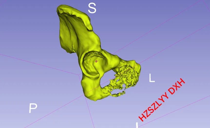
Determining the puncture point at the deepest part of the acetabulum, drilling a hole
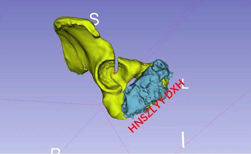
First Resection
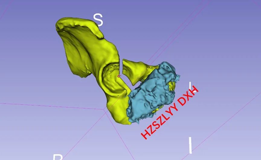
Second Resection
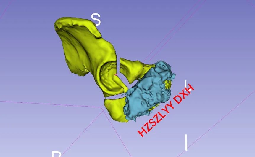
Third Resection
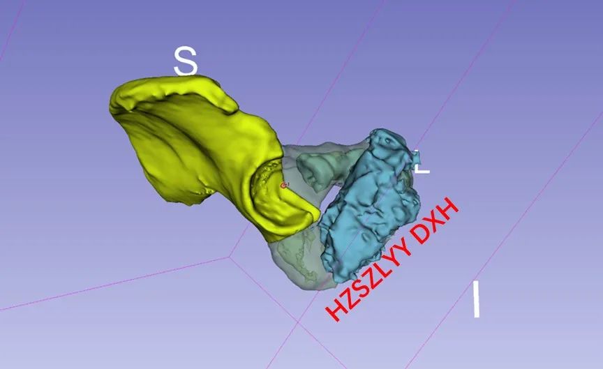
Digital Resection Plane Display
No tumor at the resection margin
Secondly, the doctors simulated the tumor resection process using the 3D model generated from image fusion, planning the boundaries for intraoperative resection.
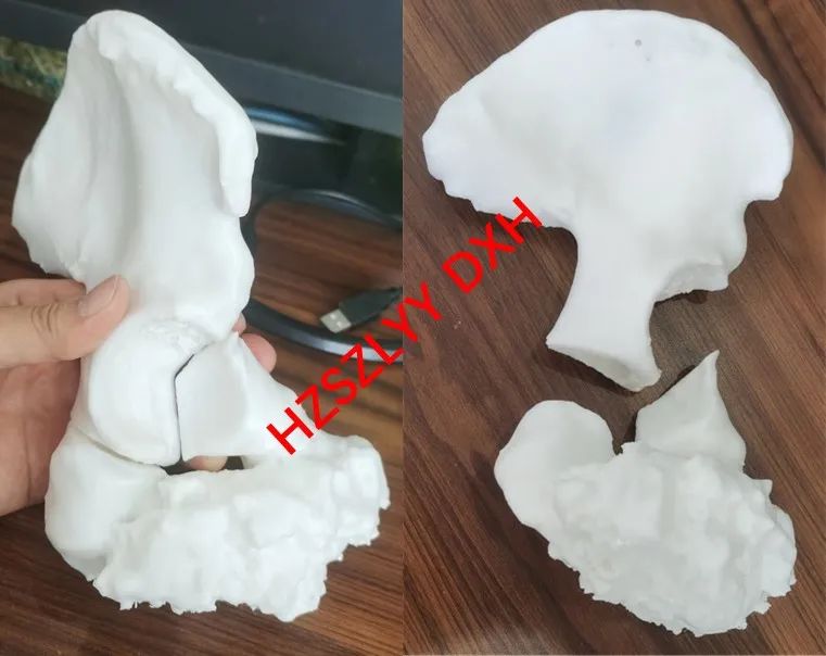
3D Printed Model Demonstrating Resection Results
During the surgery, with the aid of this technology combined with 3D printing technology, the tumor was resected and reconstructed with millimeter-level precision. The resection margin was sent for intraoperative frozen confirmation of negative margins.
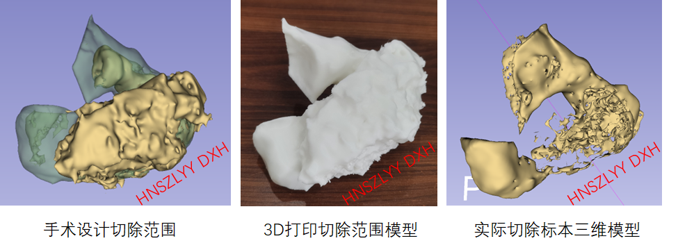
Comparison of Actual Resection Specimen with Preoperative Design Resection Range
Postoperatively, the resected specimen was modeled to evaluate whether the resection range was consistent with the preoperative plan.
Technology Empowering Surgery,
Enhancing Efficacy
This technology preserves as much bone as possible, ensuring good limb function for patients post-surgery while reducing recurrence rates.The implementation of this technology greatly promotes the surgical treatment of malignant tumors involving irregular bones or joints. Compared to traditional techniques, precise resection can reduce trauma while ensuring negative margins and preserving more normal structures and functions.
Yao Weitao, Director of the Bone and Soft Tissue Department stated that the difficulty of pelvic tumor surgeries lies mainly in the challenge of determining tumor boundaries. Due to the irregular structure of the pelvis, conventional imaging methods such as CT and MRI struggle to intuitively and three-dimensionally display tumor boundaries, making it difficult for surgeons to design resection planes in three-dimensional space using two-dimensional imaging data. Rough tumor designs often fail to achieve ideal surgical boundaries, limiting the formulation and execution of individualized surgical plans for pelvic tumors. This is also a major reason why the recurrence rate of pelvic tumors is much higher than that of similar tumors in the limbs.
This technology provides a more accurate assessment of tumors, laying the foundation for designing surgical resection boundaries. At the 2022 World Limb Preservation Conference, Du Xinhui presented this technology on behalf of the Bone and Soft Tissue Department of Henan Cancer Hospital. Currently, related results have been published in 3 SCI articles and 1 series article in China, receiving recognition from experts in related fields both domestically and internationally.
Our Bone and Soft Tissue Department is located in the Henan Provincial Bone and Soft Tissue Tumor Diagnosis and Treatment Center. Digital technology-assisted diagnosis and treatment of bone tumors is one of the center’s featured technologies. The center established two experimental platforms in 2020: the Key Laboratory for Digital Intelligent System Assessment and Surgical Assistance Tool Design for Spinal and Pelvic Surgery in Zhengzhou and the Key Laboratory for Digital Intelligent Assessment in the Perioperative Period of Bone Tumors in Henan Province, and has conducted a series of basic and clinical translational research based on this. Digital technology has transformed previously rough surgical methods, offering more precise surgical plans that provide patients with better tumor treatment outcomes and postoperative function, further enhancing the overall treatment effectiveness for patients with bone tumors and allowing them to enjoy better medical services.

Reviewed by: Pang Hongwei, Yao Weitao
Authors: Du Xinhui, Chang Rong
Editor/Proofreader: Li Huizi
Images provided by the surgeon