 Click the blue text above to subscribe

Three-dimensional (3D) printing, as a promising additive manufacturing technology, can create customized objects with precise structures, controllable components, adjustable mechanical properties, and physiological heterogeneity by computer-aided continuous deposition of biomaterials or cells, fundamentally transforming the field of biomedical engineering. In bone tissue engineering, 3D porous scaffolds provide support for the regeneration of cells and bone tissue. However, during surgery and treatment, bacteria can easily attach to the scaffold, invading the surrounding soft tissue and even affecting deep bone tissue. Furthermore, the degradation rate of the scaffold and real-time observation of its morphology after implantation are also crucial.
Recently, Professor Dai Honglian from Wuhan University of Technology and Professor Feng Haitao from Baoji University of Arts and Sciences developed a red light-emitting AIEgen molecule named 4BC, which can be loaded onto the surface of 3D printed biological scaffolds through simple surface adsorption. As shown in Figure 1, under excitation light, in situ fluorescence imaging can be performed on the implanted scaffold. Additionally, 4BC molecules exhibit a high reactive oxygen species release effect. Under light exposure, 4BC can assist the bioactive scaffold in killing infected bacteria through reactive oxygen species release, thereby preventing inflammation.
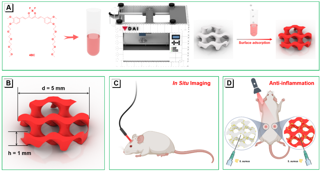
Figure 1. (A) Schematic diagram of the synthesis of multifunctional 3D printed fluorescent scaffolds.(B) Basic properties of the designed fluorescent scaffold.(C) In situ fluorescence imaging of the fluorescent scaffold in vivo.(D) The process of the fluorescent scaffold inhibiting bacterial inflammation in vivo. To demonstrate the fluorescence imaging of the scaffold obtained in vivo, the fluorescent scaffold (4BC@TMP) was implanted in the heads of 4-week-old nude mice. Photos of the mice were taken under normal light and fluorescent light. As shown in Figure 2A, a clear round bright red fluorescence was observed on the head of the mouse, and the fluorescence area corresponded to the size of the scaffold. Further examination of the in vivo scaffold 4BC@TMP fluorescence imaging was conducted using a small animal imaging system (2B). By comparing with the X-ray images, the overall size of 4BC@TMP was very consistent with the fluorescence imaging (Figure 2D and 2E). Notably, the fluorescence imaging pattern was completely opposite to the X-ray images because the light paths for X-rays and fluorescence in the small animal imaging system are located on the dorsal and ventral sides of the mouse, respectively. The results indicate that the 4BC@TMP scaffold can achieve clear in situ imaging under excitation light.
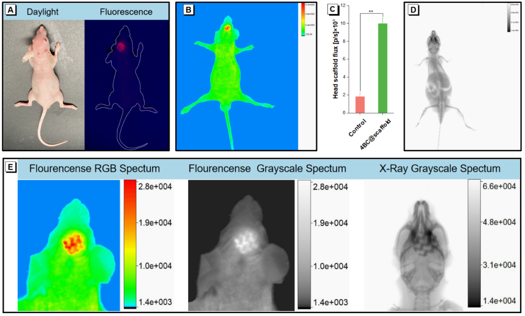
Figure 2. (A) Photos of 4BC@TMP implanted in mice under normal and fluorescent light. RGB spectrum of 4BC@TMP after implantation under a small animal imaging system (B), (C) Quantitative analysis of fluorescence intensity in the head, **P<0.01, and (D) X-ray grayscale spectrum.(E) Magnified fluorescence and X-ray images. To further evaluate the inhibitory effect of the bioactive scaffold on bacterial inflammation in vivo, they implanted the original scaffold and the fluorescent scaffold on the left and right sides of the back of the mouse (Figure 1D). Bacteria were then injected into the two scaffold implantation sites, followed by exposure to white light for 20 minutes. On days 4 and 7, the skin around the scaffold was subjected to H&E and immunofluorescence staining. As shown in Figure 3A, the control group and the original scaffold (TMP group) exhibited similar inflammatory responses. A large number of inflammatory cells infiltration was observed, possibly neutrophils or necrotic inflammatory cells. However, the fluorescent scaffold (4BC@TMP group) and the blank group exhibited almost no inflammation. From Figure 3B, it can also be seen that the inflammatory factor TNF-α (green) was abundant in the control group and TMP group, indicating a strong inflammatory invasion. The green fluorescence observed in 4BC@TMP was minimal, similar to the blank group, indicating that 4BC@TMP has anti-inflammatory effects. For IL-6, 4BC@TMP also exhibited good anti-inflammatory efficacy; quantitative indicators analyzed by ImageJ showed significant anti-inflammatory properties (Figure 3C and 3D). Compared to the control group, the expression of inflammatory factors TNF-α and IL-6 in the 4BC@TMP group accounted for only 30.3% and 16.7%, respectively. This indicates that the bioactive scaffold loaded with 4BC can help kill bacteria near the scaffold, thereby exerting anti-inflammatory effects.
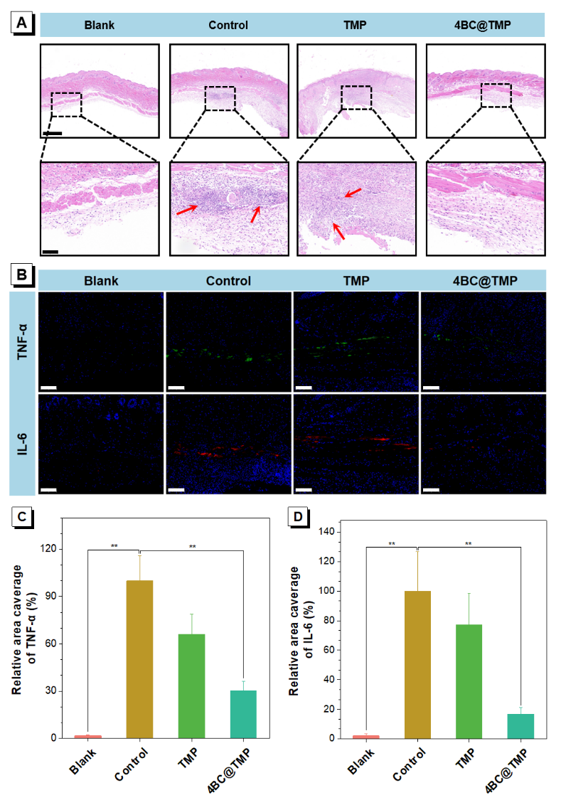
Figure 3. (A) Histological assessment of the scaffold implantation site. Scale bar = reduced image to 1000 µm, enlarged image to 100 µm. (B) Immunofluorescence staining images of TNF-α (green) and IL-6 (red). Scale bar = 100μm. Relative area coverage rates of TNF-α (C) and IL-6 (D). (n=9)**P<0.01. Related research results were published in the paper titled “In Situ Imaging and Anti-inflammation of 3D Printed Scaffolds Enabled by AIEgen in the journal ACS Applied Materials & Interfaces. The first authors of the paper are Wang Xiaohuan, a doctoral student at Wuhan University of Technology, and Chen Pu, a doctoral student at Baoji University of Arts and Sciences, with co-authors including doctoral students from Wuhan University of Technology, Yang He, Liu Jiawei, and Professor Tu Rong. The corresponding authors of the paper are Professor Feng Haitao from Baoji University of Arts and Sciences and Professor Dai Honglian from Wuhan University of Technology. This research was supported by the National Key Research and Development Program (2022YFB4601402), the National Natural Science Foundation (32201109, 52173152, 21805002), the Shenzhen Key Basic Research Program (JCYJ20200109150218836), and the Guangdong Provincial Basic and Applied Basic Research Fund (2020A1515110476).
The paper link
https://pubs.acs.org/doi/10.1021/acsami.3c03082
Related progress
Professor Dai Honglian’s research group at Wuhan University of Technology AFM: An intelligent drug delivery system for capturing bacteria combined with magnetic hyperthermia to efficiently repair infectious bone defects
Professor Dai Honglian’s team Chem. Mater.: Modular design and gel mechanism of boron-nitrogen coordination borate hydrogels with tunable gel pH and alkaline pH responsiveness
Wuhan University of Technology’s Dai Honglian/Zhejiang University Hu Yihe ACS Nano: Bio-MOF hydrogels that intelligently capture bacteria and promote neurovascular network reconstruction accelerate the repair and healing of infectious wounds
Academician Tang Benzhong/Baoji University of Arts and Sciences Professor Feng Haitao/Jilin University Professor Wang Kai “ACS Materials Lett.”: Force/thermal-induced luminescence-enhanced blue-shifted carbazole luminescent materials
Academician Tang Benzhong/Professor Feng Haitao ACS Nano: Chiral AIEgen visualization of enantiomer recognition and separation
Li Lili/Xu Wanhai/Feng Haitao Biomaterials: Activatable glycopeptide self-assembling nano-agonists that promote PKM2 tetramerization, regulate tumor glucose metabolism, and enhance chemotherapy sensitivity
Academician Tang Benzhong/Feng Haitao/Professor Zou Bo Mater. Horiz.: Binary synergistic effect to prepare high-contrast force-induced color-changing materials
Academician Tang Benzhong’s team Feng Haitao and Professor Ding Dan JACS: Regulation of photosensitizer dark toxicity through supramolecular displacement for efficient photodynamic therapy

This is an original article by Polymer Science and Technology. Personal forwarding and sharing are welcome. If publications or media need to reprint, please contact: [email protected]

Call for Papers
Experts and scholars are welcome to submit manuscripts (papers, project introductions, new technologies, academic exchanges, unit news, conference information, recruitment, and enrollment, etc.) to [email protected], and please specify detailed contact information. Polymer Science and Technology® will push it in a timely manner and also publish it on the China Polymer Network.
Welcome to Join WeChat Groups In response to the needs of colleagues from various fields of polymer industry-university-research, dozens of special exchange groups have been opened, including a group for polymer experts and scholars, as well as groups for polymer industry technology, entrepreneurs, PhD students, graduate students, media journals, and associations, covering the entire polymer industry or field. Currently, it gathers thousands of top experts, scholars, technical personnel, and entrepreneurs from domestic and foreign universities, research institutes, and corporate R&D centers.
To apply to join the group, please first add the review WeChat ID PolymerChina (or long press the QR code below), and please specify: Polymer + Name + Unit + Title (or Degree) + Field (or Industry), otherwise, it will not be processed. Qualification will be reviewed before entering the relevant professional group.

Click here to read the original text for more.