
Researchers from Chongqing Medical University in China and The Chinese University of Hong Kong have published a comprehensive review on the application of 3D printing in skeletal muscle chip (SMoC) systems. This paper, published in ScienceDirect, highlights how printing technology is utilized to manufacture microfluidic devices, arrange muscle fibers, and create biomimetic scaffolds to study skeletal muscle physiology and related diseases.
The organ-on-a-chip platform integrates microfluidic technology, bioengineering, and cell biology to replicate key functions of human tissues. While models for tissues such as lungs, heart, liver, kidneys, and brain have been developed, replicating skeletal muscle remains a challenge due to its layered fibrous structure, high metabolic demands, and reliance on neuromuscular signaling.
Additive manufacturing technology has been applied to various aspects of SMoC structures. Industrial-grade printing methods, such as Fused Deposition Modeling (FDM) and Stereolithography (SLA), have been used to construct chip scaffolds or molds. SLA utilizes ultraviolet light to cure liquid resin, achieving high precision in the fabrication of microchannel structures. Bioprinting methods further extend this technology by using live cells and hydrogels as bioinks to form contractile fibers and complex tissue environments.
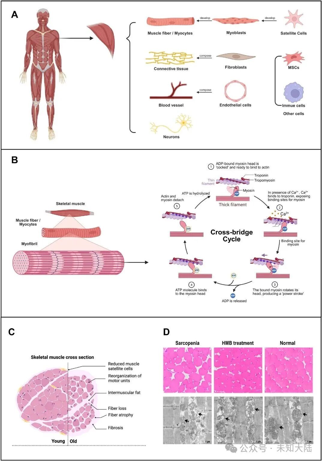
The review emphasizes that these methods can precisely deposit materials and cells, forming layered structures similar to natural skeletal muscle. For instance, researchers have combined bioprinting with electrospinning technology to align fibers neatly, promoting myotube maturation, or incorporated conductive nanomaterials to enhance cell alignment and electrical responsiveness.
Multiple studies have demonstrated that SMoCs manufactured using 3D printing technology can replicate disease states and evaluate therapies. Myoblasts from patients with Duchenne Muscular Dystrophy have been implanted into chip platforms to assess potential regenerative therapies. Other studies have utilized neuromuscular junction models combining motor neurons and skeletal muscle fibers to investigate myasthenia gravis and amyotrophic lateral sclerosis.
One approach involves constructing chips using light polymerization-based printing technology, with polyacrylamide columns serving as anchors for muscle bundle alignment. These systems can quantify passive tension and contraction force while also allowing functional assessments under injury conditions. Another study employed SLA-designed molds followed by polydimethylsiloxane (PDMS) casting to construct chip bodies for advanced glycation end products experiments, which have been shown to impair muscle contraction and structure.
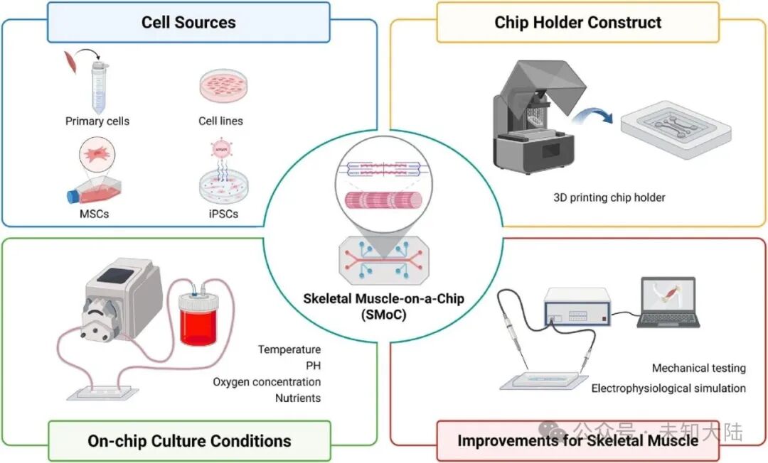
SMoC platforms have also been used for environmental research. Microphysiological systems made from 3D printed components allow researchers to expose chips simultaneously to normoxic and hypoxic conditions, providing a method to analyze oxygen-dependent signaling. Recently, printed muscle chips were sent to the International Space Station National Laboratory to study microgravity effects and screen compounds such as IGF-1 and 15-PDGH inhibitors.
Despite these advancements, several obstacles remain. Current printing technologies can only produce structures with millimeter-level resolution, insufficient to replicate cellular-level arrangements. Bioinks like hydrogels often lack the mechanical strength required for long-term experiments, and their biocompatibility can limit reproducibility. Assessing muscle contraction also requires integrated sensors, such as electrodes or strain gauges, which must be incorporated into the chip without disrupting the culture environment.
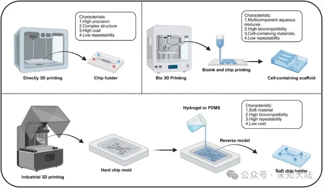
Material development is another limiting factor. While biocompatible polymers like PDMS and hydrogels are widely used, skeletal muscle requires scaffolds that possess both elasticity and strength to withstand contraction cycles. Researchers have experimented with thermoresponsive polymers, extracellular matrix-derived scaffolds, and hybrid strategies combining 3D printing and electrospinning to enhance structural and functional fidelity.
Future work emphasized in the review includes improving resolution through multi-nozzle printers, developing new bioinks with better mechanical and biological properties, and enhancing culture systems to better simulate muscle metabolism and electrophysiology. 4D printing technology, which allows printed structures to change shape or function over time in response to stimuli, has been applied in preliminary muscle studies. In one example, GelMA fibers seeded with myoblasts were induced to curl into tubular structures resembling natural muscle bundles, promoting differentiation.
Advancements in chip integration technology are also expanding SMoC research into multi-organ systems. Dual chips and modular chips have been developed to connect skeletal muscle with other tissues (such as bone or liver) through vascular flow. These platforms can be used to study systemic interactions, including off-target drug effects and metabolic regulation.
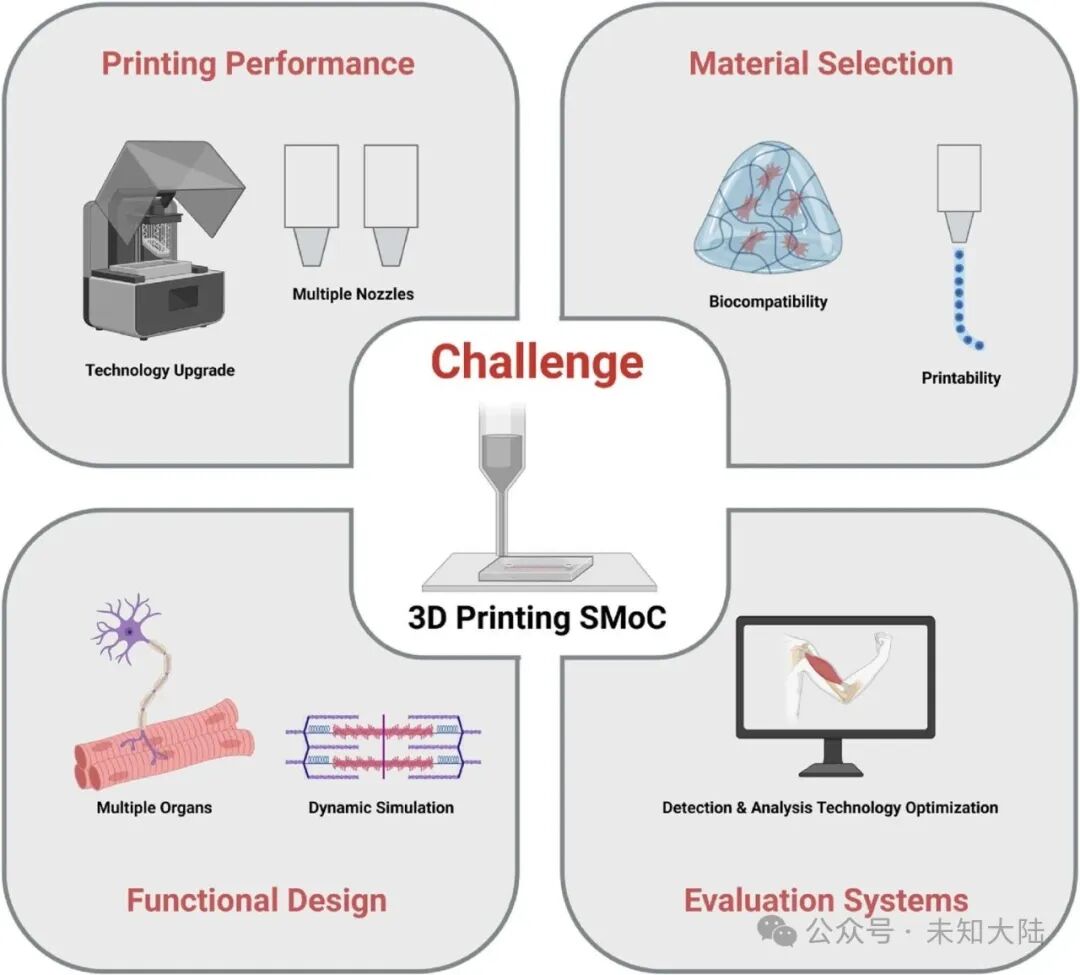
Skeletal muscle chip research is still in its early stages, but 3D printing has become central to its development. By achieving precise chip fabrication, cell alignment, and scaffold customization, additive manufacturing technology enables the replication of key structural and functional features of muscle tissue in vitro. The review points out that continuous improvements in resolution, materials, and system integration are crucial for these platforms to fully support disease modeling, drug testing, and muscle physiology research.