Abstract
This research paper focuses on the use of the Freeform Reversible Embedding of Suspended Hydrogels (FRESH) printing method to fabricate complex structures with tunable elastic modulus and porosity. Currently, three-dimensional (3D) bioprinting has been widely used to construct hard tissues such as bone and cartilage. However, constructing soft tissues with complex structures remains a challenge. The FRESH printing method can overcome the effects of gravity on printed structures and is expected to become a key technique for preparing complex soft tissues. In this study, alginate and gelatin were used as ink substrates for cell-laden printing via the FRESH method. First, bioink with tunable porosity and mechanical properties was designed and prepared, and extrusion printing was performed in a gelatin particle support bath using the FRESH method. The rheological properties of the ink were measured using a rheometer to analyze printability, and the extrusion state of the ink was further analyzed through simulations. Moreover, mechanical test results and micro-morphology showed that the printed structures have tunable elastic modulus and porosity. By optimizing the FRESH process, a series of complex structures were printed. Finally, L929 fibroblasts were used for cell-laden printing, and the cells exhibited high viability and good proliferation characteristics during a 7-day culture. All results indicate that the designed bioink has good biological and mechanical properties, and further enhancement of cell viability can be achieved by adding cell-compatible materials, demonstrating great potential in soft tissue printing.
Cite this article (Click the link at the bottom to download the PDF)
Chen Z, Huang C, Liu H, et al., 2023. 3D bioprinting of complex biological structures with tunable elastic modulus and porosity using freeform reversible embedding of suspended hydrogels. Bio-des Manuf 6(5):550–562. https://doi.org/10.1007/s42242-023-00251-5
Article Guide
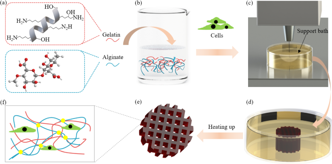
Figure 1 FRESH printing process for tissues with tunable elastic modulus and porosity. (a) Bioink monomer materials; (b) Preparation of bioink materials; (c) FRESH 3D bioprinting process; (d) Printed structure preserved in the support bath; (e) Scaffold released from the support bath at 37 °C; (f) Schematic of the microstructure of the hydrogel. FRESH: Freeform Reversible Embedding of Suspended Hydrogels
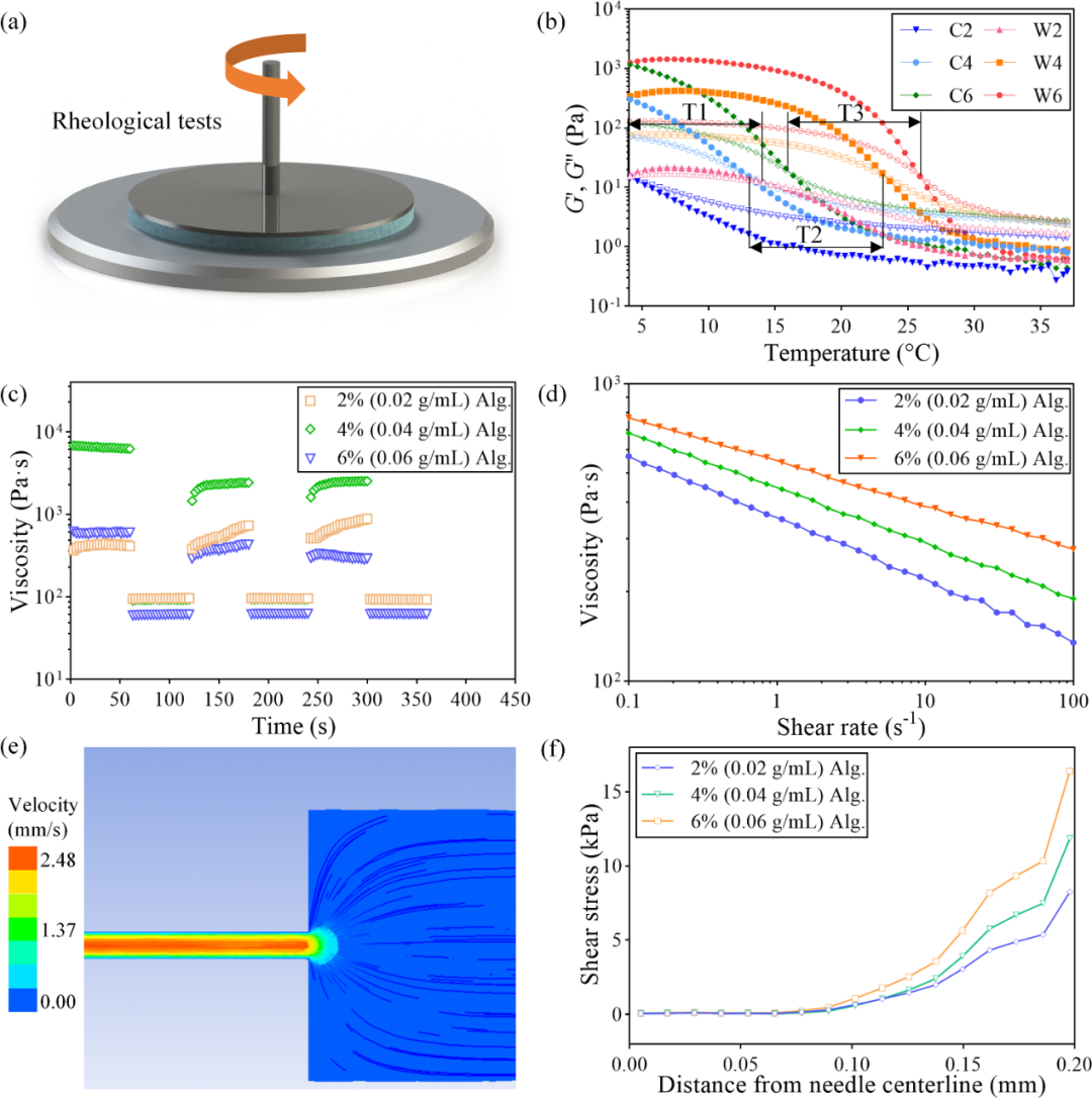
Figure 2 Rheological property testing of bioink. (a) Schematic of the rheological test; (b) Gelation dynamics curves of bioinks containing 2%, 4%, and 6% (0.02, 0.04, and 0.06 g/mL) gelatin during heating and cooling processes. Legend: Solid indicates G′, hollow indicates G′′, W indicates heating, C indicates cooling, and numbers indicate gelatin concentrations; (c) Three high (1000 s−1) and low (0.01 s−1) shear rate cycles at 37 °C; (d) Shear scanning experiments at 37 °C; (e) Velocity field of bioink flowing through the needle; (f) Shear stress at different radius positions of three ink concentrations in the needle
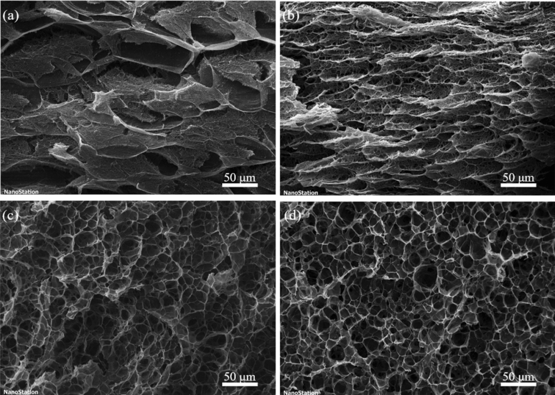
Figure 3 Micro-morphology of hydrogels at gelatin concentrations of (a) 2% (0.02 g/mL), (b) 4% (0.04 g/mL), (c) 6% (0.06 g/mL), and (d) 8% (0.08 g/mL)
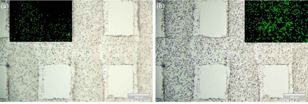
Figure 4 FRESH printing of complex structures. (a) Repeated compression test on printed cylindrical tubes; (b) Infusion test on printed elongated tubes; (c) Design and print of solid “SDU” model, size 16.8 mm × 7.08 mm × 2.00 mm, scale: 1 mm
Figure 5 Cell immunofluorescence staining. On day 1 (a) and day 7 (b) of cell culture, cells were immunofluorescently stained using AM/PI. Scale bar: 200 μm. AM: Acetoxymethyl; PI: Propidium Iodide
References
Scroll down to view more
1. Zhang B, Luo Y, Ma L et al (2018) 3D bioprinting: an emerging technology full of opportunities and challenges. Bio-Des Manuf 1(1):2–13. https://doi.org/10.1007/s42242-018-0004-3
2. Ozbolat IT, Yu Y (2013) Bioprinting toward organ fabrication: challenges and future trends. IEEE Trans Biomed Eng 60(3):691–699. https://doi.org/10.1109/TBME.2013.2243912
3. Park C, Jones MM, Kaplan S et al (2022) A scoping review of inequities in access to organ transplant in the United States. Int J Equity Health 21:22. https://doi.org/10.1186/s12939-021-01616-x
4. Hurst DJ, Potter J, Padilla LA (2022) Organ transplant and Covid-19 vaccination: considering the ethics of denying transplant to unvaccinated patients. Clin Transplant 36(5):e14589. https://doi.org/10.1111/ctr.14589
5. Langer R, Vacanti JP (1993) Tissue engineering. Science 260(5110):920–926. https://doi.org/10.1126/science.8493529
6. Xu Y, Zhang F, Zhai W et al (2022) Unraveling of advances in 3D-printed polymer-based bone scaffolds. Polymers 14(3):566. https://doi.org/10.3390/polym14030566
7. Kang Y, Xu J, Meng L et al (2023) 3D bioprinting of dECM/Gel/QCS/nHAp hybrid scaffolds laden with mesenchymal stem cell-derived exosomes to improve angiogenesis and osteogenesis. Biofabrication 15(2):024103. https://doi.org/10.1088/1758-5090/acb6b8
8. Kim DH, Kim MJ, Kwak SY et al (2023) Bioengineered liver crosslinked with nano-graphene oxide enables efficient liver regeneration via MMP suppression and immunomodulation. Nat Commun 14(1):801. https://doi.org/10.1038/s41467-023-35941-2
9. Liu N, Ye X, Yao B et al (2021) Advances in 3D bioprinting technology for cardiac tissue engineering and regeneration. Bioact Mater 6(5):1388–1401. https://doi.org/10.1016/j.bioactmat.2020.10.021
10. Lawlor KT, Vanslambrouck JM, Higgins JW et al (2021) Cellular extrusion bioprinting improves kidney organoid reproducibility and conformation. Nat Mater 20(2):260–271. https://doi.org/10.1038/s41563-020-00853-9
11. Shoulders MD, Raines RT (2009) Collagen structure and stability. Annu Rev Biochem 78:929–958. https://doi.org/10.1146/annurev.biochem.77.032207.120833
12. Kumar MN, Muzzarelli RA, Muzzarelli C et al (2004) Chitosan chemistry and pharmaceutical perspectives. Chem Rev 104(12):6017–6084. https://doi.org/10.1021/cr030441b
13. Lee KY, Mooney DJ (2012) Alginate: properties and biomedical applications. Prog Polymer Sci 37:106–126. https://doi.org/10.1016/j.progpolymsci.2011.06.003
14. Arnott S, Fulmer A, Scott WE et al (1974) The agarose double helix and its function in agarose gel structure. J Mol Biol 90(2):269–284. https://doi.org/10.1016/0022-2836(74)90372-6
15. Yu K, Zhang X, Sun Y et al (2021) Printability during projection-based 3D bioprinting. Bioact Mater 11:254–267. https://doi.org/10.1016/j.bioactmat.2021.09.021
16. Gungor-Ozkerim PS, Inci I, Zhang YS et al (2016) Bioinks for 3D bioprinting: an overview. Biomater Sci 6(5):915–946. https://doi.org/10.1039/c7bm00765e
17. He Y, Yang F, Zhao H et al (2016) Research on the printability of hydrogels in 3D bioprinting. Sci Rep 6:29977. https://doi.org/10.1038/srep29977
18. Skeldon G, Lucendo-Villarin B, Shu W (2018) Three-dimensional bioprinting of stem-cell derived tissues for human regenerative medicine. Phil Trans R Soc B 373(1750):20170224. https://doi.org/10.1098/rstb.2017.0224
19. Hölzl K, Lin S, Tytgat L et al (2016) Bioink properties before, during and after 3D bioprinting. Biofabrication 8(3):032002. https://doi.org/10.1088/1758-5090/8/3/032002
20. Pati F, Jang J, Ha DH et al (2014) Printing three-dimensional tissue analogues with decellularized extracellular matrix bioink. Nat Commun 5:3935. https://doi.org/10.1038/ncomms4935
21. Corbett DC, Olszewski E, Stevens K (2019) A FRESH take on resolution in 3D bioprinting. Trends Biotechnol 37(11):1153–1155. https://doi.org/10.1016/j.tibtech.2019.09.003
22. Balmforth NJ, Frigaard IA, Ovarlez G (2014) Yielding to stress: recent developments in viscoplastic fluid mechanics. Annu Rev Fluid Mech 46:121–146. https://doi.org/10.1146/annurev-fluid-010313-141424
23. Hinton TJ, Hudson A, Pusch K et al (2016) 3D printing PDMS elastomer in a hydrophilic support bath via freeform reversible embedding. ACS Biomater Sci Eng 2(10):1781–1786. https://doi.org/10.1021/acsbiomaterials.6b00170
24. O’Bryan CS, Bhattacharjee T, Hart S et al (2017) Self-assembled micro-organogels for 3D printing silicone structures. Sci Adv 3(5):e1602800. https://doi.org/10.1126/sciadv.1602800
25. Colly A, Marquette C, Courtial EJ (2021) Poloxamer/poly(ethylene glycol) self-healing hydrogel for high-precision freeform reversible embedding of suspended hydrogel. Langmuir 37(14):4154–4162. https://doi.org/10.1021/acs.langmuir.1c00018
26. Lee A, Hudson AR, Shiwarski DJ et al (2019) 3D bioprinting of collagen to rebuild components of the human heart. Science 365(6452):482–487. https://doi.org/10.1126/science.aav9051
27. Mirdamadi E, Tashman JW, Shiwarski DJ et al (2020) FRESH 3D bioprinting a full-size model of the human heart. ACS Biomater Sci Eng 6(11):6453–6459. https://doi.org/10.1021/acsbiomaterials.0c01133
28. Zhang Z, Wu C, Dai C et al (2022) A multi-axis robot-based bioprinting system supporting natural cell function preservation and cardiac tissue fabrication. Bioact Mater 18:138–150. https://doi.org/10.1016/j.bioactmat.2022.02.009
29. Lan X, Liang Y, Erkut EJN et al (2021) Bioprinting of human nasoseptal chondrocytes-laden collagen hydrogel for cartilage tissue engineering. FASEB J 35(3):e21191. https://doi.org/10.1096/fj.202002081R
30. Ostrovidov S, Salehi S, Costantini M et al (2019) 3D bioprinting in skeletal muscle tissue engineering. Small 15(24):1805530. https://doi.org/10.1002/smll.201805530
31. Hinton TJ, Jallerat Q, Palchesko RN et al (2015) Three-dimensional printing of complex biological structures by freeform reversible embedding of suspended hydrogels. Sci Adv 1(9):e1500758. https://doi.org/10.1126/sciadv.1500758
32. Shiwarski DJ, Hudson AR, Tashman JW et al (2021) Emergence of FRESH 3D printing as a platform for advanced tissue biofabrication. APL Bioeng 5:010904. https://doi.org/10.1063/5.0032777
33. Paxton N, Smolan W, Böck T et al (2017) Proposal to assess printability of bioinks for extrusion-based bioprinting and evaluation of rheological properties governing bioprintability. Biofabrication 9(4):044107. https://doi.org/10.1088/1758-5090/aa8dd8
34. Lindsay CD, Roth JG, LeSavage BL et al (2019) Bioprinting of stem cell expansion lattices. Acta Biomater 95:225–235. https://doi.org/10.1016/j.actbio.2019.05.014
35. Lewicki J, Bergman J, Kerins C et al (2019) Optimization of 3D bioprinting of human neuroblastoma cells using sodium alginate hydrogel. Bioprinting 16:e00053. https://doi.org/10.1016/j.bprint.2019.e00053
36. Kozlov PV, Burdygina GI (1983) The structure and properties of solid gelatin and the principles of their modification. Polymer 24:651–666. https://doi.org/10.1016/0032-3861(83)90001-0
37. Yang F, Tadepalli V, Wiley BJ (2017) 3D printing of a double network hydrogel with a compression strength and elastic modulus greater than those of cartilage. ACS Biomater Sci Eng 3(5):863–869. https://doi.org/10.1021/acsbiomaterials.7b00094
38. Sun M, Sun X, Wang Z et al (2018) Synthesis and properties of gelatin methacryloyl (GelMA) hydrogels and their recent applications in load-bearing tissue. Polymers 10(11):E1290. https://doi.org/10.3390/polym10111290
39. Hoch E, Hirth T, Tovar GEM et al (2013) Chemical tailoring of gelatin to adjust its chemical and physical properties for functional bioprinting. J Mater Chem B 1(41):5675–5685. https://doi.org/10.1039/C3TB20745E
40. Blaeser A, Duarte Campos DF, Puster U et al (2016) Controlling shear stress in 3D bioprinting is a key factor to balance printing resolution and stem cell integrity. Adv Healthc Mater 5(3):326–333. https://doi.org/10.1002/adhm.201500677
41. Engler AJ, Sen S, Sweeney HL et al (2006) Matrix elasticity directs stem cell lineage specification. Cell 126(4):677–689. https://doi.org/10.1016/j.cell.2006.06.044
42. Huebsch N, Arany PR, Mao AS et al (2010) Harnessing traction-mediated manipulation of the cell/matrix interface to control stem-cell fate. Nat Mater 9(6):518–526. https://doi.org/10.1038/nmat2732
43. Carrow JK, Kerativitayanan P, Jaiswal MK et al (2015) Polymers for bioprinting. In: Atala A, Yoo JJ (Eds.), Essentials of 3D Biofabrication and Translation. Academic Press, Boston, USA, p.229–248. https://doi.org/10.1016/B978-0-12-800972-7.00013-X
44. Leipzig ND, Shoichet MS (2009) The effect of substrate stiffness on adult neural stem cell behaviour. Biomaterials 30(36):6867–6878. https://doi.org/10.1016/j.biomaterials.2009.09.002
45. Lee W, Pinckney J, Lee V et al (2009) Three-dimensional bioprinting of rat embryonic neural cells. NeuroReport 20(8):798–803. https://doi.org/10.1097/WNR.0b013e32832b8be4
46. Hu X, Wang Y, Zhang L et al (2021) Simple ultrasonic-assisted approach to prepare polysaccharide-based aerogel for cell research and histocompatibility study. Int J Biol Macromol 188:411–420. https://doi.org/10.1016/j.ijbiomac.2021.08.034
47. Ghafari R, Jonoobi M, Amirabad LM et al (2019) Fabrication and characterization of novel bilayer scaffold from nanocellulose based aerogel for skin tissue engineering applications. Int J Biol Macromol 136:796–803. https://doi.org/10.1016/j.ijbiomac.2019.06.104
48. Shao L, Gao Q, Xie C et al (2020) Sacrificial microgel-laden bioink-enabled 3D bioprinting of mesoscale pore networks. Bio-Des Manuf 3(1):30–39. https://doi.org/10.1007/s42242-020-00062-y
49. Wang Z, Huang C, Han X et al (2022) Fabrication of aerogel scaffolds with adjustable macro/micro-pore structure through 3D printing and sacrificial template method for tissue engineering. Mater Des 217:110662. https://doi.org/10.1016/j.matdes.2022.110662
50. Chung JHY, Naficy S, Yue Z et al (2013) Bio-ink properties and printability for extrusion printing living cells. Biomater Sci 1(3):763–773. https://doi.org/10.1039/C3BM00012E
51. Moncal KK, Ozbolat V, Datta P et al (2019) Thermally-controlled extrusion-based bioprinting of collagen. J Mater Sci Mater Med 30:55. https://doi.org/10.1007/s10856-019-6258-2
52. Kreimendahl F, Kniebs C, Sobreiro AMT et al (2021) FRESH bioprinting technology for tissue engineering – the influence of printing process and bioink composition on cell behavior and vascularization. J Appl Biomater Funct Mater 19:1–11. https://doi.org/10.1177/22808000211028808
53. Ahn M, Cho WW, Kim BS et al (2022) Engineering densely packed adipose tissue via environmentally controlled in-bath 3D bioprinting. Adv Funct Mater 32:2200203. https://doi.org/10.1002/adfm.202200203
About the Journal
Bio-Design and Manufacturing (中文名《生物设计与制造》), abbreviated as BDM, is a professional English bimonthly journal sponsored by Zhejiang University, with editors-in-chief Academician Yang Huayong and Academician Cui Zhanfeng. Established in 2018, it has been indexed by SCI-E and other databases since 2019, and as of 2023, it has been changed to a bimonthly publication. The latest impact factor announced in 2023 is 7.9, ranking in the Q1 area of JCR, 14/96.
Fast Initial Review: Rapid rejection of initial submissions does not affect authors submitting to other journals.
Fast Review Speed: The average acceptance time over the past two years is about 40 days; the average rejection time is about 10 days. Articles are promptly available online on SpringerLink after acceptance. Generally, they are indexed by SCI-E in about two weeks.
Submission Directions: Mechanical engineering (3D printing and bio-processing engineering, etc.), bioinks and formulations, tissue and organ engineering, medical and diagnostic devices, biological product design, etc.
Article Types: Research Article, Review, Short Paper (including Editorial, Perspective, Letter, Technical Note, Case Report, Lab Report, Negative Result, etc.).
Journal Homepage:
http://www.springer.com/journal/42242
http://www.jzus.zju.edu.cn/ (Full text available for download in China)
Online Submission Address:
http://www.editorialmanager.com/bdmj/default.aspx
Join the Communication Group
Regarding the submission directions of the BDM journal, this public account has established an academic communication group called “Bio-Design and Manufacturing”. Add the editor’s WeChat ID icefires212 to join the communication group, or scan the following QR code