The development of ADC drugs has not been smooth sailing, with nearly 100 years of history since the concept of the “magic bullet” was proposed, but ultimately, the clouds have parted to reveal the moon. DS8201 has redefined ADC, but it is not the end of ADC drugs, rather a new beginning.
A comprehensive understanding of ADC and related technologies allows one to see further by standing on the shoulders of giants, not limited to one’s own research field, but also to understand the overall competitive landscape of ADC. This can achieve new developments and breakthroughs in the ADC drug industry.
——VIP Insights, Insights on conjugates

Author | Zhang Bo, commla
Media | VIP Insights, Insights on conjugates
Issue | Understanding ADC: Issue 1
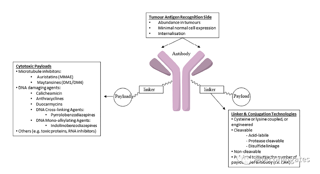
Antibody-drug conjugates (ADC) consist of linker, payload, and monoclonal antibody (mAb). They combine the advantages of high specificity targeting and strong cytotoxic effects, achieving precise and efficient killing of cancer cells, and have become a hot topic in the development of anti-cancer drugs.
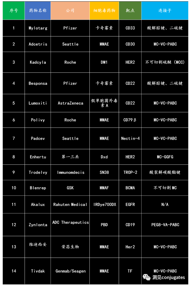
Since the first ADC drug Mylotarg® was approved by the FDA in 2000, as of December 2021, there are a total of 14 ADC drugs approved for hematologic malignancies and solid tumors worldwide. Additionally, there are currently over 100 ADC candidates in various stages of clinical trials.

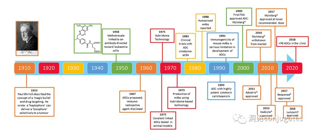
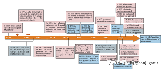
Timeline of Important Events in ADC Drug Development
As early as the early 20th century, Paul Ehrlich first proposed the concept of the “magic bullet” and hypothesized that certain compounds could enter tumor cells directly through certain targets, thereby curing diseases. Theoretically, these compounds should effectively kill cancer cells while being harmless to normal cells. In 2000, the U.S. Food and Drug Administration (FDA) first approved the ADC drug Mylotarg® (gemtuzumab ozogamicin) for adult acute myeloid leukemia (AML), marking the beginning of the era of ADC-targeted cancer therapy. Figure 1 depicts the milestones of ADC drug development from infancy to maturity over the past century. With the continuous expansion of targets and indications, ADC is leading a new era of targeted cancer therapy, and is expected to replace traditional chemotherapy drugs in the future.
 The concept of ADC was initially described by Paul Ehrlich in the early 1900s, where he referred to it as a “magic bullet.” Their development has not been smooth, and significant progress was not made until the early 1950s. In the 1980s and early 1990s, ADC also faced many challenges.
Poor Selection of Target Antigens is the most likely reason for early ADC failures, such as the KS1/4 antibody-methotrexate conjugate for non-small cell lung cancer and the BR96 antibody-docetaxel conjugate for metastatic breast cancer. Both of these ADCs entered clinical trials, but provided almost no therapeutic benefit.
Other factors that may have limited the success of these early ADCs include the use of chimeric or murine antibodies that triggered immunogenic reactions, as well as the use of payloads with low cytotoxic efficacy.
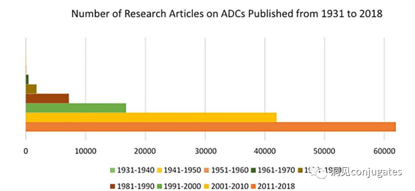
The field of ADC research has developed rapidly in recent years, as evidenced by the number of research papers published since 1970.
It is evident that the number of research articles doubled between 2000 and 2010, partly due to Kadcyla® and Adcetris® being in clinical development during this period. Over 60,000 research articles were published from 2011 to 2018.
The concept of ADC was initially described by Paul Ehrlich in the early 1900s, where he referred to it as a “magic bullet.” Their development has not been smooth, and significant progress was not made until the early 1950s. In the 1980s and early 1990s, ADC also faced many challenges.
Poor Selection of Target Antigens is the most likely reason for early ADC failures, such as the KS1/4 antibody-methotrexate conjugate for non-small cell lung cancer and the BR96 antibody-docetaxel conjugate for metastatic breast cancer. Both of these ADCs entered clinical trials, but provided almost no therapeutic benefit.
Other factors that may have limited the success of these early ADCs include the use of chimeric or murine antibodies that triggered immunogenic reactions, as well as the use of payloads with low cytotoxic efficacy.
The field of ADC research has developed rapidly in recent years, as evidenced by the number of research papers published since 1970.
It is evident that the number of research articles doubled between 2000 and 2010, partly due to Kadcyla® and Adcetris® being in clinical development during this period. Over 60,000 research articles were published from 2011 to 2018.
Mechanism of Action of ADCs
Traditional Internalization Mechanism
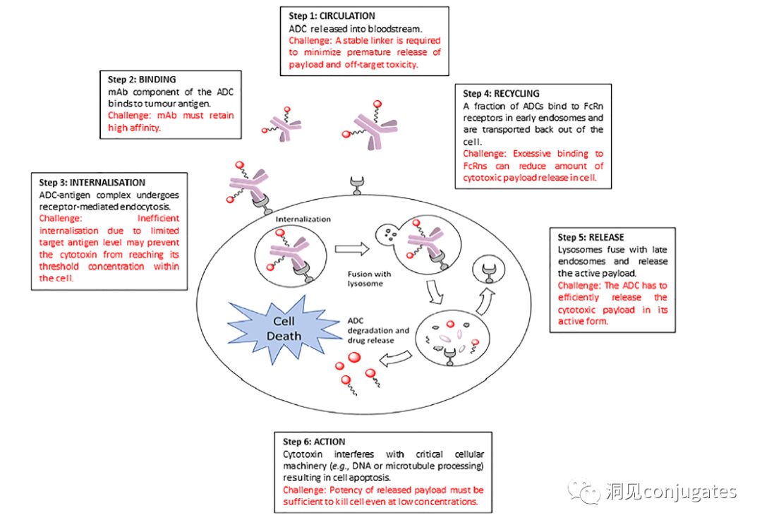
ADC is connected to mAb via linker and payload. ADC binds to the antigen on the surface of cancer cells, then internalizes, releasing highly cytotoxic payload molecules in the lysosome, usually through lysosomal cleavage.
To design a successful ADC, it is crucial to understand the potential mechanisms of action. An effective ADC needs to retain the selectivity of the monoclonal antibody while being able to release a sufficiently high concentration of payload to kill the target tumor cells. Each of these steps involves multiple unique challenges, making the design of ADCs complex.
1: Prevent ADC from Releasing in Blood
2: ADC Must Bind to Tumor Antigen
3: Internalization of ADC-Antigen Complex
4: Recycling of ADC (Fc-mediated)
5: Lysosomal Release of Payload
6: Low Concentration Payload Kills Cancer Cells (Highly Toxic)
ADC is designed to target and kill cancer cells, so the antibody must be able to recognize and bind to the corresponding antigen located on the tumor cells. Once bound to the antigen, the entire antigen-ADC complex is internalized through receptor-mediated endocytosis. The internalization process proceeds with the formation of early endosomes coated with clathrin containing the ADC-antigen complex. Once inside the lysosome, the ADC is degraded, releasing the cytotoxic payload into the cell, leading to cell death.
The mechanism of cell death will depend on the type of cytotoxic payload. For example, microtubule inhibitors, such as auristatins or maytansine, cause disruption of cytoplasmic division by interfering with microtubules; while DNA intercalators, such as PBD dimers or calicheamicin, cause DNA damage leading to apoptosis. New types of payloads currently under development will interfere with other cellular processes, such as RNA processing.
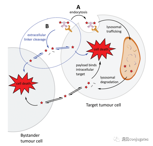
One important aspect of the ADC mechanism of action is the bystander effect, where free drugs exit tumor cells through the cell membrane into the tumor environment. This has potential therapeutic benefits in killing adjacent tumor cells, including those that may not have the relevant antigens on their surface.
Another key point is to ensure that a sufficient concentration of payload reaches inside the cell and kills the cancer cells; in fact, this is a complex process that is difficult to guarantee. It is estimated that even if the overall mechanism of action of the ADC works with 50% efficiency, only 1-2% of the payload will reach the tumor cells. Therefore, it is important that the selected payload is sufficiently cytotoxic to act at very low concentrations. It is now recognized that the choice of antigen targets, antibodies, linkers, and payload components, as well as how they effectively work together, is critical for the success of ADCs.
Non-internalization Mechanism:
Linkers break down in the extracellular tumor microenvironment to release payloads without requiring cellular endocytosis, allowing for the selection of non-internalizing antigens as targets.
One of the most critical factors in designing ADCs is the selection of the antibody. Most importantly, it must have high specificity for the antigen. Antibodies that lack high specificity and bind to other antigens may produce unpredictable effects, such as off-target toxicity due to interactions with healthy tissues or premature elimination before reaching the tumor site.
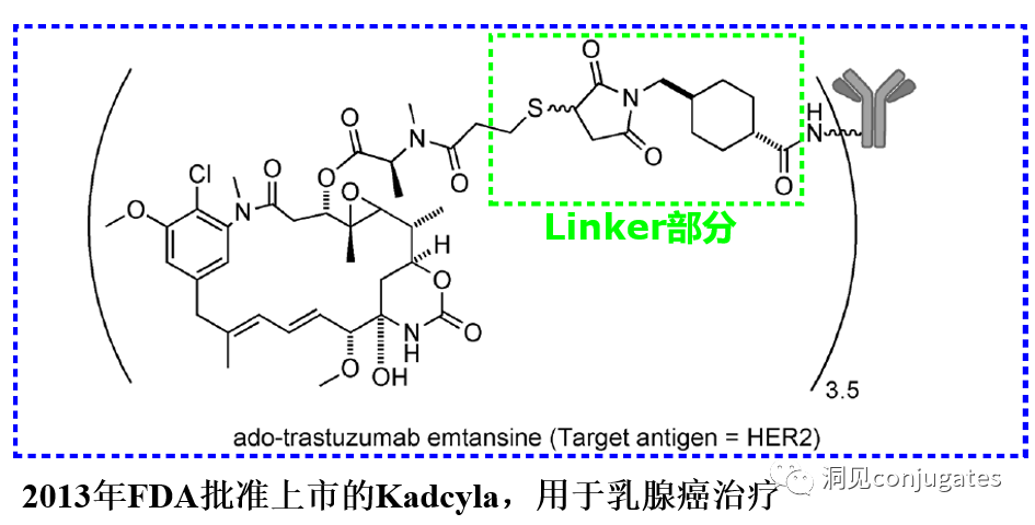
It is also important that antibodies bind to the target antigen with high affinity while maintaining low immunogenicity. Another important feature is favorable pharmacokinetic (PK) characteristics. Finally, if the mAb possesses inherent anti-tumor activity by directly modulating the biological activity of the target antigen, it would be beneficial, such as in the case of the anti-human epidermal growth factor receptor 2 (HER2) mAb trastuzumab (Herceptin®), which is the antibody component of trastuzumab emtansine (Kadcyla®).
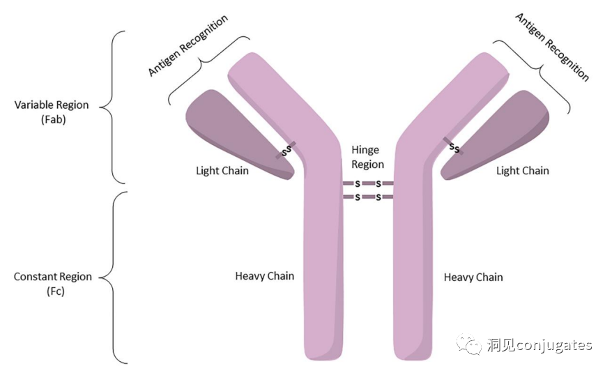
Antibodies are classified into five categories based on the sequence of their heavy chain constant regions: immunoglobulin M (IgM), IgD, IgG, IgE, and IgA.
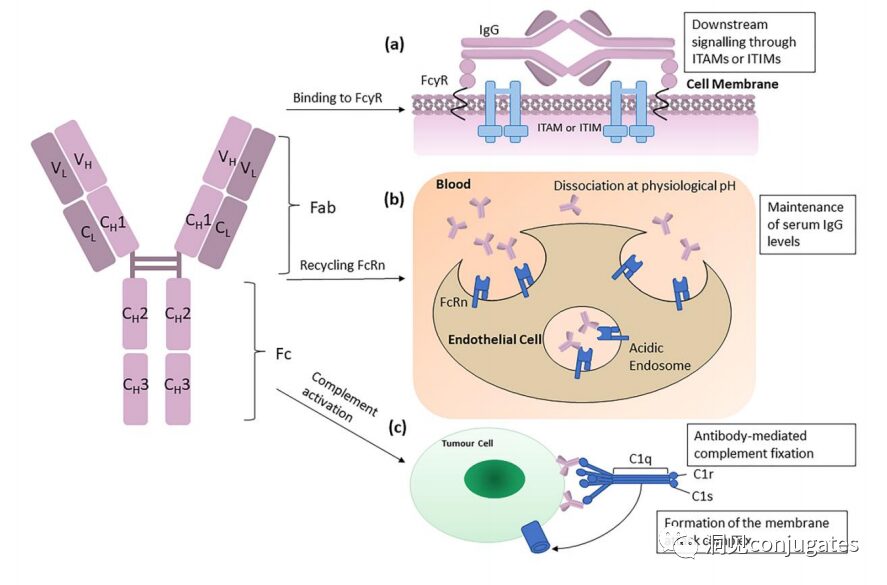
Among these five classes, IgG is the most commonly used in cancer immunotherapy. A typical IgG1 antibody consists of two heavy chains (H) and two light chains (L), which include the constant (C) region that forms the Fc domain and the variable (V) region that provides antigen specificity in the Fab domain.
The Fc region is recognized by immune cells. The Fc fragment does not recognize the corresponding antigen but binds to various cell receptors (such as T cells) and complement proteins. All antibodies have glycosylation sites at conserved positions in their constant regions. For example, there is a N-glycosylation site at the Fc region’s N297 residue.
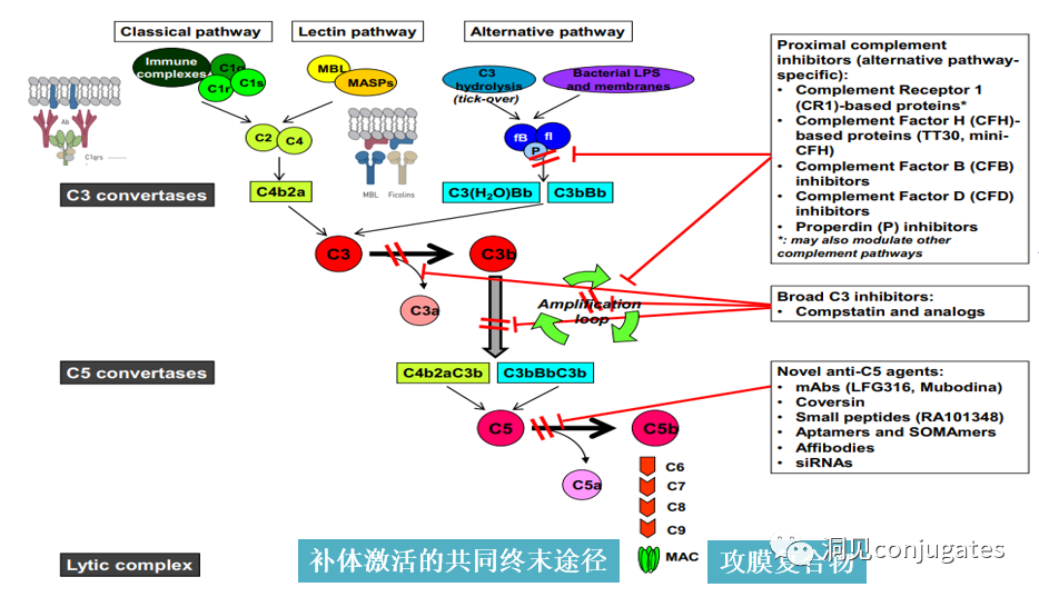
IgG subclasses, specifically IgG1 and IgG3, are effective activators of the classical complement pathway. Two or more IgG molecules bind to complement component 1q (C1q) on the cell surface, and 1q (C1q) binds with high affinity to the Fc domain, subsequently activating C1r enzyme activity, which then activates downstream complement proteins. The result of this cascade reaction is the formation of pores in the tumor cell surface by the membrane attack complex (MAC), leading to tumor cell lysis.
 To ensure effective internalization, the antigen-binding site should have as high an affinity as possible (current research finds that higher affinity is not always better; excessively high affinity can present a barrier effect in solid tumors, where if the target is expressed abnormally in tumor tissue, a high affinity may require large doses of ADC to saturate the available receptors at the tumor margin, allowing ADC molecules to continue to penetrate deeper and bind; the only adjustable factor in the barrier effect of binding sites in the tumor microenvironment is the binding affinity. Antibody drugs are more easily captured by the first encountered target in the tumor microenvironment rather than penetrating further. Thus, optimizing affinity based on tumor size to ensure sufficient affinity for drug internalization while allowing drugs to penetrate deeper into the tumor requires a balance).
The following figure illustrates how IgG antibodies bind to antigens and interact with Fc receptors to initiate signals based on immunoreceptor tyrosine activation motifs (ITAM) or immunoreceptor tyrosine inhibitory motifs (ITIM). IgG can bind to Fc receptors (FcRn) on endothelial cells to maintain serum IgG levels and bind to tumor cells, where they can recruit complement component 1q (C1q) to initiate the complement cascade, resulting in tumor cell lysis by MAC.
Considering the high specificity, affinity, and prolonged exposure required at the tumor site, ideally, antibody selection should ensure minimal interaction with healthy tissues, subnanomolar affinity for target antigens, and a longer pharmacokinetic half-life, with minimal immunogenicity.
The degree of immunogenicity of ADCs is a key factor and an important determinant of circulation half-life. In particular, antibodies lacking tumor specificity may be rapidly cleared from circulation due to immunogenicity, leading to decreased therapeutic efficacy. In the early development of ADCs, research was based on murine monoclonal antibodies, resulting in the formation of human anti-mouse antibodies within weeks after a single dose.
Thus, murine antibodies were quickly replaced by chimeric IgG antibodies, followed by humanized IgG. In recent years, ADCs have mainly been based on fully human antibodies. One of the most significant advantages of using mAbs instead of small molecule chemotherapeutics for cancer treatment is that mAb-based drugs can have favorable pharmacokinetics in terms of duration, metabolism, and elimination.
As mentioned above, once mAbs are administered into the bloodstream, they can distribute into tumor tissues through extravasation or pinocytosis. The distribution of ADCs within tumor tissues is limited by the size of the antibodies, which typically accounts for 95% of the ADC mass. However, unlike normal blood vessels, which have tightly connected monolayer endothelial cells, tumor endothelium is often characterized by excessive branching and sprouting, leading to a “leaky” monolayer. Therefore, although ADCs are larger molecules, they may still distribute into tumor tissues through the leaky vascular system, but their distribution in metabolic and clearance organs such as the liver, intestines, muscles, and skin is limited. Recently, antibody fragments are being studied, as they have smaller dimensions and stronger penetration capabilities into tumor tissues compared to full-sized antibodies, which generally have shorter half-lives.
To ensure effective internalization, the antigen-binding site should have as high an affinity as possible (current research finds that higher affinity is not always better; excessively high affinity can present a barrier effect in solid tumors, where if the target is expressed abnormally in tumor tissue, a high affinity may require large doses of ADC to saturate the available receptors at the tumor margin, allowing ADC molecules to continue to penetrate deeper and bind; the only adjustable factor in the barrier effect of binding sites in the tumor microenvironment is the binding affinity. Antibody drugs are more easily captured by the first encountered target in the tumor microenvironment rather than penetrating further. Thus, optimizing affinity based on tumor size to ensure sufficient affinity for drug internalization while allowing drugs to penetrate deeper into the tumor requires a balance).
The following figure illustrates how IgG antibodies bind to antigens and interact with Fc receptors to initiate signals based on immunoreceptor tyrosine activation motifs (ITAM) or immunoreceptor tyrosine inhibitory motifs (ITIM). IgG can bind to Fc receptors (FcRn) on endothelial cells to maintain serum IgG levels and bind to tumor cells, where they can recruit complement component 1q (C1q) to initiate the complement cascade, resulting in tumor cell lysis by MAC.
Considering the high specificity, affinity, and prolonged exposure required at the tumor site, ideally, antibody selection should ensure minimal interaction with healthy tissues, subnanomolar affinity for target antigens, and a longer pharmacokinetic half-life, with minimal immunogenicity.
The degree of immunogenicity of ADCs is a key factor and an important determinant of circulation half-life. In particular, antibodies lacking tumor specificity may be rapidly cleared from circulation due to immunogenicity, leading to decreased therapeutic efficacy. In the early development of ADCs, research was based on murine monoclonal antibodies, resulting in the formation of human anti-mouse antibodies within weeks after a single dose.
Thus, murine antibodies were quickly replaced by chimeric IgG antibodies, followed by humanized IgG. In recent years, ADCs have mainly been based on fully human antibodies. One of the most significant advantages of using mAbs instead of small molecule chemotherapeutics for cancer treatment is that mAb-based drugs can have favorable pharmacokinetics in terms of duration, metabolism, and elimination.
As mentioned above, once mAbs are administered into the bloodstream, they can distribute into tumor tissues through extravasation or pinocytosis. The distribution of ADCs within tumor tissues is limited by the size of the antibodies, which typically accounts for 95% of the ADC mass. However, unlike normal blood vessels, which have tightly connected monolayer endothelial cells, tumor endothelium is often characterized by excessive branching and sprouting, leading to a “leaky” monolayer. Therefore, although ADCs are larger molecules, they may still distribute into tumor tissues through the leaky vascular system, but their distribution in metabolic and clearance organs such as the liver, intestines, muscles, and skin is limited. Recently, antibody fragments are being studied, as they have smaller dimensions and stronger penetration capabilities into tumor tissues compared to full-sized antibodies, which generally have shorter half-lives.
 The design, structure, and chemical properties of linkers are critical to the characteristics of ADCs, influencing the specificity, efficacy, and safety of the entire ADC drug. Typically, linkers are designed to be stable in circulation and effectively release payloads at the tumor site. It is also crucial that conjugates remain stable in buffered aqueous solutions. Linkers are classified into different types based on their payload release mechanisms, with the two main types being “cleavable” or “non-cleavable” (Recommended reading: Common types of linkers in ADC drugs and their in vivo elimination mechanisms).
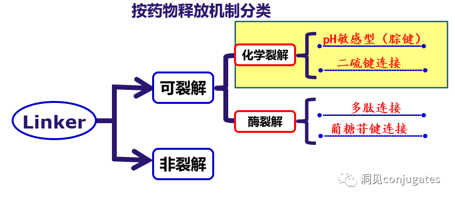
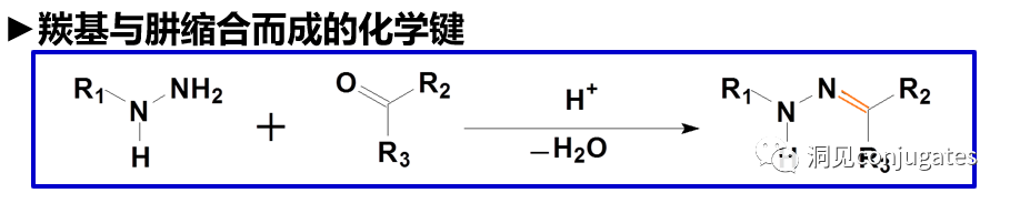
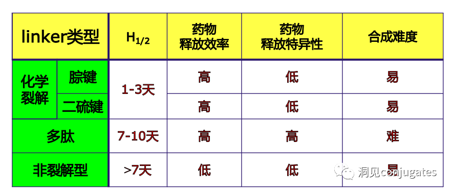 Cleavable linkers utilize the differences in conditions between the bloodstream and the cytoplasm of tumor cells. Changes in the environment after internalization of the ADC-antigen complex trigger the cleavage of the linker and the release of the active payload. Cleavable linkers are divided into three main subclasses: (1) acid-labile (e.g., hydrazone), (2) reducible (e.g., disulfide), and (3) enzyme-cleavable (e.g., peptide). The following figure shows an example of a hydrazone linker used in Gemtuzumab ozogamicin (Mylotarg®). Non-specific release of the payload can lead to systemic toxicity. This is also one of the reasons Mylotarg was withdrawn from the market in 2010.
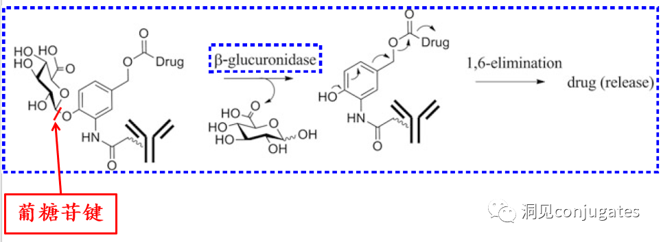
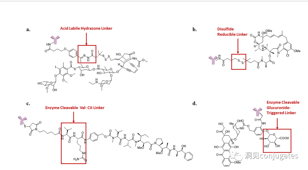 Another type of enzyme-cleavable linker is based on β-glucuronide moieties. β-glucuronidase (in lysosomes) can release payloads from linkers containing β-glucuronide, which are overexpressed in certain types of tumor cells. An important feature of β-glucuronide linkers is their hydrophilicity, which can potentially reduce aggregation during the conjugation process compared to structures based on dipeptides or other types of linkers. β-glucuronide has been used as a linker component to connect payloads, including auristatin derivatives MMAE and MMAF, as well as doxorubicin.
Chemical Cleavable Linkers:
Characteristics of Hydrazone Bonds:
In neutral blood environments (pH 7.3-7.5): approximately 6% cleavage rate; in intracellular lysosomes (pH 4.5-5.0) or endosomes (pH 5.0-6.5), easily cleavable, with a cleavage rate of about 97% at pH 4.5.
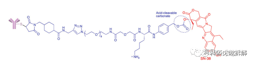
Another commonly used structure that undergoes hydrolysis under acidic conditions is carbonate linkers.
Simple carbonates have limited stability in serum and require the addition of a para-aminobenzyl (PAB) moiety to significantly enhance their half-life.
Carbonate structures release SN-38 drugs and carbon dioxide upon hydrolysis under acidic conditions.
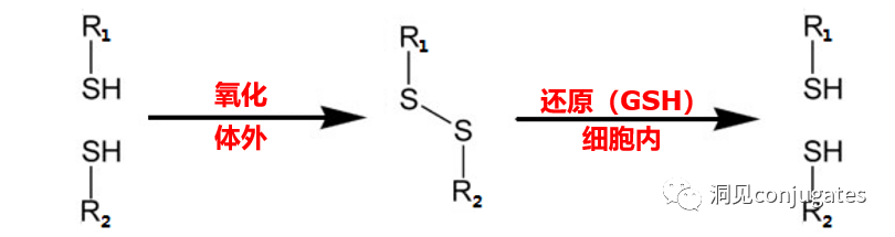
2: Disulfide Bond Chemical Cleavage
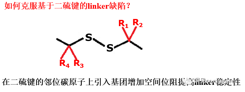
Disulfide compounds are stable under physiological pH conditions but are easily attacked by nucleophiles from thiols, thereby breaking the link (including sterically hindered disulfide bonds).
The presence of thiol-containing compounds in blood is complex, along with interference from albumin, containing small molecule substances with free thiols (GSH and Cys):
In blood environment: 0.005mM
In intracellular environment: 0.5 -10mM
Average concentrations of GSH and Cys in cancer cells are even higher.
Enzyme Cleavable Linkers:
Specific enzymes present in both the intracellular lysosome and the extracellular tumor microenvironment can selectively cleave corresponding substrates. Peptide-based linkers: utilize proteolytic enzymes such as Cathepsin and plasmin in lysosomes (these proteolytic enzymes are strictly inhibited in the blood environment, while their activity is optimal in lysosomal environments).
►Proteolytic enzyme expression is generally higher in cancer cells.
►Peptide-based linkers have higher stability in blood and cleave faster in cancer cells.
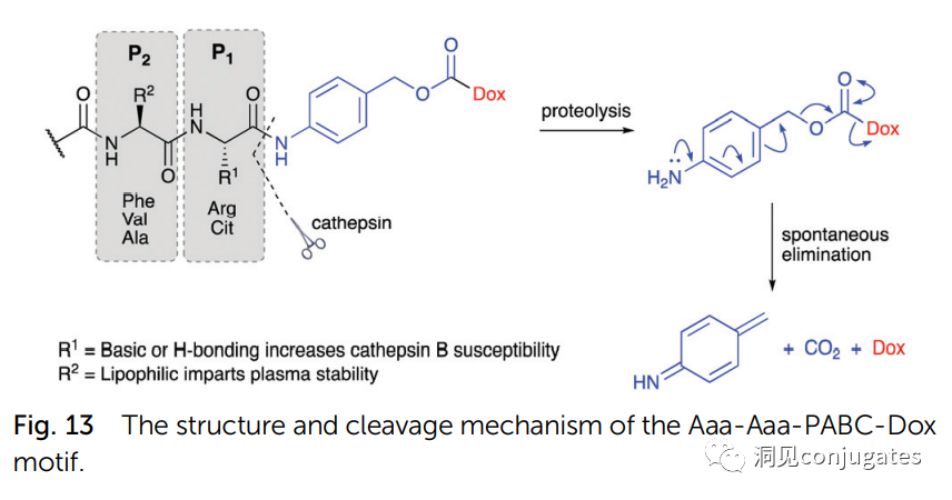
1: Cathepsin B Cleaves Dipeptides:
Linkers based on dipeptides + self-cleaving modules:
Optimal dipeptides: Val-Cit and Phe-Lys (citrulline-valine and phenylalanine-lysine), can be specifically hydrolyzed by Cathepsin B.
Self-cleaving module: PABC
Dipeptide linkers are usually targeted at Cathepsin B, which is mainly present in lysosomes, and activity is also observed extracellularly.
β-glucuronidase is a hydrolytic enzyme in the glycosidase family, which catalyzes the breakdown of β-glucuronic acid residues in polysaccharides using β-glucuronidase in lysosomes.
►β-glucuronidase is abundant in lysosomes and is overexpressed in certain cancer cells.
►β-glucuronidase activity is highest in lysosomal environments, while enzyme activity is inhibited in blood environments.
Glucuronic acid linkers have another advantage: due to their high hydrophilicity, they can significantly reduce ADC aggregation, allowing for the synthesis of ADC drugs with high payload loads (DAR=8).
β-galactosidase can cleave linkers and is similar to glucuronidase.
Acidic pyrophosphatases and acid phosphatases present in lysosomes can hydrolyze phosphoester bonds.
Another type of enzyme-cleavable linker is based on β-glucuronide moieties. β-glucuronidase (in lysosomes) can release payloads from linkers containing β-glucuronide, which are overexpressed in certain types of tumor cells. An important feature of β-glucuronide linkers is their hydrophilicity, which can potentially reduce aggregation during the conjugation process compared to structures based on dipeptides or other types of linkers. β-glucuronide has been used as a linker component to connect payloads, including auristatin derivatives MMAE and MMAF, as well as doxorubicin.
Chemical Cleavable Linkers:
Characteristics of Hydrazone Bonds:
In neutral blood environments (pH 7.3-7.5): approximately 6% cleavage rate; in intracellular lysosomes (pH 4.5-5.0) or endosomes (pH 5.0-6.5), easily cleavable, with a cleavage rate of about 97% at pH 4.5.
Another commonly used structure that undergoes hydrolysis under acidic conditions is carbonate linkers.
Simple carbonates have limited stability in serum and require the addition of a para-aminobenzyl (PAB) moiety to significantly enhance their half-life.
Carbonate structures release SN-38 drugs and carbon dioxide upon hydrolysis under acidic conditions.
2: Disulfide Bond Chemical Cleavage
Disulfide compounds are stable under physiological pH conditions but are easily attacked by nucleophiles from thiols, thereby breaking the link (including sterically hindered disulfide bonds).
The presence of thiol-containing compounds in blood is complex, along with interference from albumin, containing small molecule substances with free thiols (GSH and Cys):
In blood environment: 0.005mM
In intracellular environment: 0.5 -10mM
Average concentrations of GSH and Cys in cancer cells are even higher.
Enzyme Cleavable Linkers:
Specific enzymes present in both the intracellular lysosome and the extracellular tumor microenvironment can selectively cleave corresponding substrates. Peptide-based linkers: utilize proteolytic enzymes such as Cathepsin and plasmin in lysosomes (these proteolytic enzymes are strictly inhibited in the blood environment, while their activity is optimal in lysosomal environments).
►Proteolytic enzyme expression is generally higher in cancer cells.
►Peptide-based linkers have higher stability in blood and cleave faster in cancer cells.
1: Cathepsin B Cleaves Dipeptides:
Linkers based on dipeptides + self-cleaving modules:
Optimal dipeptides: Val-Cit and Phe-Lys (citrulline-valine and phenylalanine-lysine), can be specifically hydrolyzed by Cathepsin B.
Self-cleaving module: PABC
Dipeptide linkers are usually targeted at Cathepsin B, which is mainly present in lysosomes, and activity is also observed extracellularly.
β-glucuronidase is a hydrolytic enzyme in the glycosidase family, which catalyzes the breakdown of β-glucuronic acid residues in polysaccharides using β-glucuronidase in lysosomes.
►β-glucuronidase is abundant in lysosomes and is overexpressed in certain cancer cells.
►β-glucuronidase activity is highest in lysosomal environments, while enzyme activity is inhibited in blood environments.
Glucuronic acid linkers have another advantage: due to their high hydrophilicity, they can significantly reduce ADC aggregation, allowing for the synthesis of ADC drugs with high payload loads (DAR=8).
β-galactosidase can cleave linkers and is similar to glucuronidase.
Acidic pyrophosphatases and acid phosphatases present in lysosomes can hydrolyze phosphoester bonds.
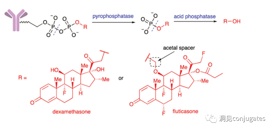 Merck designed a diphosphate linker that releases prototype glucocorticoid payloads upon hydrolysis in lysosomes; when testing monophosphate linkers, drug release was relatively slow.
►ADC containing non-cleavable linkers:
Once ADC is phagocytosed into the cell, it enters the lysosomal pathway, where drug derivatives are released after the complete degradation of the mAb by various cleaving enzymes, killing the target cells. Generally, they have better stability.
►Requirements for non-cleavable linkers:
(1) Linkers should not be degraded under extracellular conditions and factors.
(2) Drug derivatives released after intracellular degradation of ADC should also have considerable cytotoxicity, so the use of this linker has strict requirements for the drug.
►Drawbacks of non-cleavable linkers:
(1) Strict requirements for the drug, necessitating screening and validation of the cytotoxicity of drug derivatives.
(2) Relatively slow drug release rates.
(3) Generally, drug derivatives released after intracellular degradation of ADC have good water solubility, even carrying charged groups, which is not conducive to exerting the bystander effect (Bystander Kill Effect).
This issue discusses the global overview of ADCs, the mechanism of action of ADCs, the complexity of ADC drug design, the selection of antibodies, and the selection of linkers. Next issue, we will introduce how to select payloads and coupling methods.
Merck designed a diphosphate linker that releases prototype glucocorticoid payloads upon hydrolysis in lysosomes; when testing monophosphate linkers, drug release was relatively slow.
►ADC containing non-cleavable linkers:
Once ADC is phagocytosed into the cell, it enters the lysosomal pathway, where drug derivatives are released after the complete degradation of the mAb by various cleaving enzymes, killing the target cells. Generally, they have better stability.
►Requirements for non-cleavable linkers:
(1) Linkers should not be degraded under extracellular conditions and factors.
(2) Drug derivatives released after intracellular degradation of ADC should also have considerable cytotoxicity, so the use of this linker has strict requirements for the drug.
►Drawbacks of non-cleavable linkers:
(1) Strict requirements for the drug, necessitating screening and validation of the cytotoxicity of drug derivatives.
(2) Relatively slow drug release rates.
(3) Generally, drug derivatives released after intracellular degradation of ADC have good water solubility, even carrying charged groups, which is not conducive to exerting the bystander effect (Bystander Kill Effect).
This issue discusses the global overview of ADCs, the mechanism of action of ADCs, the complexity of ADC drug design, the selection of antibodies, and the selection of linkers. Next issue, we will introduce how to select payloads and coupling methods.
References:
1. Cytotoxic payloads for antibody-drug conjugates
2. Other public information compilation
Source from publicaccount Insights on conjugates
Join the fan group, add the WeChat below to enter the group~