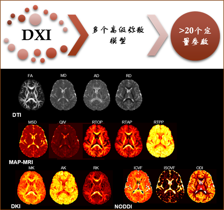
DXI is an advanced diffusion model post-processing research platform jointly developed by Siemens and East China Normal University, based on Siemens’ unique MRI diffusion spectrum imaging (DSI) scanning technology. It achieves the simultaneous application of multiple diffusion models for the first time, including Diffusion Tensor Imaging (DTI), Diffusion Kurtosis Imaging (DKI), Neurite Orientation Dispersion and Density Imaging (NODDI), and Mean Apparent Propagator Imaging (MAP-MRI), among others. This platform provides over 20 quantitative parameter images, helping researchers deeply interpret the microstructural changes and functional abnormalities of the nervous system from multiple dimensions.
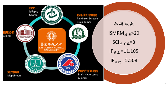
Research Achievements Overview

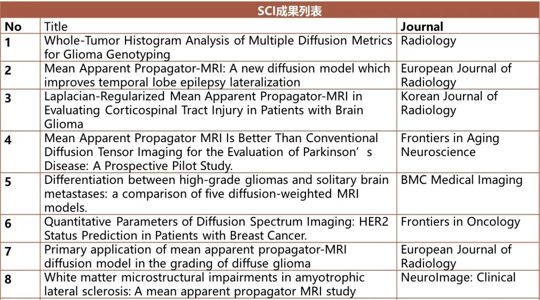
Since 2020, multiple institutions have participated in research projects based on the DXI platform. Through the collaboration of hospitals, universities, and Siemens, fruitful research cooperation results have been achieved, with over 20 ISMRM abstracts and nearly half of the work already transformed or in the process of being transformed into SCI articles. Currently, 8 high-level SCI articles have been accepted, with the highest impact factor IF 11.105 and an average IF of 5.508, creating multiple first clinical research applications of new technologies. The following sections will introduce some of these achievements.
Featured Achievements Display

[1] The Application Value of Different Diffusion Models in Gene Typing Diagnosis of Glioma (First Affiliated Hospital of Zhengzhou University)
Whole-Tumor Histogram Analysis of Multiple Diffusion Metrics for Glioma Genotyping, Radiology, 2022; 302(3):E16.DOI:10.1148/radiol.210820
In this study, the authors compared the histogram analysis of different advanced diffusion models DTI, DKI, NODDI, and MAP metrics in predicting IDH genotype and 1p/19q codeletion, and evaluated the application of MAP in glioma grading. The results showed that the whole tumor histogram analysis of multiple diffusion metrics is a promising method for glioma IDH and 1p/19q gene typing. Whether used independently or in combination, the diffusion models exhibited similarly satisfying predictive efficacy for predicting glioma IDH and 1p/19q gene typing.
[2] MAP-MRI in the Lateralization of Temporal Lobe Epilepsy (First Affiliated Hospital of Zhengzhou University)
Mean Apparent Propagator-MRI: A new diffusion model which improves temporal lobe epilepsy lateralization. European Journal of Radiology,2020; 126:108914. DOI: 10.1016/j.ejrad.2020.108914
This study compared MRI volumetric measurements, FLAIR image intensity, DTI, and MAP-MRI in distinguishing the ictal focus between the ipsilateral and contralateral hippocampal regions in patients with temporal lobe epilepsy. The results showed that the MAP parameters had the strongest effect size in differentiating the ipsilateral and contralateral hippocampal ictal foci.
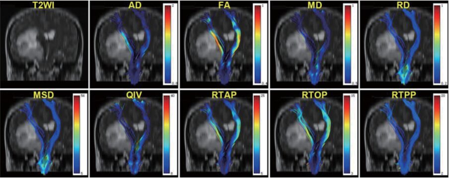
[3] MAP-MRI in Evaluating Corticospinal Tract Injury in Patients with Brain Glioma (Fujian Medical University Union Hospital)
Laplacian-Regularized Mean Apparent Propagator-MRI in Evaluating Corticospinal Tract Injury in Patients with Brain Glioma. Korean Journal of Radiology, 2021; 22(5):759-769. DOI: 10.3348/kjr.2020.0949
This study compared MAP-MRI and DTI in assessing the overall and peritumoral quantitative injury of the corticospinal tract (CST) in glioma patients. The results showed that, compared to DTI, MAP-MRI was more sensitive in detecting injury in the overall and peritumoral CST on the tumor side.

[4] MAP in the Application of Neurodegenerative Diseases PD (Sun Yat-sen Memorial Hospital of Sun Yat-sen University)
Mean Apparent Propagator MRI Is Better Than Conventional Diffusion Tensor Imaging for the Evaluation of Parkinson’s Disease: A Prospective Pilot Study. Frontiers in Aging Neuroscience,2020;12:563595. DOI: 10.3389/fnagi.2020.563595
This study applied MAP-MRI for the first time in Parkinson’s disease (PD) and evaluated its diagnostic value compared to conventional DTI. The results showed that multiple parameter AUC values of MAP-MRI in bilateral caudate nucleus, globus pallidus, putamen, and thalamus were greater than those of DTI parameters, indicating that MAP-MRI outperforms conventional DTI in diagnosing PD and assessing disease severity.

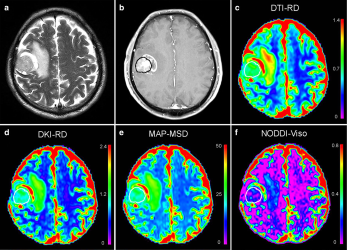
[5] DXI in the Application Value of Differentiating Gliomas and Metastatic Tumors (Sun Yat-sen Memorial Hospital of Sun Yat-sen University)
Differentiation between high-grade gliomas and solitary brain metastases: a comparison of five diffusion-weighted MRI models. BMC Med Imaging, 2020; 20(1):124. DOI: 10.1186/s12880-020-00524-w
This study applied DXI technology in differentiating high-grade gliomas (HGGs) and solitary brain metastases (SBMs), comparing the diagnostic value of DTI, DKI, NODDI, MAP-MRI, and traditional single-exponential diffusion models (DWI) in differentiating HGGs and SBMs. The results found that NODDI outperformed MAP-MRI, DKI, DTI, and DWI in differentiating HGGs and SBMs. The NODDI-based Viso had the highest discriminative performance.
[6] MAP-MRI in the Grading of Diffuse Gliomas (Inner Mongolia Medical University Affiliated Hospital)
Primary application of mean apparent propagator-MRI diffusion model in the grading of diffuse glioma.
European Journal of Radiology, 2021; 138:109622.DOI: 10.1016/j.ejrad.2021.109622
This study applied the MAP-MRI method in the differentiation of grade II, III, and IV gliomas. The results showed that MAP-MRI can differentiate between grade II and III, and between II and IV gliomas, but cannot differentiate between III and IV gliomas, demonstrating the clinical guiding value of MAP-MRI in glioma grading differentiation.
Article list as follows:
Conclusion

After clinical validation, the DXI platform obtains multiple diffusion models in a single scan, greatly saving imaging resources for clinical researchers. The application of new models and new quantitative parameters has opened up multiple valuable clinical studies, resulting in numerous research achievements. In the future, there will be more clinical applications of the DXI platform, and we will continue to introduce them to everyone.
Share this article to let more friends know about the latest advances in MRI.
Scan the QR code below to follow the [MRI Enthusiasts] public account and video account.