Recently, a study published in the prestigious international journal Advanced Healthcare Materials has brought groundbreaking progress in the treatment of spinal cord injury (SCI).
This research was conducted by a team from the Department of Mechanical Engineering, Neurosurgery, and Stem Cell Research Institute at the University of Minnesota, focusing on the development of a 3D printed spinal organoid scaffold. By combining human induced pluripotent stem cell-derived spinal neural progenitor cells (sNPCs), they successfully achieved functional recovery after spinal cord injury in rat models.
This achievement not only fills the gap in existing treatment methods but also brings new hope for the rehabilitation of spinal cord injury patients.

(DOI:10.1002/adhm.202404817)
Spinal Cord Injury: A Difficult “Medical Challenge” to Overcome
Spinal cord injury is a severe central nervous system trauma that often leads to lower limb paralysis, loss of sensation, and even lifelong disability. According to statistics, there are approximately 302,000 spinal cord injury patients in the United States, with numbers ranging from 255,000 to 383,000, and currently, there is no effective cure worldwide.
Previously, researchers attempted to transplant neural progenitor cells to repair the damage, but two key issues arose: first, directly injecting cells into the injury site lacked structural support, leading to cell loss and low survival rates; second, the transplanted cells struggled to form region-specific neural networks, failing to effectively connect with the host’s neural circuits, especially in cases of chronic spinal cord injury.
Traditional 3D printed scaffolds can provide structural support but often fail to guide cells in forming spinal tissue structures similar to those in vivo. However, organoids can address this issue. Organoids are miniature tissues that simulate the structure and function of organs in vitro, and spinal organoids can replicate the cellular diversity and neural connections of the spinal cord, allowing the research team to explore the possibility of combining “3D printed scaffolds” with “spinal organoids.”
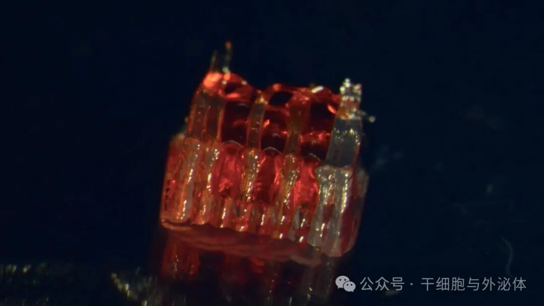
This method involves creating a unique 3D printed framework for lab-cultivated organs, known as organoid scaffolds, which contain microscopic channels. Image source: University of Minnesota
Innovative Design: How Does the 3D Printed Scaffold “Cultivate” Spinal Organoids?
To ensure the scaffold provides stable support while guiding cells to form functional neural tissue, the research team made dual innovations in material selection and structural design.
First, in terms of materials, the team chose silicone as the scaffold substrate. Silicone is widely used in the medical field due to its excellent biocompatibility (it does not provoke a strong immune response in the body), outstanding antioxidant properties, and permeability, which meets the oxygen needs of neural cells. More importantly, silicone is not easily degradable, allowing it to maintain structural stability long after cell culture and transplantation, avoiding interference with cell growth.
In terms of structural design, the scaffold adopts a “multi-layer microchannel” structure: the overall dimensions are approximately 1.6 mm wide, 0.65 mm high, and 2 mm long (suitable for the spinal cord injury site in rats), with three internal microchannels measuring 200 micrometers wide and 440 micrometers high.
These channels play a crucial role, guiding the axons of neural cells (the “wires” for neural signal transmission) to grow directionally along the channels while shortening the diffusion distance for nutrients and oxygen, ensuring cell survival. Additionally, a “sacrificial layer” (pullulan hydrogel) is laid at the bottom during scaffold printing, which will dissolve during subsequent culture, facilitating the removal of the scaffold from the culture dish without damaging the cells.
For cell selection, the team used “human induced pluripotent stem cell (iPSC)-derived sNPCs.” iPSCs are stem cells that can “transform” into any cell type in the human body. When induced to differentiate into sNPCs (spinal neural progenitor cells), they not only maintain spinal region specificity (i.e., they only develop into spinal-related neural cells) but also avoid immune rejection, laying the foundation for future clinical applications.
After printing, the team mixes sNPCs with Matrigel (a gel that simulates the extracellular matrix and contains growth factors) and injects it into the microchannels of the scaffold, then cultures it in vitro for 40 days. During this time, sNPCs gradually differentiate into various spinal-specific neural cells (such as V0, V1, V2a interneurons, which are key in regulating motor functions) and form neural networks along the microchannels, ultimately developing into a “spinal organoid scaffold.”
Experimental Validation: From In Vitro Maturation to In Vivo Repair
To validate the effectiveness of the scaffold, the team conducted research in two steps: “in vitro experiments” and “in vivo experiments,” with results exceeding expectations.
In the in vitro experiments, the team confirmed the maturity and functionality of the spinal organoids through special staining, gene sequencing, and electrophysiological testing. After 15 days of culture, the axons of the neural cells had already grown directionally along the microchannels; after 30 days, the axons extended to the surface of the scaffold, forming a complete neural network; and after 40 days, the cells successfully differentiated into various spinal neural cells such as V0, V1, and V2a, with gene sequencing showing that the gene expression patterns of these cells were highly similar to those of normal spinal tissue, indicating greater maturity than cells cultured in traditional 2D conditions. Even more surprisingly, these organoid scaffolds maintained the characteristics of neural cells after one year of in vitro culture, with neatly arranged axon bundles and long-term survival capabilities.
Electrophysiological testing further confirmed the functionality of the organoids: after 15 days of culture, the neural cells in the 3D scaffold could generate continuous electrical signals (similar to the “discharge” of normal neural cells), while 2D cultured cells could only produce weak and discontinuous signals, indicating that the neural cells in the 3D scaffold had acquired the ability to transmit neural signals.
The in vivo experiments were conducted in a rat model with a spinal cord transection. The team transplanted two spinal organoid scaffolds into a 1.8 mm injury gap in the rat’s spinal cord, while setting up a “only injury group” and an “empty scaffold group” as controls. They evaluated recovery effects using the “BBB motor function score” (a standard method for assessing lower limb motor ability in rats) and “motor-evoked potentials (MEP)” (to detect neural signal transmission from the brain to the muscles).
The results showed that 12 weeks post-transplantation, the “organoid scaffold group” rats achieved a BBB score of 8.4 (out of a maximum of 21, indicating that the rats could walk independently and had coordination), while the “empty scaffold group” scored only 3.6, and the “only injury group” scored just 2.25. MEP testing revealed that the neural signal amplitude in the organoid scaffold group was more than twice that of the other two groups, proving that the neural cells in the scaffold had connected with the host spinal cord and could effectively transmit motor signals.
More critically, 12 weeks post-transplantation, the team observed that the human neural cells in the scaffold not only survived well but also grew bidirectionally towards the “head end” and “tail end” of the rat’s spinal cord, integrating with the host neural tissue and forming synapses (the “connection points” for signal transmission between neural cells). This indicates that the organoid scaffold successfully established a “neural relay station” at the injury site, reconnecting the interrupted neural circuits.
Future Prospects: From Animal Experiments to Clinical Applications
The significance of this research lies not only in validating the effectiveness of 3D printed spinal organoid scaffolds but also in indicating the future direction of spinal cord injury treatment. The research team stated that the next steps will focus on three areas: first, combining “dorsal neural cells” and “ventral neural cells” to repair both motor and sensory circuits (current research mainly focuses on motor function, while sensory function recovery remains a challenge); second, developing biodegradable scaffolds (such as polycaprolactone materials) that gradually degrade after guiding neural repair, avoiding long-term presence in the body; and third, combining scaffolds with decellularized matrices to further enhance cell survival and integration efficiency.
Although the current research is still in the animal experiment stage, it provides a new “structure + cells + function” triad strategy for spinal cord injury treatment. With the improvement of technology, this 3D printed spinal organoid scaffold is expected to enter clinical practice in the future, bringing hope for “standing and walking again” to spinal cord injury patients and fundamentally changing the current state of spinal cord injury treatment.
【Stem Cells and Exosomes】 Public Account Editor: Wu Kaiqi
Column Chief: Wang Zheng
Text Editor: Yang Ledong. Disclaimer: All statements on this platform do not constitute medical claims or advice. If you have health issues or concerns, please consult your doctor or healthcare provider. Original articles may not be reproduced without authorization.