
Background Introduction
Can you imagine? A robot smaller than a hair strand that can “navigate itself” inside the body, report its location in real-time, and precisely “burn” tumor cells? This is not a science fiction scenario, but the latest research published in Advanced Materials. This microrobot, known as TriMag, disrupts traditional medicine with three “superpowers”: magnetic drive (free movement), magnetic particle imaging (MPI, precise positioning), and magnetic hyperthermia (targeted treatment).
The secret lies in Figure 1: The research team used two-photon lithography technology to 3D print a biocompatible hydrogel scaffold, and then embedded two types of nanoparticles—Fe₃O₄ (iron oxide) and CoFe₂O₄ (cobalt ferrite)—into the scaffold through in-situ chemical reactions. Among them, Fe₃O₄ acts as the “navigator” and “communicator,” responding to external magnetic fields to drive the robot’s movement and allowing doctors to see its location in real-time through MPI; CoFe₂O₄ serves as the “therapist,” generating heat under high-frequency magnetic fields to precisely ablate tumors. The combination of these three capabilities makes the microrobot an integrated medical tool that is “movable, traceable, and treatable.”
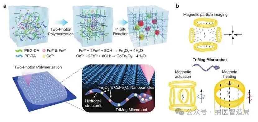
Figure 1丨Illustration of the manufacturing and functionalization of TriMag microrobots
Content Introduction
The research team used two-photon lithography technology to print a helical structure from two biocompatible materials: PEG-DA (polyethylene glycol diacrylate) and PETA (pentaerythritol triacrylate). The brilliance of this structure lies in its ability to move flexibly in bodily fluids like sperm when rotated, with a diameter of only 10-100 micrometers, thinner than human capillaries, allowing it to reach various hard-to-access areas. After printing, the hydrogel scaffold is soaked in a solution containing iron and cobalt ions, followed by the addition of an alkaline solution (such as NaOH). At this point, ions undergo chemical reactions inside the hydrogel, “growing” Fe₃O₄ and CoFe₂O₄ nanoparticles—either adhering to the surface of the hydrogel or trapped within the internal mesh, preventing them from falling off. The final robot resembles a “mini Swiss Army knife”: Fe₃O₄ allows it to be “remotely controlled” by external magnetic fields (driving function), while also serving as a “signal source” for MPI (imaging function); CoFe₂O₄ generates heat under high-frequency magnetic fields (thermal therapy function). Scanning electron microscope images (Figure 2) show that the robot’s surface is smooth and its structure precise, even capable of printing orderly “robot arrays,” proving that this manufacturing method is both precise and scalable.
The researchers injected TriMag microrobots into mice with breast cancer and used CT-MPI combined imaging for real-time tracking (Figure 3). Under the drive of an external magnetic field, the robot “crawled” from the injection point towards the tumor, with a clear and controllable route (similar to drawing a “C” shaped trajectory), monitored without any blind spots. Upon reaching the tumor site, a high-frequency magnetic field (150kHz) was activated, and the robot began to heat up. Thermal camera images showed that within 15 minutes, the local temperature rose from 35°C to 43°C—this temperature is just enough to kill cancer cells without burning surrounding healthy tissue. More importantly, when the temperature exceeds 45°C, it automatically shuts off, ensuring safety. Four days of IVIS fluorescence imaging showed that the mice receiving TriMag thermal therapy had significantly weakened tumor fluorescence signals (indicating cancer cell activity); while untreated mice or those injected only with the robot continued to have growing tumors. This indicates that the robot can not only “get to the right place” but also “treat effectively.”
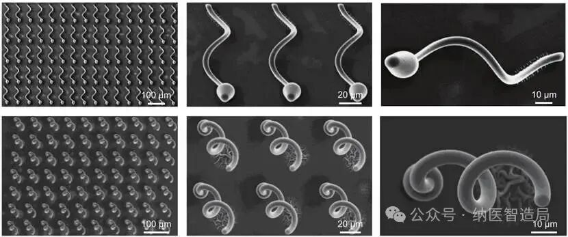
Figure 2丨Top and side views of a single helical microrobot and microrobot array
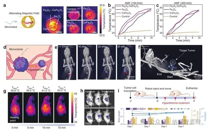
Figure 3丨Magnetic hyperthermia heating using TriMag microrobots
Conclusion
The breakthrough of TriMag microrobots addresses two major pain points in traditional medicine:
1. Manufacturing challenges: Two-photon lithography + in-situ reactions ensure the nanometer-level precision of the robots while allowing for uniform distribution of the two types of nanoparticles, ensuring stable functionality;
2. The dilemma of being “invisible and intangible”: MPI imaging technology penetrates deeply and is non-interfering, allowing for full control of the robot’s movement and treatment inside the body, breaking the depth limitations of traditional optical or ultrasound imaging.
In the future, such robots may become the main force in “minimally invasive treatment”: for example, entering the vitreous body of the eye to treat retinal diseases or crossing the blood-brain barrier to deliver drugs precisely. With advancements in materials and control technologies, perhaps one day we will truly rely on a “legion of microrobots” to perform various complex surgeries inside the body—the era of precision medicine is rapidly moving from the laboratory to the clinic.
References:
Xing, L.; Cai, Y.; Zhang, Y.; Mozel, K.; Tang, Z.; Tang, T.; Mottini, V.; Nigam, S.; Smith, B. R.; Lee, I. Y.; Nagaraja, T. N.; Wang, P.; Li, X.; Gao, T.; Li, J. TriMag Microrobots: 3D-Printed Microrobots for Magnetic Actuation, Imaging, and Hyperthermia. Advanced Materials 2025, e19708. DOI:10.1002/adma.202419708
Custom Nanomedicine,Contrast Agent Development,Medical Imaging Testing and Analysis
Contact Teacher Xu