Ultrasound examination is the preferred imaging method for thyroid nodules and plays a crucial role in clinical diagnosis and treatment decisions. The “2020 China Guidelines for Risk Stratification of Malignancy in Thyroid Nodules via Ultrasound: C-TIRADS” was published in the Chinese Journal of Ultrasound Imaging. This article shares excerpts from the section on the classification of several special case types.
Ultrasound Evaluation Classification of Thyroid Nodules (C-TIRADS) and Management Recommendations
The expert committee established C-TIRADS using a counting method, where the number of suspicious ultrasound characteristics (solid, microcalcification, very low echo, blurred margins, irregular margins, or extrathyroidal invasion, as well as vertical orientation as suspicious malignant ultrasound features, while comet tail artifacts are benign features) determines the score. If a comet tail artifact is present, the total score is reduced by 1. The final score is used to stratify the risk of the nodule.
Table 1: C-TIRADS Based on Counting Method
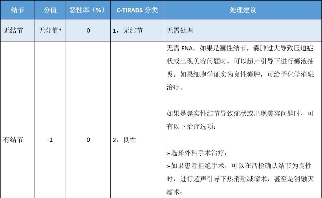
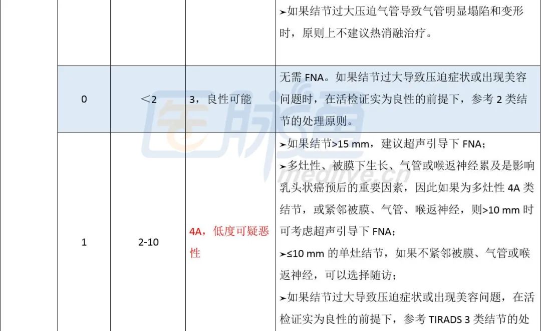
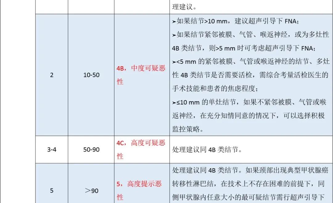

Note: *No nodules, no points assigned
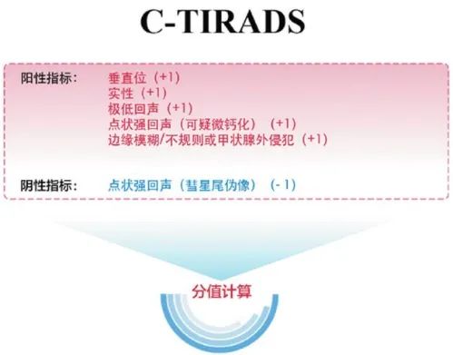
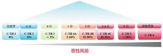
Note: The vertical orientation can be assessed in transverse or longitudinal sections; punctate strong echoes can be classified into microcalcification, comet tail artifacts, and ambiguous significance. When all three types of punctate strong echoes occur within a nodule, only microcalcification is recorded, adding 1 point in the count. Comet tail artifacts are recorded only when no microcalcification is present, deducting 1 point in the count.
Figure 1: C-TIRADS Classification Established by Counting Method
Classification of Several Special Case Types
1. Cystic Nodules and Spongiform Nodules
Cystic nodules and spongiform nodules are typically benign and can be evaluated as C-TIRADS Category 2. It is important to note the definition of spongiform nodules; there should be no solid components within the nodule.
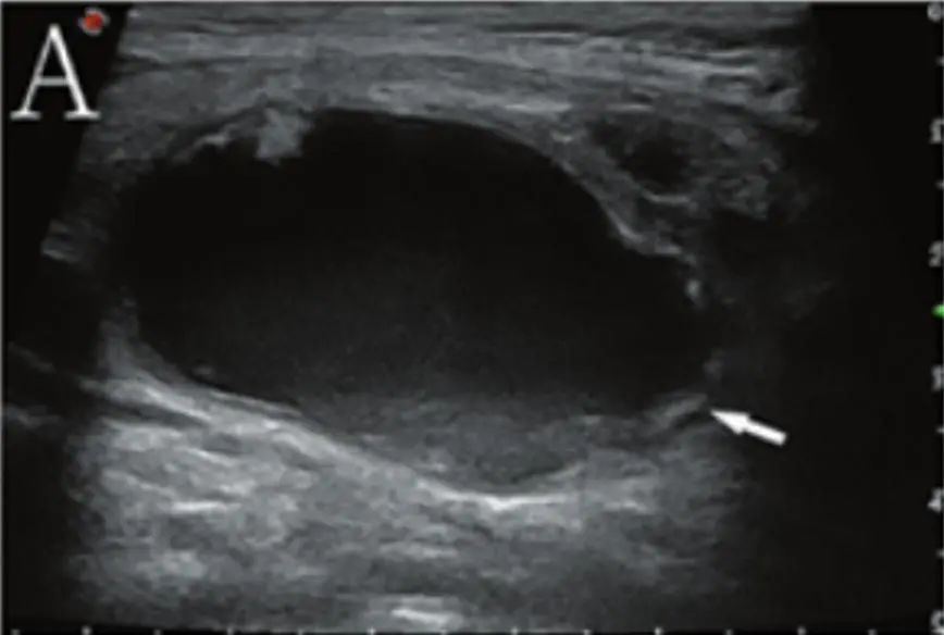
(A: Almost completely cystic nodule with debris echo, C-TIRADS 2, diagnosis: nodular goiter;)
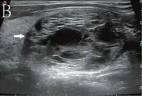
(B: Spongiform nodule, C-TIRADS 2, diagnosis: nodular goiter;)
2. Homogeneous Hyperechoic Nodules in the Context of Hashimoto’s Thyroiditis
Homogeneous hyperechoic nodules appearing in the context of Hashimoto’s thyroiditis generally do not exceed 15 mm in diameter, with typical cases being below 10 mm. These so-called “white knight” nodules are typically benign and can be evaluated as C-TIRADS Category 2.
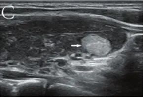
(C: White knight nodule in the context of Hashimoto’s thyroiditis, C-TIRADS 2, diagnosis: Hashimoto’s thyroiditis;)
3. Nodular Goiter
According to literature and expert opinions, when multiple solid and/or cystic nodules with similar ultrasound appearances occur within the thyroid, the corresponding pathological diagnosis is usually nodular goiter, with a low probability of malignancy. Such cases can be evaluated as C-TIRADS Category 3.
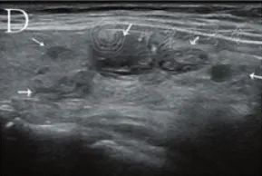
(D: Multiple solid and cystic nodules with similar ultrasound appearances, C-TIRADS 3, diagnosis: nodular goiter;)
4. Nodules with Calcification Leading to Indeterminate Internal Features
Some nodules with coarse calcification or marginal calcification may have shadowing caused by calcification, making it impossible to assess internal features. According to the ACR-TIRADS guidelines, such nodules can be evaluated as at least ACR-TIRADS Category 4, with a malignancy risk >5%. These nodules fall into C-TIRADS Category 4A.
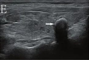
(E: Nodules with coarse calcification causing indeterminate internal features, C-TIRADS 4A, diagnosis: nodular goiter;)
5. “Snowstorm” Microcalcification Nodules
Nodules with a large number of microcalcifications that are difficult to count, known as “snowstorm” microcalcifications. This type of calcification has a specificity of 100% for diagnosing malignancy, thus can be evaluated as C-TIRADS Category 5.
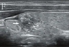
(F: Nodule with snowstorm microcalcifications, C-TIRADS 5, diagnosis: papillary carcinoma;)
6. Diffuse Microcalcifications Not Related to Nodules
Widespread diffuse distribution of numerous punctate calcifications within the thyroid parenchyma, typically appearing “snowstorm-like,” is often seen in diffuse sclerosing papillary carcinoma. If typical metastatic lymph nodes from thyroid cancer appear in the neck, it can be evaluated as C-TIRADS Category 5. If no suspicious lymph nodes are present in the neck, it can be assessed as Category 4B or even Category 5 based on clinical experience.
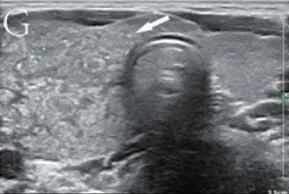
(G: Snowstorm microcalcifications unrelated to nodules in the right thyroid, C-TIRADS 5, diagnosis: papillary carcinoma)
The above content is excerpted from: The Ultrasound Medicine Branch of the Chinese Medical Association, the Chinese Thyroid and Breast Ultrasound Artificial Intelligence Alliance. 2020 China Guidelines for Risk Stratification of Malignancy in Thyroid Nodules via Ultrasound: C-TIRADS[J]. Chinese Journal of Ultrasound Imaging, 2021, 30(3): 185-200.