This Week’s Highlights
Ultrasensitive cortisol detection using Ag@Au SERS sensor with molecularly imprinted polymers
📝 Article Information

Sensors and Actuators: B.Chemical IF:7.7
🛠️ Research Background
Cortisol (Cortisol) is a key hormone in the human body that regulates blood pressure, blood sugar, and immune function. Abnormal cortisol levels can lead to various diseases, such as Cushing’s syndrome, Addison’s disease, and severe depression. Therefore, precise and dynamic monitoring of cortisol is of significant clinical value. Traditional detection methods such as enzyme-linked immunosorbent assay (ELISA), chemiluminescent immunoassay (CLIA), and liquid–chromatography-tandem mass spectrometry (LC-MS/MS) provide high sensitivity and specificity but often rely on large instruments and central laboratories, making it difficult to meet the demand for immediate testing. Although electrochemical sensors are compact, they suffer from signal drift, poor anti-interference capability, and insufficient adaptability to physiological environments, especially showing instability in long-term wearable applications. In recent years, surface-enhanced Raman spectroscopy (SERS) has become an ideal on-site rapid detection technology due to its high sensitivity and fast response characteristics, but it has poor specificity in complex biological samples. To address this, Peng et al. combined molecularly imprinted polymers (MIP) with SERS to develop a novel SMIP-SERS platform based on silver core-gold shell nanoparticles (Ag@Au NPs) and polydopamine surface-imprinted layer (PDA-SMIP).
🔍 Summary Overview
Recently, a research team developed a SERS sensor capable of rapidly and sensitively detecting cortisol, which combinesMIP with Ag@AuNPs. The MIP provides specific recognition sites for cortisol, and the surface plasmon resonance (SPR) effect amplifies the SERS signal; a stable SERS substrate is constructed using a two-step electrodeposition method, utilizing in-situ self-polymerized PDA film (~6.5 nm) for efficient recognition of cortisol, employing a “signal blocking mechanism”— where cortisol binding hinders the Raman probe from approaching the metal surface, resulting in a quantifiable signal attenuation—achieving a linear detection range of 100 pm ~0.1 mm, with a LOD of 10 pm. Additionally, it maintains stability and anti-interference capability in artificial saliva. This Ag@Au/MIPs SERS sensor features efficiency, cost-effectiveness, and ease of operation, showing great potential in clinical diagnostics, health monitoring, and point-of-care testing applications.
🔬 Research Methodology
First, silver nanoparticles (AgNPs) were electrodeposited on an ITO substrate using a two-step chronoamperometry method, followed by in-situ reduction to deposit a gold shell on the surface of AgNPs, forming a stable Ag@AuNPs structure that combines strong plasmonic resonance effects with antioxidant capabilities. On this basis, dopamine and cortisol were copolymerized in pH 8.5 Tris-HCl buffer, forming a PDA imprinted layer on the surface of Ag@AuNPs, and the template was eluted using acetic acid/methanol to obtain an MIP with specific recognition sites, namely Ag@Au/MIP sensing interface. To achieve signal recognition, 7-mercapto-4-methylcoumarin (MMC) was selected as the Raman probe molecule, which can adsorb onto the gold surface via Au–S bonds and interact with the imprinted cavity through π–π and hydrogen bonding interactions. During detection, cortisol preferentially binds to the MIP cavity, blocking MMC from diffusing to the SERS “hot spots,” leading to a linear decrease in Raman signal intensity with increasing concentration, thus enabling quantitative analysis.
📣Research Conclusion
The sensor’s detection range is from 100 pm to 0.1 mm, with a LOD of 10 pm, outperforming traditional antibody-based methods. In artificial saliva, the recovery rate ranges from 92.9% to 105.5%, demonstrating high accuracy and anti-interference capability. This Ag@Au/MIP sensor addresses key challenges in steroid hormone detection, providing higher sensitivity, selectivity, and ease of operation. Furthermore, it can detect other biomarkers by modifying the template molecule and can be integrated with microfluidic chips for real-time, multi-channel analysis, offering valuable insights for rapid clinical diagnostics and health monitoring.
📊 Figures
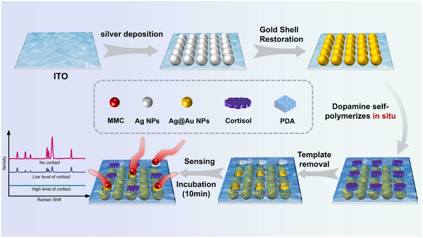
Sensor preparation process and sensing mechanism
Original Link
DOI: https://doi.org/10.1016/j.snb.2025.138034
Welcome to Follow Us


Reviewed by: Li YT
Edited by: Liu SC
Recommended by: Li YT
For submissions, please contact: [email protected]