1. Analysis of the Meaning of Ultrasound Diagnosis
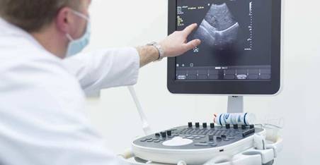
Ultrasound diagnosis mainly applies the good directionality of ultrasound and its physical characteristics similar to light such as reflection, scattering, attenuation, and the Doppler effect. By utilizing different physical parameters and using various types of ultrasound diagnostic instruments with different scanning methods, ultrasound is emitted into the human body and propagates within the tissues. When there is a certain difference in acoustic impedance between normal and pathological tissues, the interfaces they form will reflect and scatter. The echo signals are received and processed to display as waveforms, curves, or images.Due to the differences in the interface morphology of various tissues, the movement status of organs, and the degree of absorption of ultrasound, their echoes have certain commonalities and characteristics. By combining knowledge of physiology, pathological anatomy, and clinical medicine, we can observe, analyze, and summarize these different patterns to make a general and affirmative judgment about the location, nature, or degree of functional impairment of the diseased area.
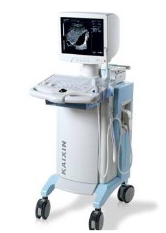
The components of medical ultrasound diagnostic equipment generally include:
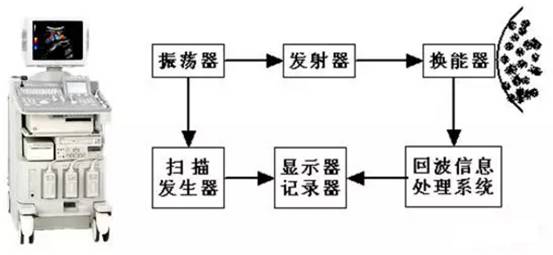
1. Oscillator: Generates synchronized trigger pulses for the control system, determining the repetition frequency of the emitted pulses.
2. Transmitter: Produces high-voltage electrical pulses to excite the ultrasound transducer after being triggered.
3. Transducer: Used to generate ultrasound pulses and receive echo pulse signals formed by targets, converting these signals into electrical signals and sending them to the echo information processing system.
4. Echo Information Processing System: This system consists of components such as RF signal receiving amplifiers, detectors, and video amplifiers, mainly used to process ultrasound echo information.
5. Display: Used to display image information.
6. Scanning Generator: Outputs scanning signals to the display, ensuring the ultrasound echo images displayed are stable.
2. Classification of Medical Ultrasound Diagnostic Equipment
Commonly used medical ultrasound diagnostic equipment can be classified into the following categories:
1. A-Mode Ultrasound Diagnostic Instrument (Amplitude)
A-Mode display is the most basic display method, where the horizontal axis of the oscilloscope represents the propagation time of ultrasound, i.e., the detection depth; the vertical axis represents the amplitude of the echo pulse (amplitude), hence the name A-Mode.
Using A-Mode diagnostic instruments, one can measure the position, size, and acoustic characteristics of various organs within the human body, and it is used for disease diagnosis.
2. M-Mode Ultrasound Diagnostic Instrument (Motion)
M-Mode ultrasound diagnostic instrument is a basic ultrasound diagnostic device developed based on A-Mode ultrasound diagnostic instrument. The brightness on its display tube indicates the amplitude of the echo, controlled by modulating the brightness of the Z-axis of the display tube with A-Mode echo amplitude; the vertical axis indicates the propagation time of ultrasound pulses, i.e., the detection depth; the horizontal deflection plate of the display tube is added with a slow time scanning voltage. Thus, when conducting human examinations, it forms a dynamic curve graph of each echo target.
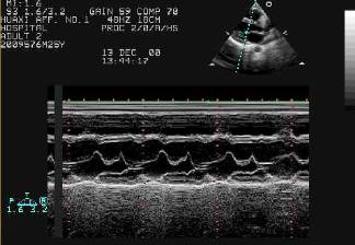
M-Mode ultrasound diagnostic instruments have several advantages when examining the heart, such as measuring the size, thickness, and valve motion of various parts of the cardiovascular system, and studying the relationship between the movements of different parts of the heart and the electrocardiogram, phonocardiogram, and pulse. Hence, it is also referred to as an ultrasound cardiograph. Additionally, it can study the conditions of other moving interfaces and, by synchronously moving the probe with slow time scanning, create some simple human cross-sectional images.
3. B-Mode Ultrasound Diagnostic Instrument (Brightness)
Also known as the B-Mode ultrasound section imaging instrument. It modulates the brightness of the display based on the amplitude of the echo pulse, with the horizontal and vertical axes of the display corresponding to the locations scanned by the sound beam, thus forming a brightness-modulated ultrasound section image.
B-Mode ultrasound diagnostic instruments are widely used in clinical applications, including cardiology, gastroenterology, urology, and obstetrics and gynecology for disease diagnosis.
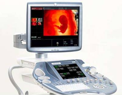
4. D-Mode Ultrasound Doppler Diagnostic Instrument
D-Mode ultrasound Doppler diagnostic instruments use the Doppler effect generated by the relative motion between ultrasound waves and the target to detect moving targets, mainly including Doppler blood flow measurement and blood flow imaging.
Current color flow imaging (CFI) is represented in real-time B-Mode ultrasound images with pseudo-color indicating blood flow in the heart or blood vessels. It utilizes multiple pulse echo correlation processing technology to obtain information on blood flow motion, hence often referred to as color Doppler flow imaging (CDFI).
Transcranial Doppler (TCD) diagnostic instruments apply low-frequency Doppler ultrasound, displaying the hemodynamic status of intracranial cerebral arteries through the temporal, occipital, foramen, and neck windows.
5. C-Mode and F-Mode Ultrasound Imaging Equipment
These are developed based on B-Mode ultrasound diagnostic instruments, mainly used to obtain echo information and imaging on planes and curves perpendicular or at an angle to the sound beam direction. Transmission-type C-mode imaging is similar to conventional X-ray imaging, reflecting the overall ultrasound characteristics of all tissues along the sound beam path, and C-mode imaging can be performed using total ultrasound attenuation and propagation time.C-Mode and F-Mode scanning imaging can provide some information that B-Mode ultrasound imaging cannot obtain.
3. Analysis of the Ultrasound Diagnosis Market Scale
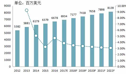
According to the “2016-2022 Global Medical Ultrasound Equipment Industry Analysis and Market Depth Investigation Report” released by market research organization QYResearch, the global medical ultrasound diagnostic equipment market has maintained steady development in recent years. Statistics show that in2016, the global medical ultrasound diagnostic equipment market size reached approximately6.7 billion USD, and it is expected to maintain an annual growth rate of around three percent in the coming years, with an estimated size of 8.1 billion USD in 2022. Europe, the United States, and Japan are the main markets for ultrasound, where advanced color ultrasound has been widely applied in these developed countries, accounting for70% of the global market. The market demand mainly focuses on product upgrades and replacements, leading to stable market growth. The international market is mainly monopolized by international giants such asGE, Philips, Siemens, and Hitachi, which firmly occupy most of the mid-to-high-end color ultrasound market share.
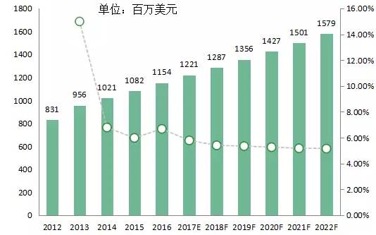
Compared to the international market, China’s ultrasound diagnostic equipment industry started relatively late, but after years of development, it has initially formed a complete industrial system with a full range of specialized categories, a complete industrial chain, and a solid foundation.In 2016, the Chinese ultrasound diagnostic equipment market reached1.15 billion USD. It is expected to maintain an annual growth rate of around six percent in the coming years, with an estimated market size of 1.58 billion USD in 2022.
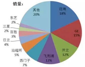
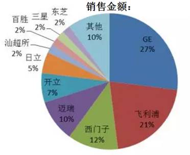
From the sales volume of the domestic color ultrasound market in 2015, the domestic brand Mindray has successfully surpassedGE, Philips, Siemens, and other foreign enterprises, ranking first. This marks the collapse of the monopoly position of imported ultrasound diagnostic equipment brands. However, in terms of sales revenue, the top three remainGE, Philips, and Siemens, while Mindray ranks fourth.
4. Overview of Major Manufacturers in the Ultrasound Diagnostic Market
Currently, the main foreign companies in medical ultrasound diagnostic equipment includeGE, PhilipsPhilips, SiemensSiemens, ToshibaToshiba, Hitachi–AlokaHitachi- Aloka, EsaoteEsaote, and Samsung–MedisonSamSung- Medison, etc., with specific company details as follows:

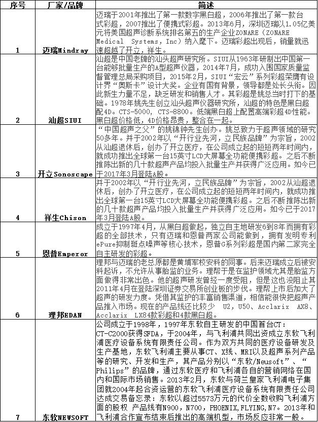
Main state-owned enterprises includeMindray, SIUISIUI, SonoscapeSonoscape, ChisonChison, EmperorEmperor, EDANEDAN, NEWSOFTNEWSOFT, etc., with specific company details as follows:
5. Conclusion
In the future, ultrasound equipment will further develop towards functional optimization, portability, and multifunctionality.
1. Functional Optimization: With the advancement of modern electronic technology and computer technology, the image quality and resolution of ultrasound diagnostic equipment are getting higher, and the diagnostic functions are becoming more comprehensive and powerful. Ultrasound imaging technology has evolved from the initial one-dimensional and grayscale two-dimensional to fully digital real-time three-dimensional and four-dimensional color ultrasound. Although there are no accurate figures yet, market research reports predict that the market scale of 3D and 4D color ultrasound has already surpassed that of conventional color ultrasound. The industry generally sees real-time digital 3D and 4D color ultrasound as the mainstream development direction of ultrasound diagnostic equipment.
2. Portability of Equipment: In 2016, handheld ultrasound was included in the national “13th Five-Year Plan” major R&D projects. In August, the first domestic handheld ultrasound device was officially put into medical application. This means that ultrasound equipment is not only lighter but can also operate without being tethered to power sources, allowing for coverage of thousands of township health centers and more village-level health institutions. Undoubtedly, portable ultrasound equipment will be an important direction in the ultrasound field in the future.
3. Multifunctionality: From market trends, multifunctional ultrasound equipment will undoubtedly be the mainstream direction, as comprehensive devices possess complete functionalities to meet complex and changing clinical diagnostic needs. In the future, more complex and comprehensive ultrasound equipment will undoubtedly emerge on the market.
In summary, like many mid-to-high-end medical devices, although the domestic ultrasound diagnostic equipment market is largely monopolized by imported products, it reveals significant market opportunities. With the introduction of policies such as the “13th Five-Year Plan,” a series of favorable policies will promote the rise of numerous medical device companies, and the research and development of new generation ultrasound imaging products will be prioritized, significantly accelerating the penetration and popularization of ultrasound equipment in primary healthcare institutions, thereby greatly contributing to the construction of a healthy China.
If reading the article is not enough and you want to chat with industry experts?
Want to discover more quality projects?
Join the investment and financing project exchange group of Siyi Research Institute!

Click above to connect with us~
To ensure the quality of communication, please submit your business card and other information.
Author: Chen Xiangqian, PhD student in Biomedical Engineering at Beihang University, research direction includes robotic surgical navigation, medical image processing, etc.

[Previous Highlights]
Hard Technology | How Insulin Pumps Manage Diabetes
Industry | In-depth Research Report on the CRO Industry
[Dry Goods] Reliable Overview of Medical Antibacterial Materials