
Please click: Nurses can easily generate or manage their electronic practice certificates using their mobile phones.
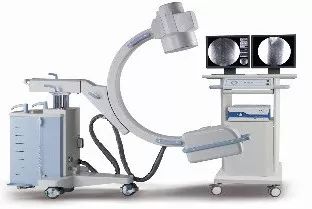
Orthopedic medical staff frequently use C-arm imaging. It is crucial to implement safety measures during surgery to minimize the harmful effects of ionizing radiation on the human body. We have compiled a C-arm protection guide for orthopedic surgery to share with everyone.
1
Are X-rays emitted by the C-arm harmful?
There are three effects of X-rays entering the human body:1. Pass through the body; 2. Be absorbed by the body; 3. Scatter (X-rays excite new rays).There are six effects of radiation entering the human body:1. Penetration effect of X-rays; 2. Fluorescent effect of X-rays; 3. Ionization effect of X-rays; 4. Thermal effect of X-rays; 5. Chemical effect of X-rays;6. Biological effect of X-rays. The biological effect on the human body is: “Due to the penetrating nature of radiation, it can enter the body and ionize cells. The ions produced by ionization can damage complex organic molecules such as proteins, nucleic acids, and enzymes, which are the main components of living cell tissues. Once they are damaged, normal chemical processes in the body can be disrupted, potentially leading to cell death.” Tissues or organs with more active cell division are more susceptible to damage. For example, children and various glands in adults such as gonads, thyroid, etc., and bone marrow. Additionally, the eyes can be damaged by the chemical effects of X-rays on the lens. Therefore, it is evident that different parts of the body have varying sensitivities to X-rays.
Radiation Exposure in Orthopedic Surgery
Radiation exposure during spinal surgery:>>Vertebroplasty (PVP) and Kyphoplasty (PKP):① Fluoroscopy time: PVP 2.7 minutes – 16.5 minutes, PKP 2.5 minutes – 7.2 minutes;② Most studies indicate that the equivalent dose to the physician’s hands ranges from 0.1mSv to 1mSv per surgery, with a maximum reported dose of 4.8mSv; the left hand receives a higher radiation dose than the right hand; eye radiation is low (0.1mSv – 0.23mSv); chest and thyroid: 0.154 – 0.526mSv;>> Lumbar or thoracolumbar fusion/screw placement/discectomy: General Orthopedics/Trauma:>> Hand/wrist and foot/ankle surgeries:① A study on foot and ankle specialists showed that during 80 surgeries within 12 months, the average fluoroscopy time was 0.62 minutes, with the surgeon’s right hand receiving an equivalent dose of 2.4mSv, averaging 0.03mSv per surgery;② Hand surgery: For hand surgeries using a mini C-arm, the average fluoroscopy time was 2.23 minutes, with external radiation exposure to the chest lead apron being 0.16mSv, and below 0.01mSv inside the apron.>> Hip, femur, and tibia:For proximal femoral fractures undergoing DHS, the radiation doses to the physician’s head, thyroid, and fingers were 0.03mSv, 0.04mSv, and 0.12mSv, respectively; the assistant’s radiation values were similar, but the fluoroscopy time was longer than that of experienced surgeons;This content is referenced from Spinal Mantra “Radiation Risks in Orthopedic Surgery: Unknowns or Known Factors” by Wang Haiqiang and Zhang Jun.2
How does the C-arm emit radiation?
1. Components of the C-arm:
 2. The process of radiation emission from the C-armGenerally, when no patients or objects are placed in the radiation beam, it can be assumed that the radiation emitted from the tube is entirely absorbed by the intensifier. The radiation exposure to nearby staff is minimal.However, once a patient is placed for exposure, the radiation situation in the operating room changes completely. The radiation emitted by the C-arm enters the patient’s body, and only 1% of the radiation passes through the patient to reach the surface of the intensifier. The remaining 80%-90% of the radiation is absorbed by the patient, and 10%-20% is scattered by the body.
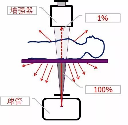 There are three types of radiation exposure for medical staff in the operating room:Direct radiation: Direct exposure to X-rays without any barriers, where the irradiated object is in direct radiation.Penetrating radiation: If X-rays pass through an object and continue to irradiate another object after attenuation, it is called penetrating radiation.Surrounding radiation: X-rays are also electromagnetic waves, which can be re-emitted after being absorbed by an object. The irradiated object becomes a secondary emission source radiating in all directions.Direct radiation has the highest intensity, penetrating radiation is next, and surrounding radiation has the least energy. If the irradiated object is a solid entity of a certain volume, such as the human body, then the intensity of surrounding radiation facing the X-ray source (tube direction) is greater. The patient mainly receives direct radiation. Medical staff mainly receive surrounding radiation.Distribution of scattered radiation:
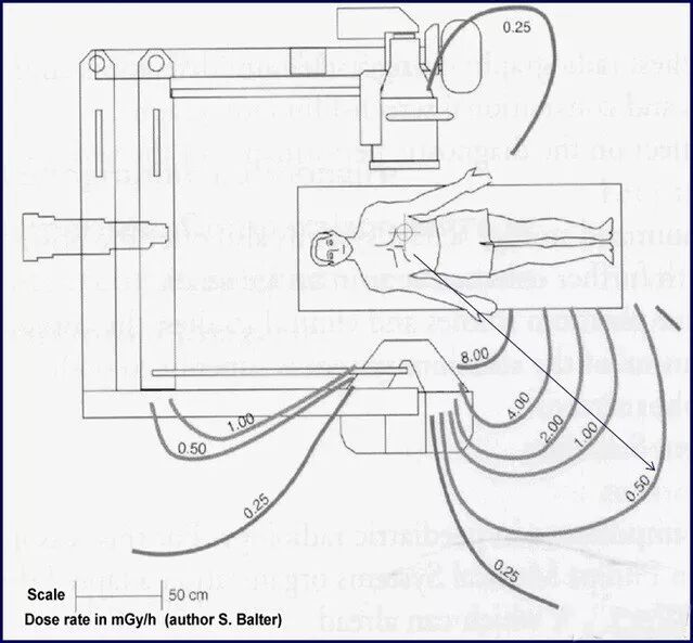
The distribution map of scattered radiation in an area near the patient is also called a positioning map. From the diagram, it can be seen that when the tube is under the bed and a lead curtain is hung beside the bed, the scattered radiation can be significantly reduced.

Tips:
1. When fluoroscopy is needed, stand on the opposite side of the C-arm tube.
-
For vertical fluoroscopy, place the tube under the bed.
-
For horizontal fluoroscopy, the operator should stand on the side of the intensifier.
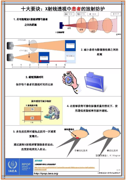
 2. Increase the distance between the X-ray tube and the patient as much as possible.3. Reduce the distance between the patient and the imaging receptor.
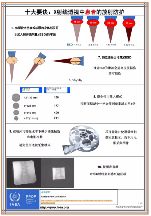
 4. Align the fluoroscopy area with the center of the intensifier.Especially when imaging the lateral view of the spine, do not simply align the spinous processes with the center of the intensifier; instead, align the vertebral body with the center of the intensifier to ensure that at least 50%-70% of the body is covered in the center position. Otherwise, there may be wasted images.5. Excessive indoor items can easily cause secondary scattering.3
How do orthopedic medical staff protect themselves?
1. Time Protection:
The greater the dose received by medical staff and patients, the greater the harm to the body. Therefore, the focus of radiation time protection is to reduce exposure time.
-
While ensuring image quality,try to minimize the time and frequency of X-ray exposure.
-
Use intermittent fluoroscopy to control the total exposure time.
-
Directly archive images to save time and avoid repetitive guidance and diagnosis.
2. Distance Protection:
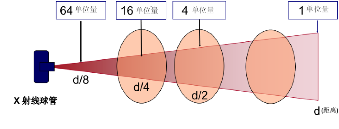
The intensity of radiation is directly proportional to the time of exposure and inversely proportional to the square of the distance. That is, if the distance is doubled, the radiation intensity is reduced to one-fourth of the original. The hands of the doctor receive the most direct radiation, up to 377μGy/min. The chest is most susceptible to surrounding radiation, while the gonads receive the least radiation. The surgical table can block some radiation. Standing on the opposite side of the patient (270°) results in the highest radiation exposure.
-
Avoid direct exposure of both hands.
-
Turning away from the patient during fluoroscopy can reduce eye radiation.
-
Stand at least 2 meters away from the C-arm for adequate protection.
- At 4 meters away, the radiation impact on the body is negligible.
3. Shielding Protection:In operating rooms without lead protective walls, hiding behind insufficiently thick concrete walls (<36cm) during C-arm fluoroscopy for psychological comfort is ineffective against penetrating radiation. Therefore,the most effective method is to use various shielding facilities and equipment to absorb radiation and reduce the radiation entering the body, including various screens in the operating room and personal protective equipment worn by staff.Lead Screens:Lead screens come in single, double, and triple configurations, with triple lead screens effectively shielding against side-scattered radiation. Lead screens can also have observation windows and are mostly mobile.Lead Curtains:Bedside lead curtains, hanging lead curtains, or lead windows.
- When the tube is under the bed, hanging lead curtains beside the bed can significantly absorb scattered radiation, using lead rubber.
Lead Glasses:Choose lead glasses with side protection to block radiation coming from the sides.Lead Neck Collars:Reduce direct radiation to the thyroid to 1.73μGy/min.Lead Aprons:Long lead aprons are heavy and not breathable; it is recommended to tighten the waist belt; two-piece lead aprons are recommended by IAEA, along with lead caps and medical lead gloves.
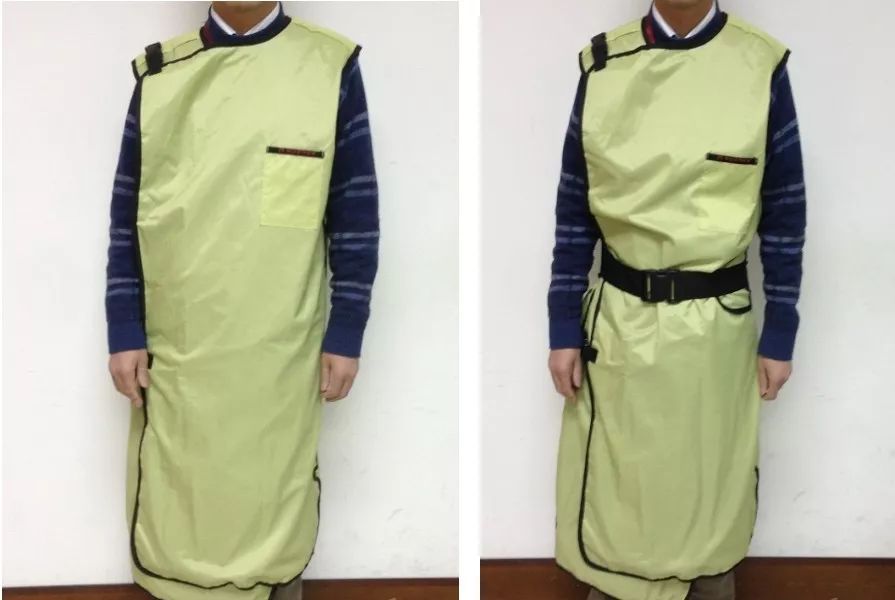
Long Lead Aprons
4. Reduce Radiation Intensity (Dose)
- Use low-dose mode.
- The exposure time for the first image should be sufficiently long.
- Switch to manual kVmA mode and lower the mA.
- In manual kVmA mode, switch to pulse mode at 1pps (one pulse per second).
- Avoid using magnification mode.
- Use collimators.
5. Personal Dose MonitoringInternational Commission on Radiological Protection data (effective dose limits)
- Public limit: 1mSv/year (uniform whole-body exposure).
- Occupational limit: 50mSv/year (uniform whole-body exposure).
- The average effective dose over five consecutive years should not exceed 20mSv/year (uniform whole-body exposure).
- Eye lens limit: 150mSv/year.
- Other individual organ or tissue annual limit: 500mSv/year.
- The dose limit is the sum of internal and external exposure but does not include natural background radiation and medical exposure to the eye lens.
Attachments
Posters on medical radiation protection from the International Atomic Energy Agency: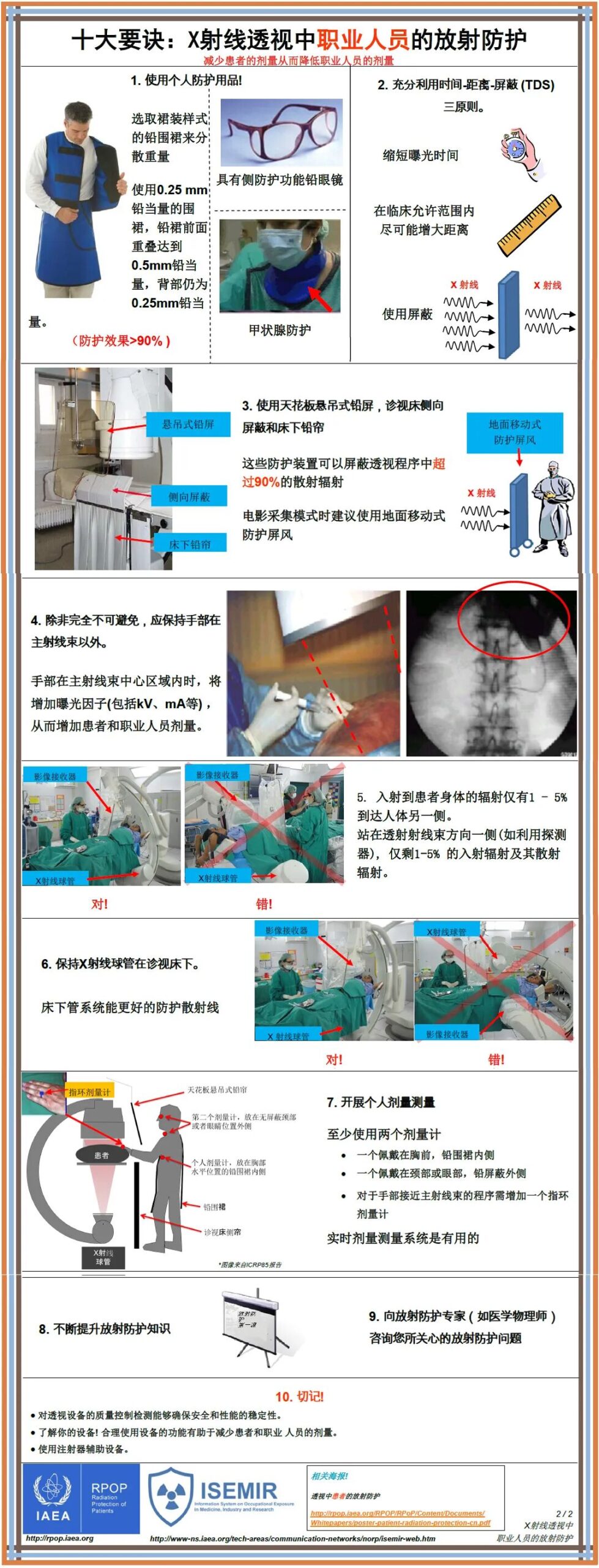 Radiation Protection for Patients During X-ray Fluoroscopy
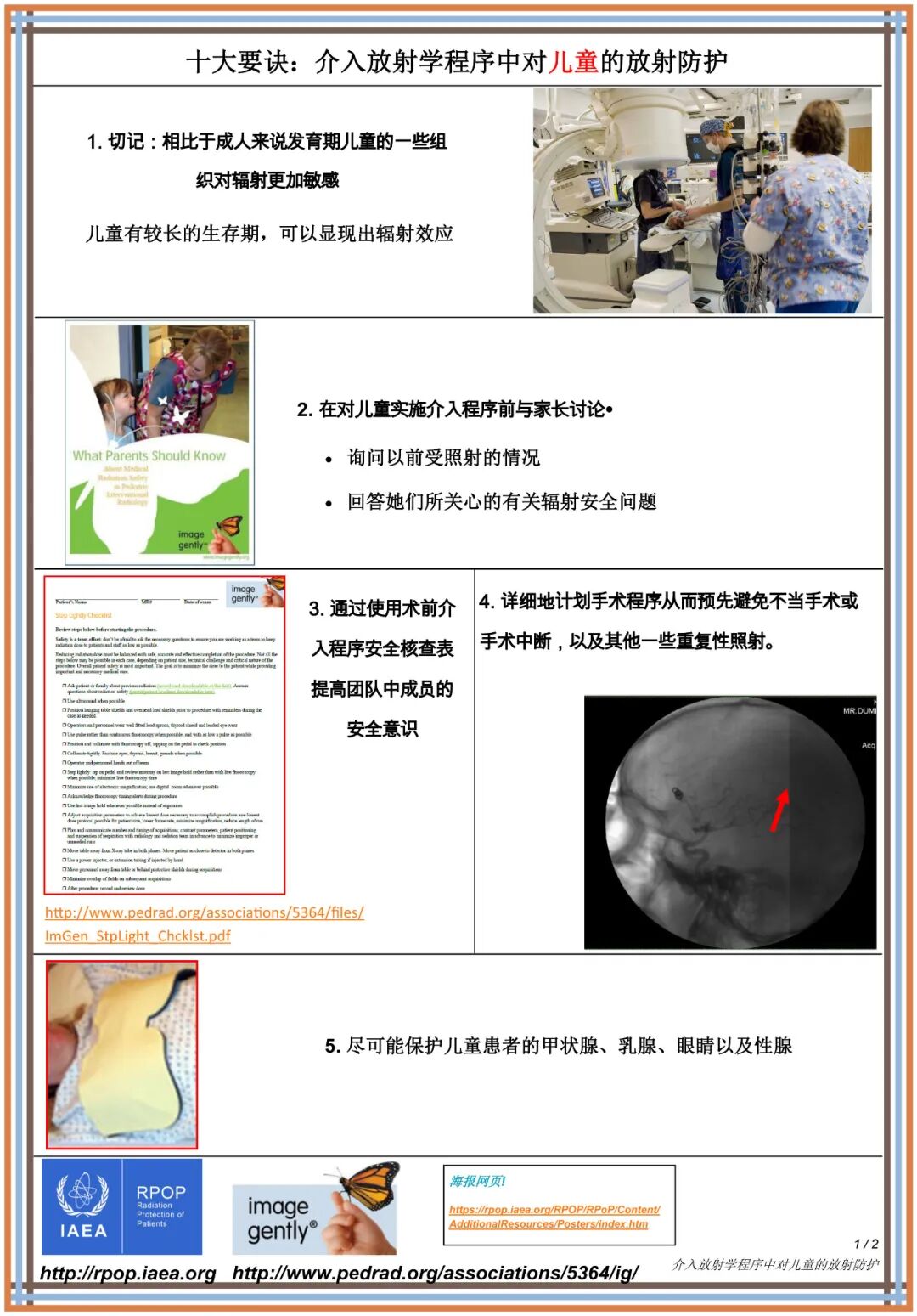
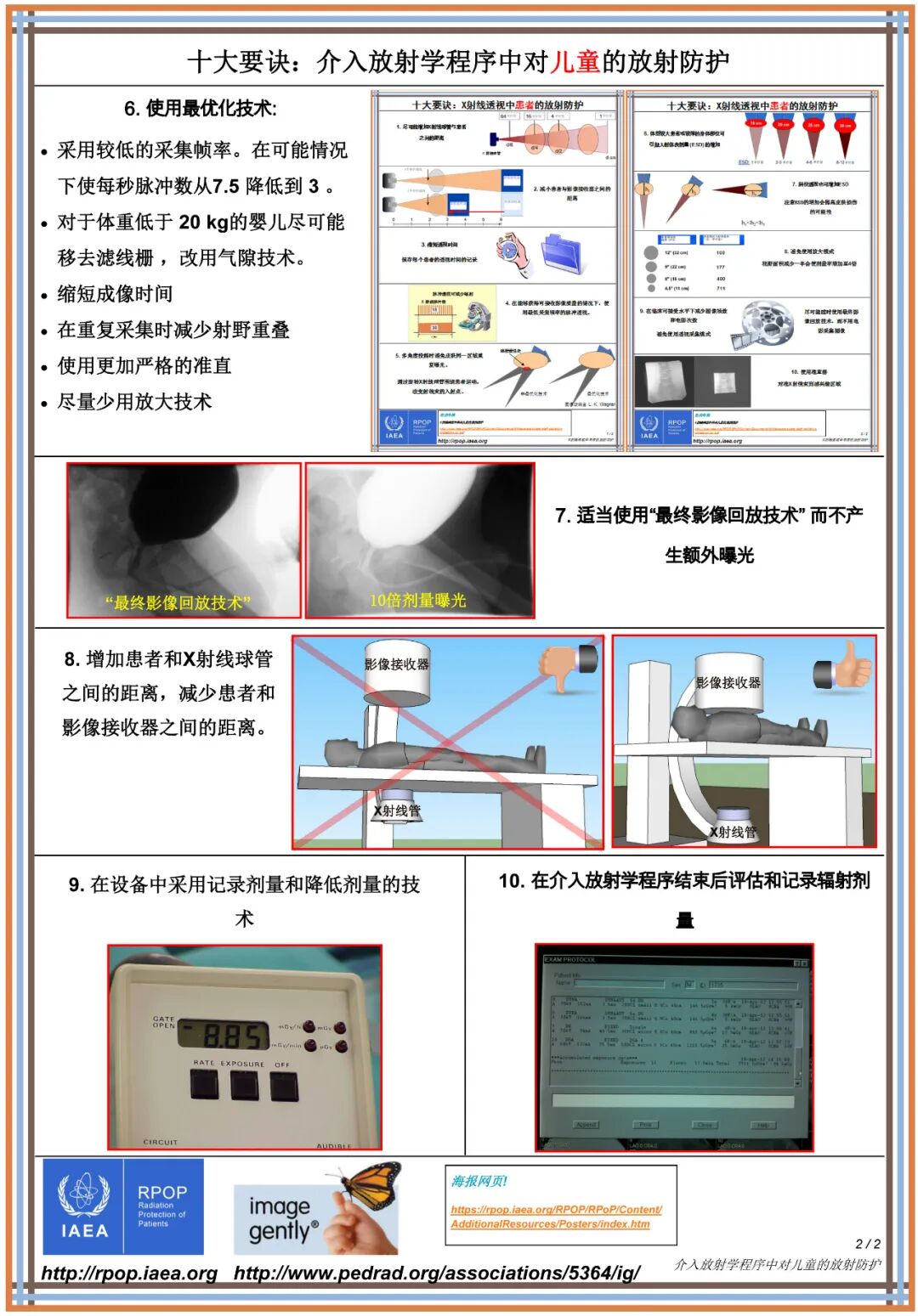
Radiation Protection for Children in Interventional Radiology Procedures
Source: Bone Today
For submissions, please email [email protected]

Click “Read the original text” below to follow various WeChat public accounts categorized by nursing departments.