NBT | MulTI-Tag: Multi-Factor Epigenetic Mapping at Single-Cell Resolution

Written by | Yi

The chromatin maps at gene locus resolution can reflect gene regulatory features, helping researchers define cell types and developmental trajectories. In recent years, genomic localization methods for epigenetic-related proteins have been applied at the single-cell level, such as CUT&Tag (Cleavage Under Targets & Tagmentation) based on Tn5 transposase【1,2】. However, mapping and comparing chromatin-related proteins within the same sample has always been a challenge, and a method that can accurately and simultaneously characterize multiple epigenetic targets at scale is still lacking.
Recently, a team led by Steven Henikoff from the Howard Hughes Medical Institute published an article titledMultifactorial profiling of epigenetic landscapes at single-cell resolution using MulTI-Tag in Nature Biotechnology. Theydeveloped a method calledMulTI-Tag (Multiple Target Identification by Tagmentation) that utilizes antibody barcoding to simultaneously map multiple chromatin features at the single-cell level.
NBT | MulTI-Tag: Multi-Factor Epigenetic Mapping at Single-Cell Resolution
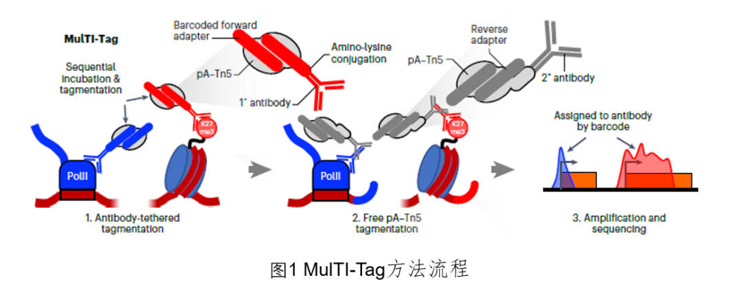
The authors tested various experimental conditions for antibody-barcode associations using mutually exclusive H3K27me3 and PolIIS5P antibodies as controls to achieve experimental efficiency and target recognition fidelity. Contrary to previous studies, the authors found that pre-incubation of the barcode protein A-Tn5 (pA-Tn5) complex, as well as simultaneous incubation and labeling of all antibodies, resulted in high levels of false cross-enrichment between targets. They also noted that starting labeling from predicted low-abundance targets slightly reduced off-target read distribution, and the primary antibody binding showed higher target differentiation than secondary antibodies, but with greater data quality fluctuations, possibly due to fewer pA-Tn5 complexes accumulating at each target site in the absence of secondary antibodies. To address these issues, the authors loaded pA-Tn5 onto the i5 forward adapter linked to primary antibodies, sequentially labeled target chromatin, and added secondary antibodies, followed by loading the i7 reverse adapter onto pA-Tn5 for final labeling. Experiments showed that MulTI-Tag provided highly accurate detection of H3K27me3 and PolIIS5P, indicating that MulTI-Tag can enrich targets without cross-contamination. Furthermore, in human embryonic stem cells (hESCs), MulTI-Tag accurately detected H3K27me3, H3K4me2, and H3K36me3, demonstrating that sequentially labeling targets does not hinder co-enrichment detection of two targets at the same site.
NBT | MulTI-Tag: Multi-Factor Epigenetic Mapping at Single-Cell Resolution
Next, the authors aimed to apply MulTI-Tag at the single-cell level. They utilized Takara’s ICELL8 microfluidic system for single-cell barcode labeling through combinatorial indexing. In human K562 cells and mouse NIH3T3 cells, when detecting H3K27me3 and H3K36me3 separately or in combination, the proportion of species cross-contamination was around 10%, comparable to ATAC-seq levels (7–12%)【3,4】. When mixing K562 and H1 hESCs, MulTI-Tag accurately distinguished cells from the same species. Additionally, in terms of representative site coverage, input cell recovery, and library complexity, MulTI-Tag performed similarly or better than current single-cell chromatin mapping technologies. Furthermore, in K562, H1, and K562-H1 mixed cells, the three-factor combinatorial analysis (H3K27me3–PolIIS5P, H3K27me3–H3K9me3, H3K27me3–H3K4me1) demonstrated that MulTI-Tag could distinguish cell types through multi-target enrichment analysis, showcasing its flexibility.
The authors then sought to understand the relative target abundance and instances of their co-occurrence at the same site in single cells. To achieve this cross-labeling analysis, they integrated transcription-related markers (H3K27me3–H3K4me2–H3K36me3) in individual H1 and K562 cells. They found that H3K27me3 was the most abundant, and within the window from 1kb upstream of transcription start sites (TSS) to the gene end, the authors observed co-enrichment of different targets within the same cell, such as enrichment of H3K4me2 or H3K36me3 in NR5A2 and H3K27me3 in HOXB3. Additionally, the authors could classify genes based on the frequency of individual or co-enrichment with specific targets in single cells, and quantify the co-enrichment levels between pairs of target genes within the same gene. For instance, the co-enrichment level of H3K27me3 and H3K36me3 in H1 was higher, which contradicts previous studies observing an antagonistic effect of the two both in vivo and in vitro【5,6】. This indicates that MulTI-Tag can observe chromatin enrichment patterns at single-cell, single-site resolution.
So how do histone modifications co-occur within single cells during continuous developmental states? The authors differentiated H1 hESCs into three germ layers (endoderm, mesoderm, ectoderm), collecting nuclei at corresponding time points for MulTI-Tag, and after quality control, they obtained data on H3K27me3, H3K4me1, and H3K36me3 from 7,727 cells. The authors found that UMAP based on H3K36me3 could not distinguish cell types, while UMAP based on H3K27me3 and H3K4me1 could separate the main two groups corresponding to endoderm and mesoderm. They discovered that the pseudotime based on H3K27me3-H3K4me1 data fitted better with known differentiation, revealing two important branches of hESCs tri-lineage differentiation: ectoderm and mesoderm lineages defined by TGF-β and WNT signals, and endoderm and mesoderm separated by BMP and FGF signals【7,8】. This demonstrates that multi-factor data integration is crucial for accurately characterizing chromatin states during continuous development.
Furthermore, the authors explored the relationship between chromatin enrichment transitions and gene expression. They quantified the changes in enrichment of H3K27me3, H3K4me1, and H3K36me3 among transcription factors (TFs), discovering trajectory-specific differences. For TFs that expressed lower levels during differentiation, H3K36me3 enrichment decreased in mesoderm and endoderm, with H3K27me3 and H3K4me1 levels also being lower, while in ectoderm only H3K4me1 enrichment decreased. So do global changes in histone modification enrichment differ as well? The authors found that H3K27me3 in ectoderm rapidly decreased along the pseudotime trajectory, resulting in a significantly lower proportion of H3K27me3 in terminal ectoderm compared to other cell types. Notably, hESCs predicted to participate in the ectoderm trajectory also had a lower proportion of H3K27me3 than those in the mesoderm trajectory. Strangely, the authors found that the decrease of H3K27me3 in most genes was not significant, indicating that low H3K27me3 in hESCs is associated with a unique developmental state. Finally, the authors discovered that the H3K27me3-H3K4me1 “bivalency” in ectoderm was significantly lower than in hESCs and mesoderm/endoderm. This indicates that global changes in chromatin modification enrichment and co-enrichment can be detected even before differentiation and are related to specific developmental endpoints.
In summary, the articleproposes an efficient tool for understanding chromatin regulation at single-cell, single-site resolution—MulTI-Tag, which holds great promise for comprehensively elucidating cell-specific gene regulation landscapes in development and disease.
Original link:
https://doi.org/10.1038/s41587-022-01522-9

Editor: Eleven

References

1. Kaya-Okur, H. S. et al. CUT&Tag for efficient epigenomic profiling of small samples and single cells. Nat. Commun. 10, 1930 (2019).
2. Kaya-Okur, H. S., Janssens, D. H., Henikoff, J. G., Ahmad, K. & Henikoff, S. Efficient low-cost chromatin profiling with CUT&Tag. Nat. Protoc. 15, 3264–3283 (2020).
3. Cusanovich, D. A. et al. Multiplex single cell profiling of chromatin accessibility by combinatorial cellular indexing. Science 348, 910–914 (2015).
4. Cusanovich, D. A. et al. A single-cell atlas of in vivo mammalian chromatin accessibility. Cell 174, 1309–1324 (2018).
5. Yuan, W. et al. H3K36 methylation antagonizes PRC2-mediated H3K27 methylation. J. Biol. Chem. 286, 7983–7989 (2011).
6. Lu, C. et al. Histone H3K36 mutations promote sarcomagenesis through altered histone methylation landscape. Science 352, 844–849 (2016).
7. Gifford, C. A. et al. Transcriptional and epigenetic dynamics during specification of human embryonic stem cells. Cell 153, 1149–1163 (2013).
8. Tsankov, A. M. et al. Transcription factor binding dynamics during human ES cell differentiation. Nature 518, 344–349 (2015).
NBT | MulTI-Tag: Multi-Factor Epigenetic Mapping at Single-Cell Resolution

NBT | MulTI-Tag: Multi-Factor Epigenetic Mapping at Single-Cell Resolution

Reprint Notice

[Original Article] BioArt original article, welcome personal forwarding and sharing, unauthorized reproduction is prohibited, all works published are owned by BioArt. BioArt reserves all legal rights, and violators will be prosecuted.

NBT | MulTI-Tag: Multi-Factor Epigenetic Mapping at Single-Cell Resolution

Leave a Comment