
1 (Supplementary Print) Recommended Neuroscience Practical Books – “Application Anatomy of the Mouse Brain” [Science Press]
2 Research Tool Book “Rodent Models of Mental Disorders and Behavioral Experiments” by Cui Donghong/Xu Lin
3 Major New Book Recommendation | “Neuropsychiatry” (3rd Edition)
Written by: He Dahong, Qiu Wenli
Edited by: Hu Yuting, Li Hao
Adenosine triphosphate (ATP) was first discovered in 1929, and is the common energy currency of all living cells[1].The field of ATP signaling has made numerous advances over the past century.ATP serves as an ideal extracellular messenger with the following 4 characteristics:① High signal-to-noise ratio: extracellular concentration is close to zero;② High hydrophilicity, allowing unrestricted diffusion in the aqueous interstitial environment[2];③ Multiple cell receptors that can accurately decode signals[3];④ Rapid degradation, terminating signals and preventing desensitization[4].
Neuronal activity is the basis for information encoding and processing in the brain. During neuronal excitation, ATP must be produced to meet high energy demands. At the same time, ATP can also be secreted extracellularly as a central neurotransmitter, inhibiting neuronal activity to prevent excessive excitation of the nervous system[5].ATP can be released by all brain cells, widely diffuse, and target different types of purinergic receptors on neurons and glial cells, coordinating neuronal activity in the brain and participating in various physiological processes such as sleep and wakefulness[6, 7], learning and memory[8, 9], and feeding[10-12]. Furthermore, dysregulation of extracellular ATP can lead to abnormal nervous system function, which has been found in the pathophysiology of mental disorders such as depression[13, 14], anxiety[15, 16], schizophrenia[17, 18], and autism spectrum disorder (ASD)[19].
On April 26, 2024, Academician Gao Tianming’s team from Southern Medical University / Guangdong-Hong Kong-Macao Greater Bay Area Brain Science and Brain-like Research Center was invited by National Academy of Medicine member John H. Krystal to publish a review article titled “Extracellular ATP Is a Homeostatic Messenger That Mediates Cell-Cell Communication in Physiological Processes and Psychiatric Diseases” in Biological Psychiatry.
The article was co-authored by Academician Gao Tianming from Southern Medical University, Professor Chen Yihua from Southern Medical University, and Professor Lin Song from Jinan University, with illustrations by Dr. Jin Shiyang from Southern Medical University.This review summarizes the latest progress on the mechanism by which extracellular ATP regulates neuronal activity as an intercellular signaling molecule, focusing on how ATP maintains the homeostasis of neural networks. The article outlines the abnormal changes in neuronal activity caused by dysregulation of extracellular ATP, emphasizing the important role of extracellular ATP dysregulation in the pathophysiology of certain mental disorders, clarifying the role and mechanism of extracellular ATP in intercellular communication and mental diseases, and prospects for the application of ATP as a potential therapeutic target in the treatment of mental disorders.
1. Overview of ATP Signaling
ATP is produced through glycolysis and mitochondrial oxidative phosphorylation and can be released from almost all types of cells in the central nervous system (Figure 1A). After release, ATP can be hydrolyzed by at least four ecto-nucleotide triphosphate diphosphohydrolases to generate adenosine diphosphate (ADP), adenosine monophosphate (AMP), and adenosine (ADO) (Figure 1B).ATP receptors are also known as P2 receptors (P2Rs), which can be pharmacologically divided into two subtypes: P2XR and P2YR (Figure 1C).P2XR is a cation channel, including P2X1R ~ P2X7R; P2YR is a G protein-coupled receptor (GPCR), including P2Y1R, P2Y2R, P2Y4R, P2Y6R, and P2Y11R ~ 14R.ADO receptors are also known as P1 receptors, including A1R, A2AR, A2BR, and A3R, all of which are GPCRs (Figure 1D). Different ATP receptors exhibit sensitivity to ATP from the nmol level to the mmol level, allowing ATP to elicit various responses. In summary, ATP receptors can function throughout the central nervous system and produce different effects depending on the receptors involved.
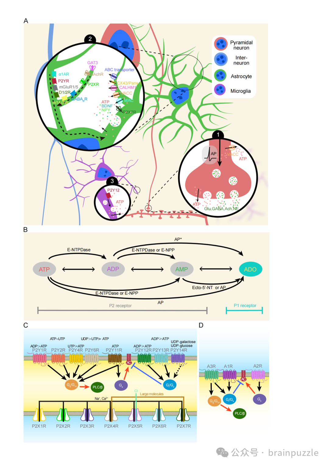
Figure 1 Schematic of the purinergic system2. Extracellular ATP as a Homeostatic Messenger Regulating Neuronal Activity and Brain Circuits
In 1992, researchers first discovered that ATP could act as a neurotransmitter in the medial habenula. However, electron microscopy showed that the two main fast-responding receptors to ATP, P2X2R and P2X4R, are mostly located around synapses and postsynaptic sites, far from the presynaptic terminal neurotransmitter release area. Furthermore, ATP-mediated excitatory postsynaptic currents are often small and rare, detectable only in specific brain regions with strong electrical stimulation of certain neuronal subpopulations. Therefore, current experimental evidence does not support that ATP is a fast neurotransmitter; rather, ATP may act as a neuromodulator (Figure 2).
It has been reported that endogenous ADO can reduce the excitability of hippocampal CA1 pyramidal neurons by activating the A1 receptor, or inhibit the presynaptic release of glutamate to reduce excitatory synaptic transmission in hippocampal pyramidal neurons. Additionally, in the hippocampus CA3, neurons containing P2X2Rs form synapses with inhibitory interneurons and are activated by ATP during theta oscillations, causing the release of glutamate, which in turn excites interneurons and increases inhibition of the neuronal network.
In the hippocampus, activation of P2X2R increases the activity of phosphatases or CaMKII (calcium/calmodulin-dependent protein kinase II) after calcium influx, leading to the internalization of AMPA receptors on the membrane surface, resulting in synaptic inhibition. In the paraventricular nucleus, endogenous ATP activates the extrasynaptic P2X7R, increasing postsynaptic effects by inserting AMPA receptors into dendritic spines. However, P2X7Rs are very rare in neurons, so this mechanism is not common in the central nervous system. Moreover, activation of postsynaptic P2Y1R or P2XR in pyramidal neurons of the prefrontal cortex (PFC) has an inhibitory effect on NMDA receptors.
In neural networks, inhibitory synaptic transmission is crucial for brain activity. ATP mainly participates through the following mechanisms:
1) Interneurons in the PFC and hippocampus express P2Y1Rs, and activation of these receptors induces inward non-selective cation currents, depolarizing the interneuron membrane and increasing its firing frequency, leading to increased synaptic inhibition of pyramidal neurons.
2) Extracellular ATP activates P2X2R, promoting the release of presynaptic GABA (gamma-aminobutyric acid).3) Astrocytes respond to endogenous GABA release by releasing ATP. Subsequently, ATP is broken down into ADO, activating postsynaptic A1R, increasing synaptic inhibition of pyramidal cells.
4) The enhancement of inhibitory transmission may also be due to the increased function of GABAA receptors in postsynaptic neurons.
It is noteworthy that extracellular ATP can also reduce neuronal activity in a circuit-dependent manner. For example, extracellular ATP induces the activation of GABAergic interneurons in the PFC through P2X2R, thereby inhibiting the activity of the medial PFC (mPFC) – lateral habenula (LHb) circuit. In the amygdala, extracellular ATP inhibits excitatory synaptic transmission to the basolateral amygdala and activates A1R and A2AR through adenosine, increasing inhibitory synaptic transmission in the lateral central amygdala.
The axon initial segment is the site of action potential generation and is susceptible to external signals. One study showed that extracellular ATP inhibits the generation of action potentials in highly active neurons by acting on P2X7R. Astrocyte-derived ATP may also regulate signal transmission along the axon, where its hydrolysis product ADO slows down axonal conduction through A2R at the nodes of Ranvier. Additionally, the release of astrocytic ATP is regulated by synaptic activity, and synaptic activation can inhibit neuronal activity through paracrine and autocrine ATP, while ATP released by surrounding cells can increase astrocytic [Ca2+]i, further triggering ATP release, forming a positive feedback loop. These feedback mechanisms may work together to inhibit local circuits.
ATP/ADP released by microglia can activate microglial P2Y12R, leading to a rapid increase in contact points between neuronal cell bodies and microglial processes to prevent excitotoxicity. Consistent with these findings, microglia-specific knockout of P2Y12R increased the excitability of CA1 pyramidal neurons. Furthermore, microglia can hydrolyze ATP into ADO and further activate A1R. A1R is expressed at presynaptic sites, and activation of A1R reduces neurotransmitter release and decreases neuronal excitability in a G protein-dependent manner.
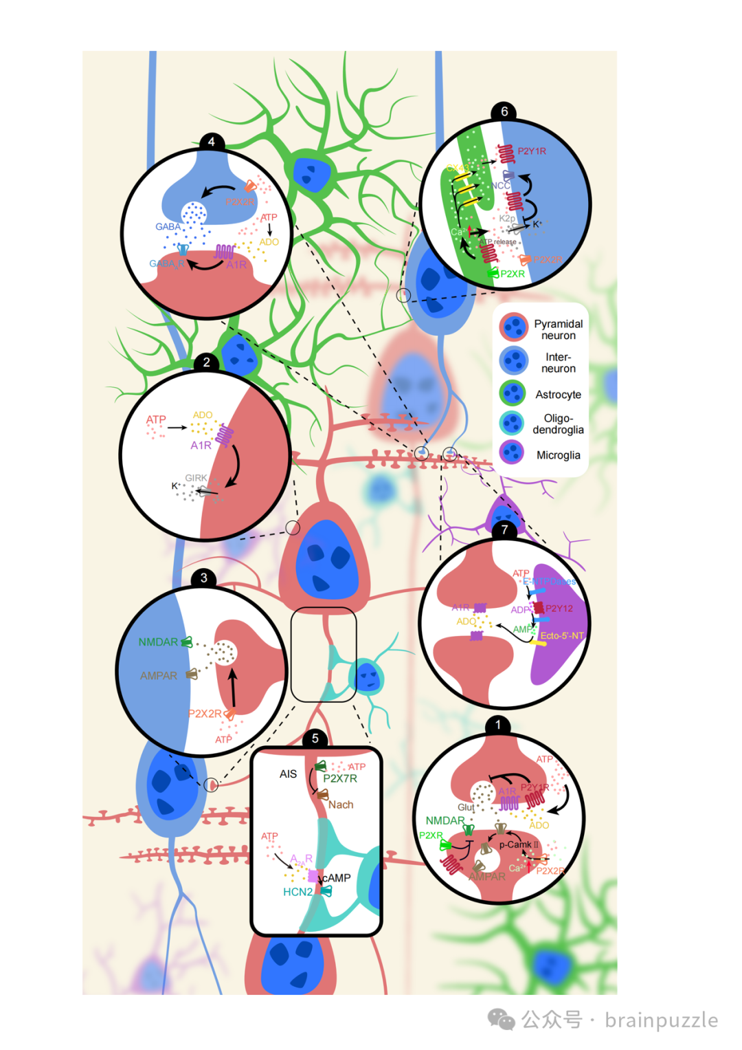
Figure 2 Extracellular ATP Modulates Neuronal Activity
3. Physiological Effects of Extracellular ATP
Extracellular ATP plays a key role in coordinating neuronal activity in the brain and is involved in various physiological processes, including sleep and wakefulness, learning and memory, and feeding (Figure 3).
3.1 Sleep and WakefulnessThe basal forebrain (BF) plays a crucial role in sleep-wake regulation. Studies have found that ATP levels in the BF are stable during wakefulness but surge during sleep, correlating with δ activity during non-rapid eye movement (NREM) sleep. Other studies have shown that extracellular ATP levels increase during wakefulness and rapid eye movement (REM) sleep, while decreasing during NREM sleep, suggesting a function related to promoting wakefulness. Supporting this view, activation of P2R in the BF promotes wakefulness, while inhibition of P2R promotes sleep. ADO inhibits glutamatergic input to GABAergic neurons to promote sleep. In contrast, extracellular ATP depolarizes GABAergic neurons in the BF, promoting wakefulness. In summary, these observations provide important insights into the role and mechanism of extracellular ATP in the homeostatic regulation of sleep and wakefulness.3.2 Learning and MemoryExtracellular ATP signaling significantly affects learning and memory. For example, the lack of astrocytic ATP release or the absence of receptors leads to learning and memory impairments. Synaptic plasticity is the basis of learning and memory, including long-term potentiation (LTP) and long-term depression (LTD). It is well known that extracellular ATP signaling has significant effects on both LTP and LTD. For instance, local administration of ATP can cause small or large amounts of Ca2+ influx in hippocampal CA1 neurons, leading to LTP or LTD, respectively. The lack of astrocytic ATP release impairs the induction of LTP. Mice with knockout of the P2X3R gene show impaired LTD, while having no effect on LTP. In contrast, mice with knockout of P2X4R show impaired induction of LTP. Furthermore, activation of P2YR weakens glutamate release from hippocampal CA1 neurons, leading to the induction of heterosynaptic LTD. These findings emphasize the role of extracellular ATP signaling in regulating learning and memory as well as hippocampal synaptic plasticity.3.3 FeedingThe hypothalamus is recognized as a brain region involved in controlling feeding, consisting of complex structures made up of small nuclei, such as the arcuate nucleus, lateral hypothalamus, and paraventricular nucleus. Anatomically, P2XRs are abundantly expressed in the arcuate nucleus, paraventricular nucleus, and other hypothalamic nuclei, while P2Y1Rs are expressed in the ventromedial nucleus and lateral hypothalamus, indicating that ATP may be involved in feeding regulation. Functionally, administration of ATP in hypothalamic brain slices can induce excitatory responses in neurons of the lateral hypothalamus, paraventricular nucleus, dorsomedial nucleus, and ventromedial nucleus. In summary, ATP appears to have an excitatory effect on hypothalamic neurons.So far, only a few studies have directly shown how extracellular ATP affects feeding. Injection of ATP/ADP analogs into the lateral ventricle increased food intake in rats, which could be blocked by pretreatment with a selective P2Y1R antagonist, while pharmacological activation of P2Y1R in the ventromedial nucleus and lateral hypothalamus enhanced food intake. Additionally, non-selective pharmacological inhibition of P2 receptors in the nucleus accumbens reduced food intake and eating time. Mice with restricted inactivation of P2Y6 receptors in AgRP neurons showed reduced food intake. Furthermore, a recent study indicated that optogenetic activation of hypothalamic elongation cells could induce acute binge eating by activating purinergic receptors on appetite-promoting neuropeptide-positive neurons in the arcuate nucleus. However, the specific role and potential mechanisms of P2R in hypothalamic feeding regulation across different cell types remain unclear.
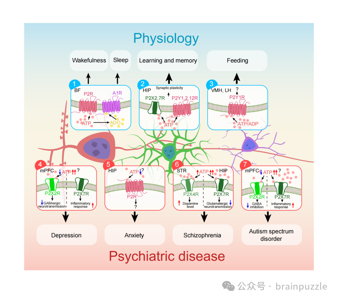
Figure 3 Extracellular ATP Involvement in Physiological Processes and Psychiatric Disorders4. The Role of Dysregulation of Extracellular ATP in the Pathogenesis of Mental Disorders
A large number of studies have shown that the physiological processes of various behaviors require the involvement of extracellular ATP, while dysregulation of extracellular ATP is associated with the pathogenesis of many mental disorders, including depression, anxiety, schizophrenia, and autism spectrum disorder (ASD).
4.1 Depression
The relationship between extracellular ATP and the pathogenesis of major depressive disorder (MDD) and its successful treatment has been widely studied. Previous studies have shown that ATP synthesis is reduced in the basal ganglia and frontal cortex of patients with MDD. Further research indicates that genes related to ATP biosynthesis and utilization are altered in the PFC of suicidal MDD patients. Moreover, effective treatment of MDD is associated with normal levels of ATP biosynthesis. Studies on rodents have found that a clinically fast-acting antidepressant, ketamine, is accompanied by an increase in ATP levels while producing rapid antidepressant effects. These studies suggest that the reversal of depressive symptoms is related to the restoration of ATP-related bioenergetic states.
Studies on depressive animal models show that reduced extracellular ATP levels in the PFC and hippocampus inhibit the release of astrocytic ATP, leading to the development of depressive-like behaviors in mice. In contrast, enhancing the release of astrocytic ATP has antidepressant effects, which can be blocked by ATPase in the mPFC. Recent studies have shown that reduced release of ATP weakens stimulation of P2X2R, thereby decreasing the excitability of GABAergic interneurons in the mPFC, which in turn weakens the inhibitory effect of GABAergic neurons on mPFC neurons projecting to the LHb, enhancing the activity of this neural circuit and inducing depressive-like behaviors.
The concentration of extracellular ATP is influenced by its release and enzymatic degradation. Recent studies have confirmed the impact of ATP release and degradation on depression. For instance, astrocytic eicosapentaenoic acid signaling and glucocorticoid receptors are involved in regulating vesicular-dependent ATP release and the expression of depressive-like behaviors. Additionally, non-vesicular release factors of ATP, such as Calhm2, also play a role in depressive symptoms. Regarding the enzymatic degradation of ATP, upregulation of ecto-nucleotide triphosphate diphosphohydrolase-1 (which hydrolyzes extracellular ATP) in the hippocampus is associated with social avoidance and despair behaviors, but not with anhedonia induced by chronic social defeat stress. These findings collectively advance our understanding of the complex roles of ATP release and enzymatic degradation in depression.
P2X7R is activated by high concentrations of ATP (EC50≥100 mmol/L) in immune activation and tissue damage, effectively triggering inflammasome activation, which may be involved in the pathogenesis of depression. Notably, some studies have shown that restraint stress can lead to a transient increase in extracellular ATP concentrations. Furthermore, P2X7R knockout mice exhibit an antidepressant-like phenotype and enhanced resilience to stress. However, whether the accumulation of extracellular ATP in these depressed animals can activate P2X7R remains to be further studied.
4.2 Anxiety
Research on the role of extracellular ATP signaling in anxiety is becoming increasingly common. The specific single nucleotide polymorphism (rs1718119) of P2X7R is associated with an increased risk of anxiety attacks in patients with anxiety disorders. However, some studies have failed to find an effect of P2X7R inactivation on anxiety-like behavior in mice. This discrepancy may be attributed to the presence of active P2X7R splice variants in the knockout mouse models used, allowing them to escape gene inactivation. Conversely, some studies have shown that ATP signaling also has anxiolytic effects. A recent study utilized a transgenic mouse model with increased surface density of P2X4R in the hippocampus, demonstrating anxiolytic effects. Additionally, non-specific P2YR agonists can produce anxiolytic effects, while knockout of P2Y12R can induce anxiety-like effects. Furthermore, optogenetic activation of astrocytes in the hippocampus can induce the release of ATP and produce anxiolytic-like behaviors, which can be blocked by non-specific P2R antagonists.
4.3 Schizophrenia
Schizophrenia is characterized by various symptoms, including reduced emotional responses, impaired social cognition, and cognitive deficits. Research has linked increased ATP signaling to the development of schizophrenia, with higher levels of ATP biosynthesis observed in the left hemisphere of patients with schizophrenia. Moreover, recent reports have shown elevated mRNA levels of P2X7R in the dorsolateral PFC of schizophrenia patients, which is associated with inflammatory markers such as SERPINA3. Additionally, blocking P2X7R through pharmacological and genetic methods in rodents can alleviate schizophrenia-like behaviors. Notably, P2X7R plays a key role in regulating excitatory neurotransmission in the hippocampus. Given the role of glutamatergic neurotransmission in the pathophysiology of schizophrenia, these findings suggest a potential link between P2X7R-mediated inflammatory signaling, glutamatergic neurotransmission, and the pathogenesis of schizophrenia.
The genes for P2X7R and P2X4R are located close to each other on human chromosome 12, reflecting a close relationship between the two, suggesting that P2X4R may also play a role in schizophrenia. Researchers have observed elevated mRNA levels of P2X4R in the dorsolateral PFC of schizophrenia patients. Notably, in the striatum of P2X4R knockout mice, both presynaptic and postsynaptic dopaminergic markers were altered, indicating that P2X4R plays an important role in maintaining dopamine homeostasis. Furthermore, dopamine receptor antagonists can alleviate schizophrenia-like behaviors induced by activation of P2X4R. Since hyperactivity of the dopaminergic system is associated with schizophrenia, these findings suggest that activation of P2X4R may promote the progression of schizophrenia to some extent by inducing dopaminergic system hyperactivity. Considering the close proximity of the P2X7R and P2X4R genes, when these two receptors function as a P2X4-P2X7R multiprotein complex, they may be activated by high concentrations of ATP.
Evidence suggests that application of selective P2Y1R agonists in the mPFC can induce schizophrenia-like behaviors, while activation of P2Y1R can increase dopamine release in the nucleus accumbens (NAc). Future research is needed to determine the relationship between P2Y1R, dopamine activity, and schizophrenia.
4.4 Autism Spectrum Disorder
ASD is characterized by social and communication impairments, along with restricted and repetitive behaviors. Currently, there are few clinical studies on the role of extracellular ATP in ASD, but animal studies have suggested its involvement. Our recent research indicates that impaired Ca2+ signaling in astrocytes leads to ASD-like behaviors, including social interaction deficits and repetitive behaviors. Furthermore, administration of ATP reversed social interaction deficits without affecting repetitive behaviors, suggesting that dysfunction of extracellular ATP signaling may primarily impact social behavior. Another study found that ablation of the P2X4R gene led to disordered social communication functions, confirming the above hypothesis. Recent studies have indicated that defects in repetitive behaviors may arise from impaired Ca2+ signaling in astrocytes in the striatum, and whether ATP signaling is involved remains to be further explored. Additionally, elevated extracellular ATP levels may act as a “pathogen,” triggering inflammatory responses that lead to ASD. For example, high concentrations of ATP can activate P2X7R and induce ASD-like behaviors in mice, and inflammatory signaling mediated by activation of P2X7R is also associated with autism animal models. These studies collectively suggest that disruption of extracellular ATP homeostasis may be related to the pathogenesis of ASD and depression. In fact, there is a high comorbidity rate between ASD and depression.
5. The Role of ATP Signaling in the Treatment of Mental Disorders
We have discussed the key role of ATP signaling in mental disorders, emphasizing that ATP and its receptors are potential therapeutic targets. In fact, our team and other researchers have demonstrated that increasing extracellular ATP levels can improve depressive-like and autistic behaviors. Moreover, other researchers have found that an ecto-nucleotide triphosphate diphosphohydrolase inhibitor, ARL67156, alleviates depressive-like behaviors by reducing the extracellular metabolism of ATP. Additionally, soluble cyclooxygenase inhibitors, TPPU, and caloric restriction can produce antidepressant-like effects, potentially achieved by increasing the release of astrocytic ATP.
Among the P2Rs, P2X7R is a promising target for mental disorders, as it can be specifically activated by abnormally high levels of extracellular ATP, ensuring that its blockade does not affect the physiological release of ATP acting on other P2XR subtypes. P2X7R antagonists include Abbott’s A-804598 and A-438079, as well as Janssen’s JNJ-47965567; these antagonists have been shown to improve depressive and anxiety behaviors in rodents. A clinical study reported that JNJ-54175446 (from Janssen) can alleviate anhedonia in patients with major depressive disorder after complete sleep deprivation. Furthermore, JNJ-47965567 also improves schizophrenia-like and autism-like behaviors.
Related clinical trials are also underway. Notably, a phase II double-blind, placebo-controlled clinical study aims to assess the safety and efficacy of combining ATP with fluoxetine for rapid improvement of depression (NCT05431413). Moreover, two clinical studies are evaluating the efficacy of P2X7R antagonists JNJ-54175446 (NCT04116606) and JNJ-55308942 (NCT05328297) in the treatment of depression. Additionally, a small sample phase I and II clinical trial is underway for children with ASD to assess the effects of suramin (a non-specific P2R antagonist) (NCT02508259) against ASD. Results show significant improvement in symptoms of children with ASD receiving low-dose suramin treatment. Despite these advances, further research, including larger clinical trials, is needed to elucidate the potential of ATP signaling-related therapies in the treatment of mental disorders.
6. Conclusion
In summary, there is credible evidence that ATP released by neurons, astrocytes, and microglia can activate P2R, and that ADO produced from the breakdown of ATP can activate P1R in different regions of the central nervous system.ATP mediated signaling has a minor effect on postsynaptic membrane depolarization, but it contributes to the slow and diffuse regulation of synaptic homeostasis and plasticity.ATP can inhibit neural networks in different physiological environments; disruptions in this signaling are associated with the occurrence of some mental disorders.
Although progress has been made in this field, there are still several points that need further exploration.
1) Due to differences in concentration, extracellular ATP may have different or even contradictory effects. The exact mechanisms of ATP homeostasis regulation need further investigation.2) Given the importance of extracellular ATP concentration, it is necessary to monitor the concentration of ATP and its degradation products ADO in real-time under physiological and pathological conditions. Developing specific tools for recognizing ATP and ADO, such as gene-encoded fluorescent indicators, will provide new directions for this field.3) A better understanding of the specific functions of purinergic receptors in different cell subtypes under physiological and pathological contexts is needed to provide a basis for drug intervention. Fortunately, the emergence of new technologies is driving progress in this field.References[1] Fiske C H, Subbarow Y. PHOSPHORUS COMPOUNDS OF MUSCLE AND LIVER[J]. Science, 1929, 70(1816): 381-382.[2] Di Virgilio F, Vultaggio-Poma V, Falzoni S, et al. Extracellular ATP: A powerful inflammatory mediator in the central nervous system[J]. Neuropharmacology, 2023, 224: 109333.[3] Burnstock G, Kennedy C. Is there a basis for distinguishing two types of P2-purinoceptor?[J]. Gen Pharmacol, 1985, 16(5): 433-440.[4] Zimmermann H. Biochemistry, localization and functional roles of ecto-nucleotidases in the nervous system[J]. Prog Neurobiol, 1996, 49(6): 589-618.[5] Lalo U, Pankratov Y. ATP-mediated signalling in the central synapses[J]. Neuropharmacology, 2023, 229: 109477.[6] Peng W, Liu X, Ma G, et al. Adenosine-independent regulation of the sleep-wake cycle by astrocyte activity[J]. Cell Discov,2023, 9(1): 16.[7] Yang C, Larin A, McKenna J T, et al. Activation of basal forebrain purinergic P2 receptors promotes wakefulness in mice[J]. Sci Rep, 2018, 8(1): 10730.[8] Chi S, Cui Y, Wang H, et al. Astrocytic Piezo1-mediated mechanotransduction determines adult neurogenesis and cognitive functions[J]. Neuron, 2022, 110(18): 2984-2999.e2988.[9] Domingos L B, Hott S C, Terzian A L B, et al. P2X7 purinergic receptors participate in the expression and extinction processes of contextual fear conditioning memory in mice[J]. Neuropharmacology, 2018, 128: 474-481.[10] Kittner H, Franke H, Harsch J I, et al. Enhanced food intake after stimulation of hypothalamic P2Y1 receptors in rats: modulation of feeding behaviour by extracellular nucleotides[J]. Eur J Neurosci, 2006, 24(7): 2049-2056.[11] Kittner H, Krügel U, Hoffmann E, et al. Modulation of feeding behaviour by blocking purinergic receptors in the rat nucleus accumbens: a combined microdialysis, electroencephalographic and behavioural study[J]. Eur J Neurosci, 2004, 19(2): 396-404.[12] Steculorum S M, Timper K, Engström Ruud L, et al. Inhibition of P2Y6 Signaling in AgRP Neurons Reduces Food Intake and Improves Systemic Insulin Sensitivity in Obesity[J]. Cell Rep, 2017, 18(7): 1587-1597.[13] Cao X, Li L P, Wang Q, et al. Astrocyte-derived ATP modulates depressive-like behaviors[J]. Nat Med, 2013, 19(6): 773-777.[14] Lin S, Huang L, Luo Z C, et al. The ATP Level in the Medial Prefrontal Cortex Regulates Depressive-like Behavior via the Medial Prefrontal Cortex-Lateral Habenula Pathway[J]. Biol Psychiatry,2022, 92(3): 179-192.[15] Erhardt A, Lucae S, Unschuld P G, et al. Association of polymorphisms in P2RX7 and CaMKKb with anxiety disorders[J]. J Affect Disord, 2007, 101(1-3): 159-168.[16] Basso A M, Bratcher N A, Harris R R, et al. Behavioral profile of P2X7 receptor knockout mice in animal models of depression and anxiety: relevance for neuropsychiatric disorders[J]. Behav Brain Res, 2009, 198(1): 83-90.[17] Deicken R F, Calabrese G, Merrin E L, et al. Asymmetry of temporal lobe phosphorous metabolism in schizophrenia: a 31phosphorous magnetic resonance spectroscopic imaging study[J]. Biol Psychiatry, 1995, 38(5): 279-286.[18] Trubetskoy V, Pardiñas A F, Qi T, et al. Mapping genomic loci implicates genes and synaptic biology in schizophrenia[J]. Nature,2022, 604(7906): 502-508.[19] Wang Q, Kong Y, Wu D Y, et al. Impaired calcium signaling in astrocytes modulates autism spectrum disorder-like behaviors in mice[J]. Nat Commun, 2021, 12(1): 3321.