Xu Shufei 1Zeng Yunying 1Zhu Huangxin 1  Liu Qilin 2Fang Chengjun 2Kong Fande 1Cai Shaoxin 3Lin Shuangqing 1
(1. Xiamen Customs Technical Center, Xiamen 361026; 2. Dongshan Customs Comprehensive Technical Service Center, Dongshan 363401; 3. Xiamen Customs, Xiamen 361000)
DOI:10.13326/j.jea.2023.1659
Abstract Based on the RNA polymerase fragment 1 of the Tilapia Lake Virus (TiLV) and the capsid protein gene fragment of the Nervous Necrosis Virus (NNV), two sets of specific primers were designed to establish a duplex RT-PCR (dRT-PCR) detection method for simultaneous detection of TiLV and NNV, and the reaction system and conditions of the established method were optimized. The results showed that the optimized 25 µL reaction system for dRT-PCR was 12.5 µL of 2 × One-Step Reaction Solution B, 1 µL of One-Step Enzyme Mix, 0.5 µL of NNV F (20 µmol/L), 0.5 µL of NNV R (20 µmol/L), 0.5 µL of TiLV F (20 µmol/L), 0.5 µL of TiLV R (20 µmol/L), 0.5 µL of template 1, 0.5 µL of template 2, and RNase-free water added to a total volume of 25 µL. The optimized reaction conditions were 50 ℃ for 30 min, 94 ℃ for 2 min; 94 ℃ for 30 s, 56 ℃ for 30 s, 72 ℃ for 1 min, 30 cycles; and 72 ℃ for 5 min. The sensitivity was 1×10⁻⁵ ng/µL, and the specificity was good. This method can simultaneously detect two common viruses TiLV and NNV in tilapia and can also detect TiLV or NNV individually, providing technical support for the monitoring and prevention of TiLV and NNV.Keywords Tilapia Lake Virus; Nervous Necrosis Virus; Specific Primers; dRT-PCRTilapia is the second largest freshwater fish farming species in the world, with a global production of 6.4 million tons in 2015, and a production of 1.8 million tons in China. Tilapia has been known for its ease of farming and strong disease resistance. However, the emergence of the Tilapia Lake Virus (TiLV) has caused significant losses in the tilapia farming industry. TiLV can infect both farmed and wild tilapia, with an optimal survival temperature of 25 ℃, and can replicate at temperatures ranging from 24 to 33 ℃, causing disease from May to October each year. Infected fish exhibit black coloration, skin ulcers, and noticeable eye lesions, with cloudy lenses in the early stages and ruptured, inflamed lenses in the later stages, leading to loss of normal vision and mass mortality in infected populations. TiLV poses a serious threat to global tilapia farming. The Nervous Necrosis Virus (NNV) is highly infectious and has a high mortality rate, and tilapia are also susceptible to this virus. NNV primarily causes brain and retinal lesions in tilapia, severely infecting the retina and causing vacuolation in neurons of the sympathetic ganglia and spinal ganglia. Massive deaths occur in Egyptian tilapia infected with NNV, with clinical symptoms and pathological changes consistent with traditional NNV. NNV RNA has been detected. Currently, there are no molecular biological methods for simultaneously detecting these two viruses infecting tilapia. This study designed two pairs of specific primers based on the RNA polymerase fragment 1 of TiLV and the RNA2 gene sequence of NNV to establish a duplex RT-PCR method for simultaneous detection of TiLV and NNV.
1 Materials and Methods
1.1 Sample Source
Clinical samples of tilapia were collected from various aquaculture farms in Fujian Province, including Meichuan Shuangtian Reservoir, Changshan Jixiang Aquaculture Farm, Hongkeng Reservoir, and others. Samples of grouper were obtained from samples retained for quarantine inspection by Xiamen Customs.
1.2 Nucleic Acids Used for Specificity Testing
Nucleic acids of various viruses were provided by the Shenzhen Customs Technical Center, including Red Sea Bream Iridovirus (RSIV), Koi Herpesvirus (KHV), Epizootic Haematopoietic Necrosis Virus (EHNV), Goldfish Haematopoietic Necrosis Virus (GFHNV), Aphanomyces invadens, Spring Viremia of Carp Virus (SVCV), Infectious Haematopoietic Necrosis Virus (IHNV), Infectious Salmonid Alphavirus (ISAV), Salmonid Alphavirus (SAV), and Viral Haemorrhagic Septicaemia Virus (VHSV).
1.3 Main Reagents and Instruments
MagSi Total RNA Kit (M7930-02) from OMEGA; Evo M-MLV One-Step RT-PCR Kit (AG11705) from Aikuirui Biological Products (including 2 × One-Step Reaction Solution B, One Step Enzyme Mix, RNase-free water). Multi-functional Gradient PCR Instrument (Veriti) from ABI; Fully Automated Nucleic Acid Protein Analysis System (Qsp100) from Bioptic; Magnetic Bead Purification System (Auto-Pure32A) from Hangzhou Aosheng; Benchtop Refrigerated Centrifuge (Allegra64R) from Beckman.
1.4 Primer Design and dRT-PCR Reaction System
1.4.1 Primer DesignSpecific primers for simultaneous detection of TiLV and NNV were designed based on the TiLV strain EC-2012 fragment 1 (GenBank accession number MK392372) and the Nile tilapia NNV strain ET Egypt 2018 (GenBank accession number MN701084). NNV F 5′-AACGATCACACCTTCGACGC-3′ (20 µmol/L), NNV R 5′-CTTTCCCGACGAGGTCCAG-3′ (20 µmol/L), TiLV F 5′-AACCCCACTTACACAACGAGG-3′ (20 µmol/L), TiLV R 5′-TGACTCTGATACGAGGAGCCTATG-3′ (20 µmol/L). 1.4.2 Duplex RT-PCR Reaction System12.5 µL of 2 × One-Step Reaction Solution B, 1 µL of One-Step Enzyme Mix; 0.5 µL of NNV F (20 µmol/L), 0.5 µL of NNV R (20 µmol/L), 0.5 µL of TiLV F (20 µmol/L), 0.5 µL of TiLV R (20 µmol/L), and 0.5 µL of each template 1 and 2; add RNase-free water to a total of 25 µL. If only one template is present, add 0.5 µL of that template per tube. Unless otherwise specified, all subsequent experiments were conducted according to this reaction system. 1.4.3 Reaction ConditionsReverse transcription at 50 ℃ for 30 min, pre-denaturation at 94 ℃ for 2 min; denaturation at 94 ℃ for 30 s, annealing at 56 ℃ for 30 s, extension at 72 ℃ for 1 min, 30 cycles; and extension at 72 ℃ for 5 min. Unless otherwise specified, all subsequent experiments were conducted according to this reaction condition.
1.5 TiLV and NNV Standard Plasmids and Lentivirus Sequences
Based on synthesized TiLV standard plasmids and lentivirus sequences, both sequences were 976 bp, including the TiLV fragment sequence amplified in this study, which is 255 bp; synthesized NNV standard plasmids and lentivirus sequences were 524 bp, including the TiLV fragment sequence amplified in this study, which is 120 bp. TiLV and NNV plasmids were synthesized by Xiamen Punopu Biotechnology Co., Ltd. TiLV lentivirus and NNV lentivirus were synthesized by Xiamen Anti-Hailar Biotechnology Co., Ltd., simulating true viruses. Nucleic acid quality concentrations: TiLV plasmid 5 ng/µL, NNV plasmid 5 ng/µL, TiLV lentivirus 25 ng/µL, NNV lentivirus 25 ng/µL. RSIV nucleic acid 20 µg/mL, KHV 20 µg/mL, EHNV 20 µg/mL, GFHNV 30 µg/mL, Aphanomyces invadens 22 µg/mL, SVCV 20 µg/mL, IHNV 23 µg/mL, ISAV 15 µg/mL, SAV 20 µg/mL, VHSV 20 µg/mL.
1.6 RNA Extraction
Tissues from the liver, spleen, kidney, and brain of adult tilapia and brain tissues of grouper were collected and RNA was extracted according to the MagSi Total RNA Kit operation manual.
1.7 Screening of Primer Concentration for dRT-PCR Detection Method
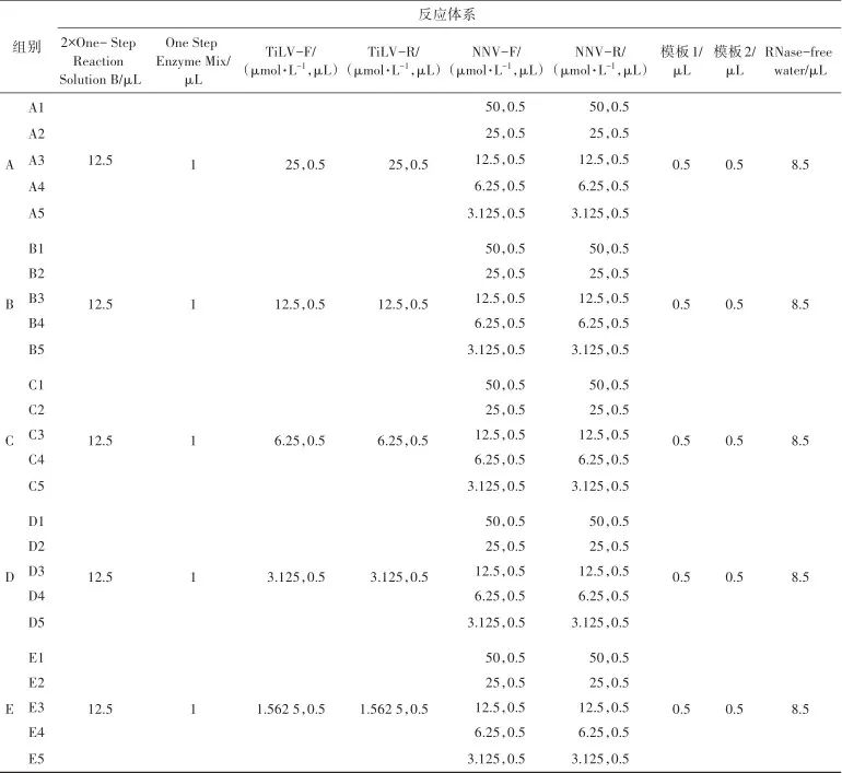
The initial primer concentrations of TiLV F, TiLV R, NNV F, and NNV R were set to 50 µmol/L and subjected to 2-fold gradient dilution, with concentrations of 50, 25, 12.5, 6.25, 3.125, and 1.5625 µmol/L. Various combinations of TiLV and NNV primers at different concentrations were tested. Group A had 5 tubes, each with 0.5 µL of TiLV F and TiLV R at a concentration of 25 µmol/L (final concentration of 0.5 µmol/L); Group B had 5 tubes with 0.5 µL of TiLV F and TiLV R at a concentration of 12.5 µmol/L (final concentration of 0.25 µmol/L); Group C had 5 tubes with 0.5 µL of TiLV F and TiLV R at a concentration of 6.25 µmol/L (final concentration of 0.125 µmol/L); Group D had 5 tubes with 0.5 µL of TiLV F and TiLV R at a concentration of 3.125 µmol/L (final concentration of 0.0625 µmol/L); Group E had 5 tubes with 0.5 µL of TiLV F and TiLV R at a concentration of 1.5625 µmol/L (final concentration of 0.03125 µmol/L). Eppendorf tubes A1, B1, C1, D1, E1 each contained 0.5 µL of NNV F and NNV R at a concentration of 50 µmol/L (final concentration of 1 µmol/L); Eppendorf tubes A2, B2, C2, D2, E2 each contained 0.5 µL of NNV F and NNV R at a concentration of 25 µmol/L (final concentration of 0.5 µmol/L); Eppendorf tubes A3, B3, C3, D3, E3 each contained 0.5 µL of NNV F and NNV R at a concentration of 12.5 µmol/L (final concentration of 0.25 µmol/L); Eppendorf tubes A4, B4, C4, D4, E4 each contained 0.5 µL of NNV F and NNV R at a concentration of 6.25 µmol/L (final concentration of 0.125 µmol/L); Eppendorf tubes A5, B5, C5, D5, E5 each contained 0.5 µL of NNV F and NNV R at a concentration of 3.125 µmol/L (final concentration of 0.0625 µmol/L). The remaining reaction system followed the method described in section “1.4.2” (Table 1).
Table 1 Optimal concentration of dRT-PCR primers for TiLV and NNV detection simultaneously
1.8 Screening of Annealing Temperature for dRT-PCR Detection Method
Reagents were prepared according to the optimized reaction system and evenly distributed into 7 PCR reaction tubes. The annealing temperatures were set to 54, 56, 58, 60, 62, 64, and 66 ℃ in sequence.
1.9 Sensitivity Test of dRT-PCR Detection Method
The original quality concentrations of TiLV and NNV plasmids were both 5 ng/µL, diluted by a factor of 10 to obtain concentrations ranging from 5×10⁻¹ to 5×10⁻⁷ ng/µL. Aside from the templates, reagents were prepared according to the optimized reaction system and distributed into 7 PCR reaction tubes. Eppendorf tubes 1 to 7 were added with 0.5 µL of the gradient diluted TiLV and NNV plasmids, with corresponding final quality concentrations of 1×10⁻⁸ to 1×10⁻² ng/µL. Reverse transcription amplification was conducted according to the optimized reaction conditions.
1.10 Interference Test of RT-qPCR Method for TiLV and NNV
Reagents were prepared according to the optimized reaction system (excluding templates) and evenly distributed into 8 PCR reaction tubes. Tube 1 was added with 0.5 µL of NNV lentivirus nucleic acid, tube 2 with 0.5 µL of NNV plasmid, tube 3 with 0.5 µL of NNV virus nucleic acid, tube 4 with 0.5 µL of NNV lentivirus and 0.5 µL of TiLV lentivirus, tube 5 with 0.5 µL of NNV plasmid and 0.5 µL of TiLV plasmid, tube 6 with 0.5 µL of TiLV lentivirus, tube 7 with 0.5 µL of TiLV plasmid, and tube 8 without any template. The optimized reaction conditions were selected for the mutual interference test of TiLV and NNV.
1.11 Specificity Test of RT-qPCR Method
Reagents were prepared according to the optimized reaction system (excluding templates) and evenly distributed into 12 PCR reaction tubes. TiLV plasmid 0.5 µL and NNV plasmid 0.5 µL, RSIV 1 µL, KHV 1 µL, EHNV 1 µL, GFHNV 1 µL, Aphanomyces invadens 1 µL, SVCV 1 µL, IHNV 1 µL, ISAV 1 µL, SAV 1 µL, VHSV 1 µL were added, with tube 12 containing no template.
1.12 Detection of Clinical Samples
To evaluate the practicality of the dRT-PCR method for detecting TiLV and NNV, 300 samples of tilapia and grouper were collected from routine testing samples and aquaculture reservoirs for dRT-PCR detection.
2 Results and Analysis
2.1 Optimization of dRT-PCR Method for Detecting TiLV and NNV
Results of the optimization experiment for the two sets of primers are shown in Figure 1. Figure 1 shows lanes 1 to 5 corresponding to A1 to A5, lanes 6 to 10 corresponding to B1 to B5, lanes 11 to 15 corresponding to C1 to C5, lanes 16 to 20 corresponding to D1 to D5, and lanes 21 to 25 corresponding to E1 to E5, with lane 26 as the negative control. The 255 bp band is the TiLV amplification product, and the 120 bp band is the NNV amplification product. The results indicate that the concentration of TiLV amplification bands in groups A, B, C, D, and E was generally not affected by the concentration changes of NNV primers, and vice versa. It can be seen that the concentration of one pair of primers does not significantly affect the amplification effect of the other pair. The usage concentration of NNV primers was between 12.5 and 50.0 µmol/L, and the usage concentration of TiLV primers was between 6.25 and 25.00 µmol/L, both achieving good target bands. In subsequent experiments, the concentrations of TiLV F, TiLV R, NNV F, and NNV R primers were set to 20 µmol/L, with each primer added at 0.5 µL in a 25 µL reaction system, resulting in a final concentration of 0.4 µmol/L.
Figure 1 Results of concentration screening of primers in the two groups of dRT-PCR primersFigure 1 Results of concentration screening of primers in the two groups of dRT-PCR primers
M. DNA Marker DL 1000; 1~5. TiLV F, TiLV R final concentration of 0.5 µmol/L; 6~10. TiLV F, TiLV R final concentration of 0.25 µmol/L; 11~15. TiLV F, TiLV R final concentration of 0.125 µmol/L; 16~20. TiLV F, TiLV R final concentration of 0.0625 µmol/L; 21~25. TiLV F, TiLV R final concentration of 0.03125 µmol/L; 26. Negative control. 1, 6, 11, 16, 21. NNV F, NNV R final concentration of 1 µmol/L; 2, 7, 12, 17, 22. NNV F, NNV R final concentration of 0.5 µmol/L; 3, 8, 13, 18, 23. NNV F, NNV R final concentration of 0.25 µmol/L; 4, 9, 14, 19, 24. NNV F, NNV R final concentration of 0.125 µmol/L; 5, 10, 15, 20, 25. NNV F, NNV R final concentration of 0.0625 µmol/L
The optimal annealing temperature test results are shown in Figure 2. Annealing temperatures of 54 to 66 ℃ produced two specific bands of 255 and 120 bp, demonstrating good amplification results.
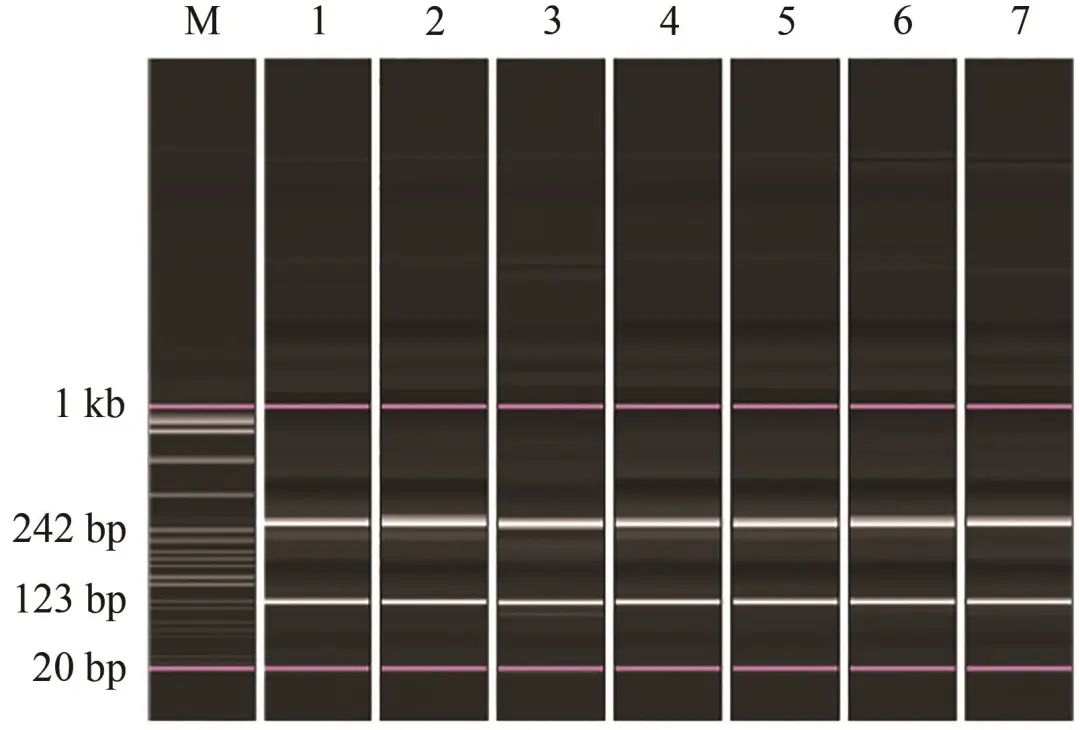
Figure 2 Results of screening of optimal annealing temperature of the dRT-PCRFigure 2 Results of screening of optimal annealing temperature of the dRT-PCR
M. DNA Marker DL 1000; 1~7. 54, 56, 58, 60, 62, 64, 66 ℃
2.2 Mutual Interference Test
Results of the mutual interference test for TiLV and NNV templates are shown in Figure 3. Lanes 1 to 3 contained only NNV templates, lanes 6 to 7 contained only TiLV templates, and lanes 4 to 5 contained both TiLV and NNV templates. The amplification test results showed that the presence of TiLV or NNV templates did not affect the other specific band, indicating no mutual interference.
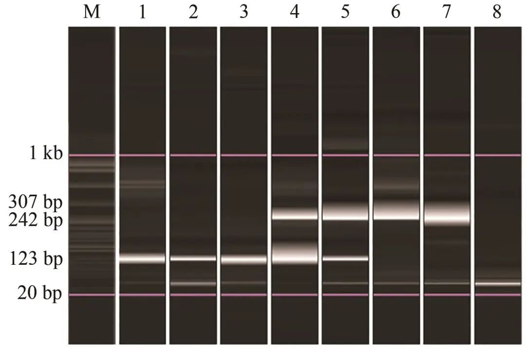
Figure 3 TiLV and NNV mutual interference test of the dRT-PCRFigure 3 TiLV and NNV mutual interference test of the dRT-PCR
M. DNA Marker DL 1000; 1. NNV lentivirus; 2. NNV plasmid; 3. NNV virus; 4. NNV lentivirus + TiLV lentivirus; 5. NNV plasmid + TiLV plasmid; 6. TiLV lentivirus; 7. TiLV plasmid; 8. Negative control
2.3 Sensitivity Analysis
The sensitivity test results are shown in Figure 4. Samples 4 to 7 yielded good specific bands and signal peaks, while samples 2 and 3 showed faint amplification bands with weak signal peaks, thus determining the sensitivity of the dRT-PCR for detecting TiLV and NNV to be at sample 4, with final quality concentrations of 1×10⁻⁵ ng/µL.
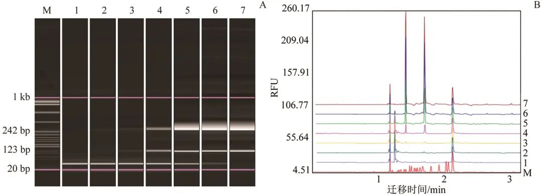
Figure 4 Sensitivity test of the dRT-PCRFigure 4 Sensitivity test of the dRT-PCR
A. Gel electrophoresis image; B. Signal graph; M. DNA Marker DL 1000; 1~7. Final quality concentrations of TiLV and NNV plasmids ranged from 1×10⁻⁸ to 1×10⁻² ng/µL, corresponding to usage quality concentrations of 5×10⁻⁷ to 5×10⁻¹ ng/µL
2.4 Specificity Analysis
In the specificity test, RSIV, KHV, EHNV, and GFHNV are all DNA viruses of fish; SVCV, IHNV, ISAV, SAV, and VHSV are all RNA viruses of fish; and epizootic ulcerative syndrome (EUS) is a parasitic disease caused by a species of Aphanomyces. The specificity test results are shown in Figure 5. None of the pathogens from 10 common fish diseases, including RSIV, showed specific bands.
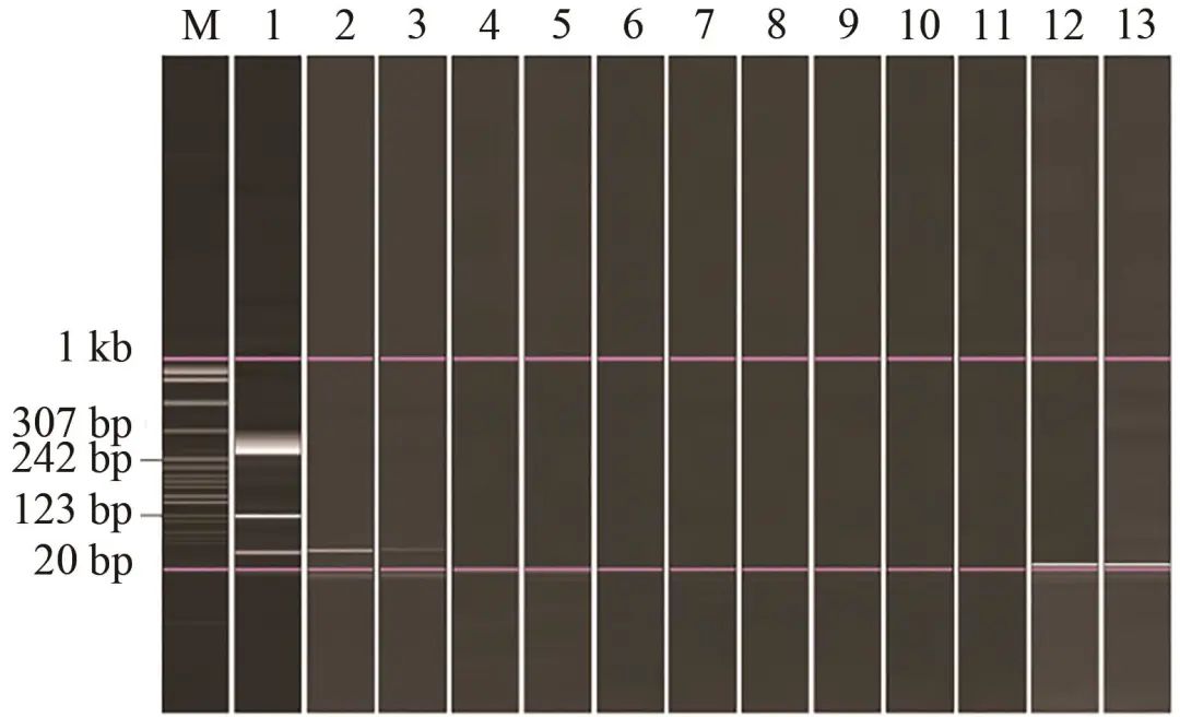
Figure 5 dRT-PCR specificity resultsFigure 5 Specificity test of the dRT-PCR
M. DNA Marker DL 1000; 1. VNNV plasmid + TiLV plasmid; 2. RSIV; 3. KHV; 4. EHNV; 5. GFHNV; 6. Aphanomyces; 7. SVCV; 8. IHNV; 9. ISAV; 10. SAV; 11. VHSV; 12. Negative control; 13. Blank control
2.5 Preliminary Application
The dRT-PCR method established in this study for simultaneous detection of TiLV and NNV was applied to 300 routine testing samples and reservoir samples, with no TiLV detected and 30 cases of NNV detected.
3 Discussion
Research has found that TiLV exhibits characteristics of a positive-sense RNA virus, suggesting that TiLV may be a new type of positive-sense RNA virus. TiLV has a total of 10 gene fragments, with the largest being gene fragment 1, which has a weak homology to the PB1 subunit sequence of the influenza virus. Tilapia are easily infected by TiLV and NNV, with 31 gene fragments of TiLV virus strain 1 found in GenBank; 4 NNV virus strains n-27 (GenBank accession number GQ857479), ET Egypt 2018 (GenBank accession number MN701084), ET (GenBank accession number MN698297), and ETB Egypt KFS 2019 (GenBank accession number MN698298) were identified. This study designed duplex RT-PCR primers based on the TiLV fragment 1 gene and the NNV capsid protein gene. This study established a dRT-PCR method capable of simultaneously detecting TiLV and NNV, optimizing the reaction system and conditions. The sensitivity for detecting TiLV and NNV was both 1×10⁻⁵ ng/µL (final nucleic acid quality concentration), with good specificity. When using this method to test 300 clinical samples, only 30 cases of NNV positive were detected, and no TiLV was detected, but this does not indicate a failure of the method for TiLV identification. Lei Yan et al. reported TiLV from tilapia samples in Hainan, and there have been no official reports of TiLV from other research teams. Based on literature reports and annual monitoring results from customs and aquaculture technology promotion stations, the incidence of TiLV in domestic tilapia is low, which is why it was not detected. The dRT-PCR method established in this study for simultaneous detection of TiLV and NNV has good specificity, high sensitivity, and is convenient and quick, and can also be used for individual detection of TiLV or NNV. It has certain clinical application value and shows good prospects for the diagnosis of tilapia lake disease and viral nervous necrosis disease epidemiological investigation.
Cite this article: Xu Shufei, Zeng Yunying, Zhu Huangxin, et al. Establishment of Duplex RT-PCR for Detection of Tilapia Lake Virus and Viral Nervous Necrosis Virus [J]. Journal of Economic Animal, 2023, 27(3): 182-188. (XU Shufei, ZENG Yunying, ZHU Huangxin, et al. Establishment of Duplex RT-PCR for Detection of Tilapia Lake Virus and Viral Nervous Necrosis Virus [J]. Journal of Economic Animal, 2023, 27(03): 182-188.)
Author Information: Xu Shufei, female, master’s student, mainly engaged in research on aquatic animal diseases.
Corresponding Author: Zhu Huangxin, E-mail: [email protected]
Funding Information: Fujian Province Guidance Project (2019N0030)
Classification Number: S943
Article Number: 1007-7448 (2023) 03-0182-07
Literature Identification Code: A
Submission Date: 2021-11-07
Publication Date: 2023-09-25
Online Publication Date: 2023-11-09

Journal of Economic Animal
Editor:|Yu Jing
Reviewer:|Ju Shan Hong
Scan the QR code to follow us