
Abstract
Objective To establish a capillary zone electrophoresis method to detect the polysaccharide content and molecular size distribution of A, C, Y, and W135 group meningococcal polysaccharide vaccines.
Methods A capillary zone electrophoresis method was established to detect four types of polysaccharides, exploring the optimal separation voltage and temperature. The method characterizes the four polysaccharides simultaneously in a single experiment, measuring their content and molecular size distribution, and validating the method’s linearity, accuracy, repeatability, and specificity.
Results When the content of the four polysaccharides ranged from 0.0813 to 0.4875 μg/μL, the capillary zone electrophoresis method showed good linearity with a coefficient of determination > 0.98; for low, medium, and high concentration samples, the recovery rate was between 95% and 110%, with a relative standard deviation < 2.0% in six experiments, and no interfering peaks appeared at the target peak position.
Conclusion The capillary zone electrophoresis method for detecting the four types of meningococcal polysaccharides demonstrates good linearity, accuracy, repeatability, and specificity, making it suitable for measuring the polysaccharide content and molecular size distribution of A, C, Y, and W135 group meningococcal polysaccharide vaccines.
Main Text
The Neisseria meningitidis, also known as meningococcus, includes the A, C, Y, and W135 groups, which are the main pathogens of epidemic meningitis (meningococcal meningitis). The mortality rate among infected individuals is high, and the sequelae can be severe, especially in children. Currently, vaccination with polysaccharide vaccines that use the meningococcal capsular polysaccharide as a protective antigen is the primary preventive measure. The determination of polysaccharide content and molecular size is an important basis for assessing vaccine quality: a certain standard of polysaccharide content must be reached to elicit an immune response; an appropriate molecular size distribution ensures effective stimulation of the immune system. There are two conventional methods for polysaccharide detection: converting polysaccharides to monosaccharides under high temperature and strong acid conditions for detection, such as measuring phosphorus content to determine group A polysaccharide content, and measuring sialic acid content for groups C, Y, and W135; and combined detection using rocket electrophoresis and normal pressure liquid chromatography. These methods have significant safety risks or are heavily dependent on the stability of serum potency. The capillary electrophoresis method can simultaneously separate and detect the content of four polysaccharides, with high resolution, short time consumption, and economic safety. This study aims to establish and optimize a capillary zone electrophoresis method for detecting the content and molecular size distribution of four polysaccharides in A, C, Y, and W135 group meningococcal polysaccharide vaccines, and to investigate the method’s linearity, accuracy, repeatability, and specificity.
1
Materials and Methods
1.1
Instruments and Reagents
The PA800 PLUS capillary electrophoresis instrument was purchased from SCIEX, USA; the fused silica capillary (inner diameter 50 μm, total length 69 cm, effective length 59 cm) was purchased from Beckman Coulter, USA; the AKTA PURE 25 normal pressure liquid chromatography system and Sepharose-CL4B agarose gel column (void volume 193.28 mL, bed volume 62.28 mL) were purchased from Cytiva, USA; the A, C, Y, and W135 group meningococcal polysaccharide vaccine (lyophilized powder) and polysaccharide standards for A, C, Y, and W135 groups were provided by Beijing Institute of Biological Products Co., Ltd.
1.2
Preparation of Solutions
1.2.1 Stock Solution Preparation Mix 16.2 mL of A group polysaccharide standard (5.30 g/L), 16.5 mL of C group polysaccharide standard (5.21 g/L), 15.8 mL of Y group polysaccharide standard (5.42 g/L), 27.2 mL of W135 group polysaccharide standard (3.16 g/L), and 56.3 mL of ultrapure water to prepare a 0.65 g/L stock solution of 132.0 mL.
1.2.2 Preparation of Linear and Accuracy Solutions According to the 2020 edition of the Chinese Pharmacopoeia, the qualification standard for the polysaccharide content of A, C, Y, and W135 group meningococcal polysaccharide vaccines is 35-65 μg of A, C, Y, and W135 group polysaccharides per human dose. After dissolving in 200 μL of ultrapure water, this corresponds to 0.175-0.325 g/L. Five concentration standard solutions were prepared at 25%, 40%, 100%, 125%, and 150% of the upper limit concentration (0.325 g/L), which are 0.0813, 0.1300, 0.3250, 0.4063, and 0.4875 g/L, covering the qualification standard range. Among them, 0.0813 and 0.1300 g/L cover 50% and 75% of the lower limit concentration of the qualification standard, and the standard solutions at concentrations of 0.1300, 0.3250, and 0.4063 g/L were also used for accuracy testing.
1.3
Electrophoresis Analysis Conditions
The electrophoresis buffer was 50 mmol/L boric acid-borax buffer (pH 9.0); column temperature: 20 °C; detector type: UV detector; detection wavelength: 206 nm; injection method: constant pressure injection (138 kPa); injection time: 20 s; separation voltage: 30 kV. Rinsing steps: before each sample measurement, rinse with 0.1 mol/L NaOH solution, ultrapure water, and electrophoresis buffer for 3 minutes each.
1.4
Sample Preprocessing
In normal pressure liquid chromatography, separate the A, C, Y, and W135 group meningococcal polysaccharide vaccine, collect the polysaccharide elution, and label the number. The flow rate is 0.3 mL/min, UV 206 nm, and the mobile phase is 0.2 mol/L sodium chloride solution (pH 7.0).
1.5
Optimization of Electrophoresis Conditions
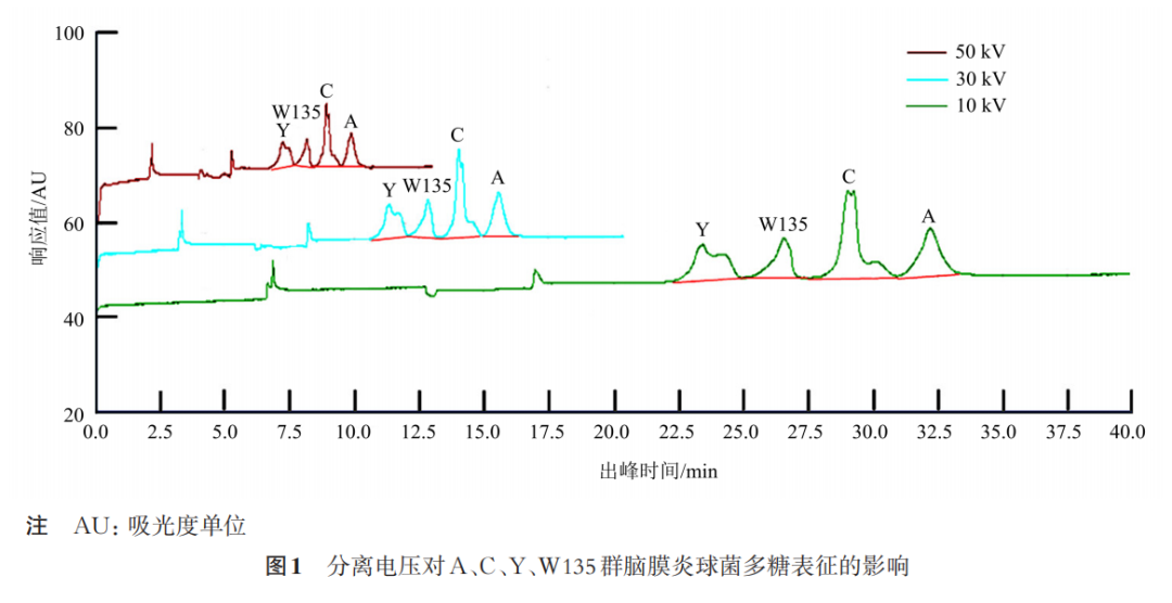
1.5.1 Separation Voltage According to the electrophoresis separation conditions in 1.3, investigate the separation effect of the four polysaccharides in the stock solution at separation voltages of 10, 30, and 50 kV.
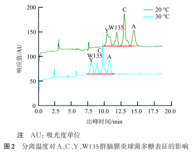
1.5.2 Separation Temperature According to the electrophoresis separation conditions in 1.3, investigate the separation effect of the four polysaccharides in the stock solution at separation temperatures of 30 and 20 °C.
1.6
Method Validation
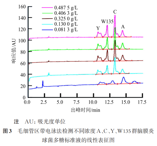
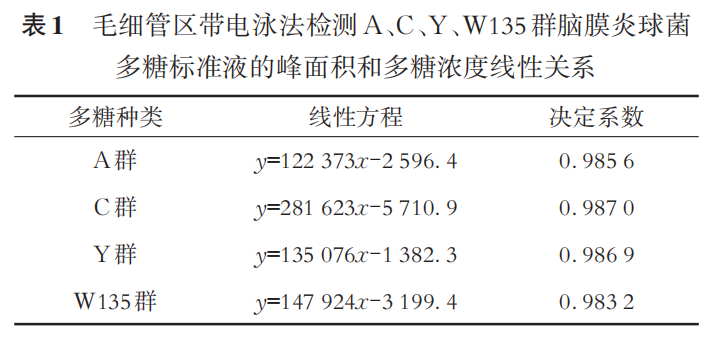
1.6.1 Linearity Detect the content of the four polysaccharides in the standard solutions of 0.0813, 0.1300, 0.3250, 0.4063, and 0.4875 g/L, using the concentration of the standard solution as the x-axis and the peak area of each polysaccharide as the y-axis, performing linear fitting to calculate the coefficient of determination (R2). The linear fitting curves for the four polysaccharides should all have an R2 greater than 0.98.
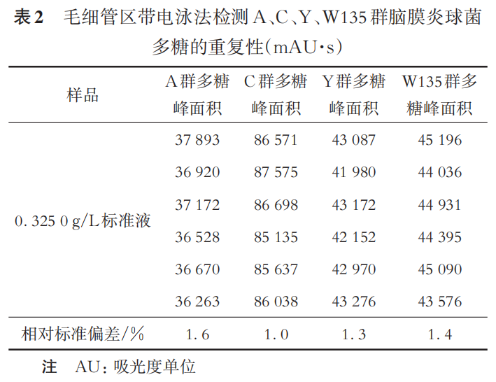
1.6.2 Repeatability Repeat the detection of the 0.3250 g/L polysaccharide standard solution six times, calculating the relative standard deviation (RSD) of the content of the four polysaccharides. The RSD of the content of the four polysaccharides should all be less than 2.0%.
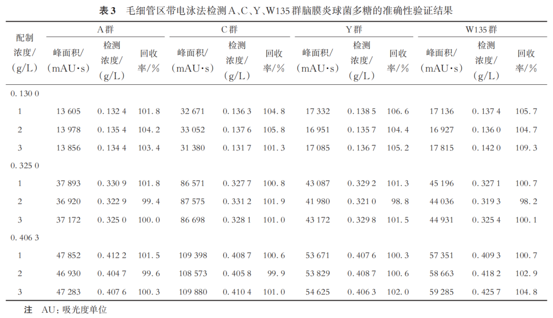
1.6.3 Accuracy Detect the standard solutions of 0.1300, 0.3250, and 0.4063 g/L three times each, calculating the recovery rate of the content of the four polysaccharides. The recovery rate of the four polysaccharides should all be between 95% and 110%.
1.6.4 Specificity Investigate the peak area of the electrophoresis buffer at the retention time of the A, C, Y, and W135 group polysaccharides (if any), and the peak in the electrophoresis buffer at the peak position of the four polysaccharides should be less than 0.02% of the target peak area in the polysaccharide standard solution.
1.7
Method Application
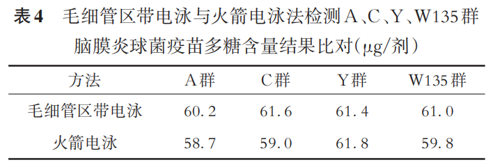
1.7.1 Detection of Polysaccharide Content For each batch, take one dose of the A, C, Y, and W135 group meningococcal polysaccharide vaccine sample, dissolve it in 200 μL of ultrapure water, and perform capillary zone electrophoresis detection according to the electrophoresis analysis conditions in 1.3. At the same time, use rocket electrophoresis to detect the content of the four polysaccharides in the same sample.
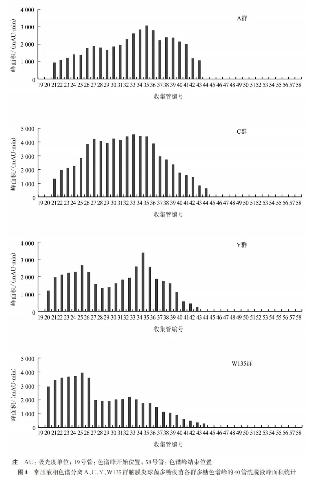
1.7.2 Detection of Polysaccharide Molecular Size and Recovery Rate Take 50 bottles of the vaccine lyophilized powder to be tested, dissolve them, and use the normal pressure liquid chromatography system for separation, collecting one tube of elution liquid every 3 mL and labeling them. The content of the four polysaccharides in the collected elution liquid of the collection tubes was detected using two methods. Using the collection tube number as the x-axis and the peak area detected in the elution liquid of the tube as the y-axis, a bar chart was drawn to find the collection tube number of the main peak, and the distribution coefficient was calculated. When the distribution coefficient is 0.5, the height of the rocket electrophoresis band before the collection tube number divided by the total peak area of the capillary zone electrophoresis equals the total height of all rocket electrophoresis bands divided by the total peak area of the capillary zone electrophoresis, thus obtaining the recovery rate.
2
Results
2.1
Optimization of Electrophoresis Conditions
2.1.1 Optimization of Separation Voltage As shown in Figure 1, at separation voltages of 10, 30, and 50 kV, the four polysaccharides can be completely separated. At a separation voltage of 10 kV, the peak width is larger, and the separation speed is lower; at a separation voltage of 50 kV, the peak area decreases, and the detection sensitivity decreases. Therefore, 30 kV was chosen as the separation voltage.
2.1.2 Optimization of Separation Temperature As shown in Figure 2, the four polysaccharides can achieve complete baseline separation at 20 °C. When the separation temperature is 30 °C, although the migration rate of the four polysaccharides is slightly accelerated, complete baseline separation cannot be achieved. Therefore, 20 °C was chosen as the separation temperature.
2.2
Method Validation
2.2.1 Linearity The electrophoretic characterization of the five concentration polysaccharide standard solutions is shown in Figure 3, and the linear equations are shown in Table 1. The R2 values are all greater than 0.98, indicating that the linearity validation is qualified.
2.2.2 Repeatability The results of the repeatability validation are shown in Table 2. The RSD of the content of the four polysaccharides is less than 2.0%, indicating that the repeatability validation is qualified.
2.2.3 Accuracy The results are shown in Table 3. The recovery rates of the four polysaccharide standard solutions are all between 95% and 110%, indicating that the accuracy validation is qualified.
2.2.4 Specificity No electrophoretic peaks appeared in the electrophoresis buffer at the peak times of the A, C, Y, and W135 group polysaccharides, indicating that the specificity validation is qualified.
2.3
Method Application
2.3.1 Detection of Polysaccharide Content The content of the four polysaccharides in the same A, C, Y, and W135 group meningococcal polysaccharide vaccine sample was detected using two methods, and the results are shown in Table 4. The paired sample t test showed that t=1.98, P=0.142, indicating no statistical significance, and the polysaccharide content detected by the capillary zone electrophoresis method and the rocket electrophoresis method is equivalent.
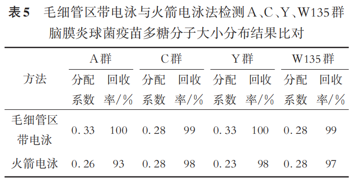
2.3.2 Detection of Polysaccharide Molecular Size Distribution The four polysaccharides in the vaccine were separated using normal pressure liquid chromatography. The collection tube number of the main peak elution was determined through Figure 4, and the distribution coefficient and recovery rate were calculated, with results shown in Table 5. The distribution coefficient and recovery rate were subjected to paired sample t tests. The distribution coefficient t=1.68, P=0.191; recovery rate t=2.21, P= 0.114, indicating no statistical significance, and the molecular size distribution of the four polysaccharides detected by the capillary zone electrophoresis method and the rocket electrophoresis method is equivalent.
3
Discussion
In capillary electrophoresis, factors such as separation voltage, separation temperature, type of buffer, and pH can affect the migration time and peak shape of the components to be tested. This study used fused silica capillaries and boric acid-borax buffer. Although higher voltage increases the electric field force on the four polysaccharides in the capillary, accelerating their migration speed, excessively high voltage can lead to increased Joule heating and baseline noise. Selecting an appropriate separation voltage in capillary electrophoresis can shorten detection time and stabilize the background baseline. The separation temperature affects the viscosity of the electrophoresis buffer, which in turn affects the diffusion, migration time, and peak shape of the A, C, Y, and W135 group polysaccharides. Although higher separation temperatures accelerate component diffusion and migration speed, excessively high temperatures can also increase baseline noise. Therefore, selecting an appropriate separation temperature in capillary electrophoresis can shorten detection time, stabilize the background baseline, and reduce noise interference. This study compared the effects of three separation voltages (10, 30, and 50 kV) and two separation temperatures (20 and 30 °C) on the separation of the four polysaccharides, and the results showed that the separation effect was best at 30 kV and 20 °C.
This study established a capillary zone electrophoresis method for analyzing the A, C, Y, and W135 group polysaccharides in meningococcal polysaccharide vaccines, validating the method’s linearity, accuracy, repeatability, and specificity. The results indicate that the capillary zone electrophoresis method is suitable for detecting the content and molecular size distribution of the four polysaccharides. The capillary zone electrophoresis method and the rocket electrophoresis method yield equivalent results for the content and molecular size distribution of the four polysaccharides.
Compared to the method in the third part of the pharmacopoeia 3419 for detecting the polysaccharide content and molecular size distribution of the A, C, Y, and W135 group meningococcal polysaccharide vaccines, this method is simple to operate, does not require organic reagents, requires a small sample volume, avoids the influence of serum potency, and addresses issues such as poor experimental repeatability and long experimental cycles due to insufficient serum quantity or unstable reference serum potency during the quality control of A, C, Y, and W135 group meningococcal polysaccharide vaccines, thus improving detection efficiency and providing a new method for enterprises to assess the polysaccharide content and molecular size distribution of A, C, Y, and W135 group meningococcal polysaccharide vaccines.
Authors
Wang Huimin, Li Yan, Liu Jing, Yang Bing, Hou Qianqian, Ding Ling, Zhu Xiujuan, Liu Yicheng
Quality Control Department, Beijing Institute of Biological Products Co., Ltd., Beijing 100176
Corresponding Author: Liu Yicheng,
Email: [email protected]
Reference: Wang Huimin, Li Yan, Liu Jing, et al. A, C, Y, and W135 Group Meningococcal Polysaccharide Vaccine Polysaccharide Content and Molecular Size Distribution Determined by Capillary Zone Electrophoresis [J]. International Journal of Biological Products, 2025, 48(5): 337-342.
DOI: 10.3760/cma.j.cn311962-20241215-00087