Skip to content
Utilizing neuronal regeneration to treat neurodegenerative diseases remains a key challenge in the field of regenerative medicine. The direct conversion of local glial cells into neurons has become a viable alternative to replace functional neurons【1,2】. Considering that the increase in reactive oxygen species generation significantly hinders the process of glial cell conversion into neurons, and that neurons rely on oxidative phosphorylation(Oxphos), the importance of mitochondrial activity during neuronal conversion is evident. Specific mitochondrial proteins may be required to meet the cell type-specific metabolic demands【3,4】. However, how mitochondria adapt to a new fate during neuronal reprogramming remains unclear.
Recently, the research group led by Magdalena Gtöz from Ludwig Maximilian University in Germany published an article titled “CRISPR-Mediated Induction of Neuron-Enriched Mitochondrial Proteins Boosts Direct Glia-to-Neuron Conversion” in Cell Stem Cell. In this study, the authors assessed the similarities and differences in the mitochondrial composition of cultured neurons and astrocytes, aiming to regulate their respective genes through CRISPRa(clustered regularly interspaced short palindromic repeat activation) mediated transcriptional engineering to improve the mismatch issues during the reprogramming process. Early activation of mitochondrial protein-coding genes mediated by dCas9 can significantly enhance the conversion efficiency of glial cells to neurons and the survival rate of neurons, especially for antioxidant proteins enriched in neurons rather than astrocytes, indicating that mitochondrial proteins play a driving role in fate conversion.
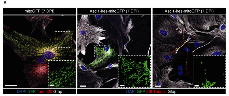
First, the authors isolated astrocytes from the cortex of mice at postnatal day five, transducing them with a retrovirus carrying a green fluorescent protein targeted to mitochondria, along with (or without) previously proven reprogramming precursor factors that can convert astrocytes into GABAergic neurons(Ascl1-ires-mitoGFP)【5】, to monitor mitochondrial morphological changes during the reprogramming process. The successfully converted neuronal cells exhibited smaller mitochondrial volume and shorter, rounder morphology, consistent with high fission characteristics; in contrast, astrocytes had longer mitochondrial morphology and stronger fusion characteristics(see Figure 1).
Figure 1. Left: Astrocytes (mitochondria labeled by mitoGFP); Middle: Asc1-non-reprogrammed astrocytes; Right: Mitochondrial morphology images of Asc1-induced neurons.
So, what causes this difference? The authors used liquid chromatography-tandem mass spectrometry(LC-MS/MS) to identify proteins in the mitochondria of neurons and astrocytes. A total of 757 and 738 mitochondrial proteins were detected in astrocytes and neurons, respectively, with 164 being specifically enriched in astrocytes(22%), and 141 enriched in neurons(19%), indicating that about one-fifth of the mitochondrial proteome is significantly different between these cell types. GO analysis suggested that fatty acid catabolic pathways are enriched in astrocytes, and the authors wanted to know whether this pathway has direct functional relevance to reprogramming. In the early stages of conversion, using inhibitors of key regulators of fatty acid β-oxidation to block this pathway significantly improved the reprogramming conversion rate compared to controls. For mitochondrial proteins enriched in neurons, GO analysis suggested important pathways related to RNA metabolism and function, further highlighting the previously proposed notion that tRNA biogenesis is an important activity in neuronal mitochondria, and its dysfunction is associated with neurodevelopmental diseases【6】.
Next, to determine whether and when astrocytes downregulate their characteristic mitochondrial proteins and how they increase the expression of neuron-enriched mitochondrial proteins during conversion to neurons, the authors selected functionally relevant candidate proteins with differential enrichment detectable by immunostaining and found that changes in mitochondrial proteins enriched in astrocytes(Sfxn5 and Cpox) or neurons(Prdx2 and Gls) were relatively late during the Advl1-mediated reprogramming process. Notably, these changes correlated with the degree of conversion, suggesting a functional relevance hypothesis for these proteins.
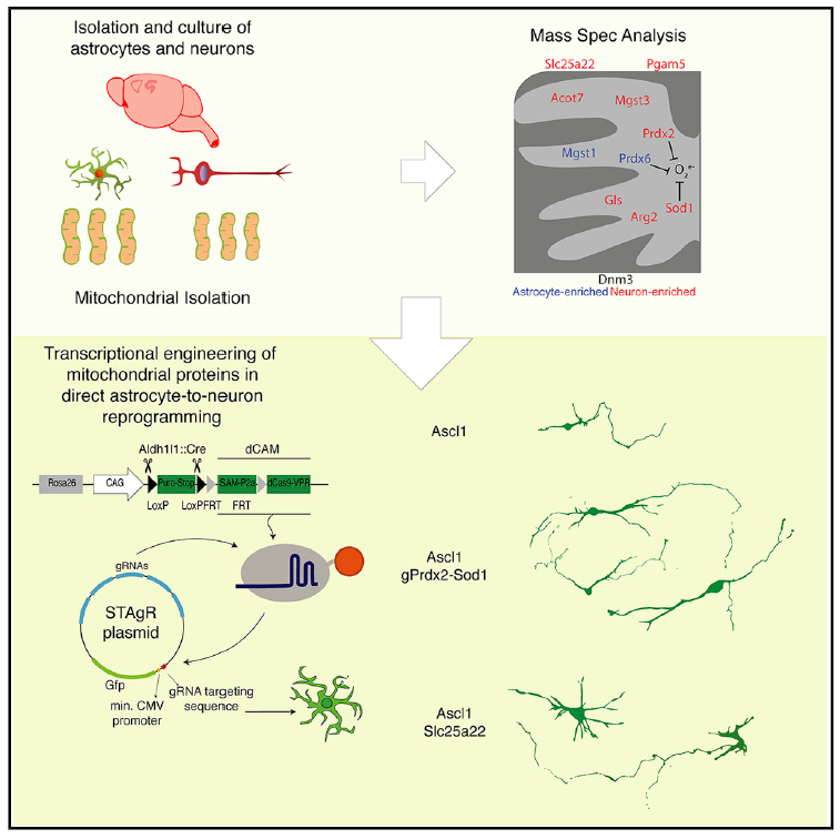
Finally, to validate the above hypothesis, the authors selected eight candidate proteins enriched in neuronal mitochondria, transfected them with dCas9-VPR encoding plasmids and non-targeting control gRNA or gRNA targeting the promoter regions for 48 hours, and the qRT-PCR results of FACS-sorted cells showed different induction levels for the candidates. After cloning the gRNA of selected candidates into plasmids with GFP reporter modules, primary cultures of astrocytes from transgenic mouse lines were performed, resulting in these products accelerating the conversion of glial cells to neurons. Notably, they also increased the complexity of axons and dendrites of neurons; the above results indicate that neuron-specific mitochondrial proteins are particularly important during the conversion of glial cells to neurons, and their early induction and high expression can improve reprogramming. Subsequently, the authors confirmed through real-time imaging of single cells that these neuron-enriched mitochondrial proteins(Prdx, Sod1) not only accelerate the conversion process but also protect neurons from apoptosis.
Figure 2. Early induction of neuron-enriched mitochondrial encoding genes mediated by CRISPRa enhances the speed and efficiency of neuronal reprogramming.
In summary, this study elucidated the mitochondrial proteome of cortical astrocytes and neurons, revealing that astrocyte-specific mitochondrial proteins are typically only partially downregulated during the direct reprogramming of astrocytes to neurons, while neuron-specific mitochondrial proteins are upregulated later. Therefore, early induction of neuron-enriched mitochondrial encoding genes mediated by CRISPRa can enhance the speed and efficiency of direct neuronal reprogramming, importantly providing new insights for the functional study of cell type-specific mitochondrial proteins.
https://doi.org/10.1016/j.stem.2020.10.015
1, Barker, R.A., Götz, M., and Parmar, M. (2018). New approaches for brain repair-from rescue to reprogramming. Nature 557, 329–334.
2, Vignoles, R., Lentini, C., d’Orange, M., and Heinrich, C. (2019). Direct Lineage Reprogramming for Brain Repair: Breakthroughs and Challenges. Trends Mol. Med. 25, 897–914.
3, Harris, J.J., Jolivet, R., and Attwell, D. (2012). Synaptic energy use and supply. Neuron 75, 762–777.
4, Calvo, S.E., and Mootha, V.K. (2010). The mitochondrial proteome and human disease. Annu. Rev. Genomics Hum. Genet. 11, 25–44.
5, Heinrich, C., Blum, R., Gasco ´ n, S., Masserdotti, G., Tripathi, P., Sánchez, R., Tiedt, S., Schroeder, T., Götz, M., and Berninger, B. (2010). Directing astroglia from the cerebral cortex into subtype specific functional neurons. PLoS Biol. 8, e1000373.
6, Schaffer, A.E., Pinkard, O., and Coller, J.M. (2019). tRNA Metabolism and Neurodevelopmental Disorders. Annu. Rev. Genomics Hum. Genet. 20, 359–387.