Professor Xu Haibo’s team from the Department of Radiology at Zhongnan Hospital of Wuhan University recently published a paper titled “Lifespan trajectories of fornix volume and tractography: a 5.0 T MRI study” in the renowned neuroscience journal Cerebral Cortex.
In 1937, neuroanatomist James Papez proposed a closed neural circuit composed of the hippocampus, mammillary bodies, and anterior thalamic nuclei, known as the Papez circuit. This circuit is not only a core component of the limbic system but is also referred to as the “emotional brain“ due to its critical role in emotional regulation and memory formation.
As the core conduction pathway of this circuit, the fornix connects the hippocampus to the hypothalamic mammillary bodies via arcuate white matter fiber bundles, while also playing important roles in information exchange between the left and right hemispheres and the integration of hypothalamic and medial temporal lobe functions. With breakthroughs in diffusion magnetic resonance imaging technology, scientists can finally visualize the morphological characteristics of this mysterious structure non-invasively.
Clinical studies have shown that damage to the fornix caused by hypoxia, trauma, or inflammation can lead to significant memory impairments. Notably, changes in the integrity of fornix fiber bundles during normal aging occur even before structural changes in the hippocampus, making it a sensitive indicator for predicting cognitive decline. Consequently, deep brain stimulation of the fornix has emerged as a new direction for the treatment of Alzheimer’s disease.
Despite existing technologies achieving visualization of the fornix, the developmental patterns of its entire lifecycle and its dynamic relationship with memory function remain unclear. This is primarily limited by three technical bottlenecks: significant individual anatomical variation, severe imaging artifacts, and difficulties in precise segmentation. With the introduction of domestically developed 5.0T ultra-high-field magnetic resonance equipment, along with the application of machine learning algorithms in brain segmentation, new solutions to these issues have emerged.
This study innovatively employed 5.0T ultra-high-field MRI to collect multimodal brain imaging data from 376 healthy individuals aged 18-85, focusing on: (1) age-related changes in fornix volume and fiber bundle properties; (2) the dynamic relationship between these structural features and memory ability. The study hypothesizes that fornix fiber bundle parameters are more sensitive to age-related changes than volume metrics and can more effectively predict memory function.
The data from this study indicate that the volume of the fornix exhibits a nonlinear decline trend, with a significant inflection point around 50 years of age; fiber bundle density continues to accumulate during young adulthood (18-40 years) but shows a dramatic decline after age 40—this may be a key neural marker for cognitive decline in the elderly. Additionally, the lateralization of fornix volume and fiber bundles shows dynamic evolution throughout the lifespan, suggesting complex interactions between brain network reorganization and specific cognitive domain regression. Regression analysis further reveals that fornix structural features directly modulate, rather than mediate, age-related cognitive differences, indicating their potential as independent predictive factors. These high-resolution imaging results provide new insights into the role of fornix morphology and structural connectivity in individual cognitive differences and the aging process.
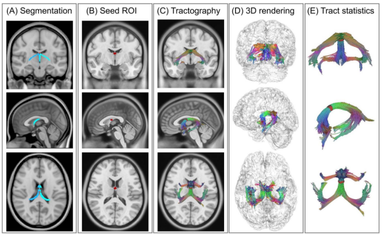
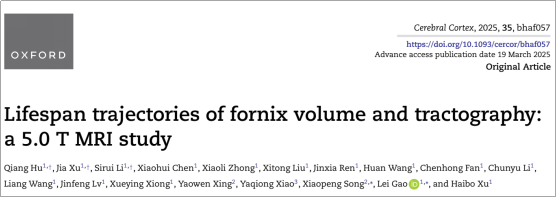
Figure 1. Illustration of data preprocessing.
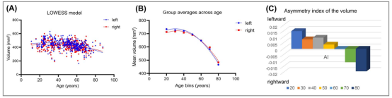
Figure 2.
Life trajectory of fornix volume.
Figure 3. Life trajectory of fornix fiber bundles.
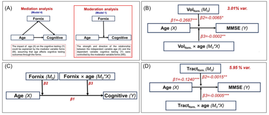
Figure 4. Mediation analysis. (A) Illustration of mediation and moderation analysis. The fornix’s (B) volume and (D) number of fiber bundles significantly modulate the relationship between age and overall cognitive MMSE test scores. (C) Regression equations for moderation analysis.
Additionally, to compare the advantages of 5.0T in characterizing the fornix, this study also collected a batch of young healthy volunteers (n=21) to simultaneously conduct retests of 3.0T and 5.0T diffusion imaging. The results showed that under the same resolution and main parameters, 5.0T has advantages in visualizing the fornix.
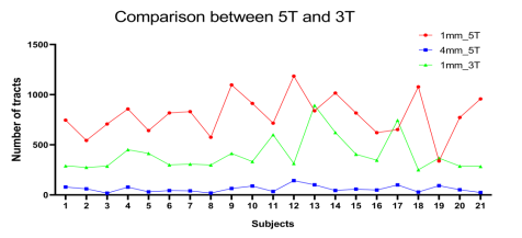
Figure S2. Differences in fornix visualization between 3.0T and 5.0T imaging.
Hu Qiang, chief technician of the Department of Radiology at Zhongnan Hospital of Wuhan University, Xu Jia, chief technician, and Li Sirui, associate chief physician, are co-first authors of this paper. Professor Xu Haibo, associate chief technician Gao Lei, and Dr. Song Xiaopeng from United Imaging are co-corresponding authors.
Research Team Introduction
First Author

Hu Qiang, chief technician of the Department of Medical Imaging at Zhongnan Hospital of Wuhan University, focuses on new technologies in neuroimaging. He has published 3 SCI papers in recent years, including one as the first author, and has participated as a key researcher in a project funded by the Hubei Provincial Natural Science Foundation.
Xu Jia, chief technician of the Department of Medical Imaging at Zhongnan Hospital of Wuhan University, focuses on neuroimaging and has published 3 SCI papers in recent years, including 2 as the first author.
Li Sirui, MD, associate chief physician, has over ten years of experience in imaging diagnosis, specializing in neuroimaging, with rich experience in imaging diagnosis of central nervous system diseases such as cerebrovascular diseases, brain tumors, and inflammatory demyelinating diseases. He has led and participated in multiple national and provincial research projects and has published over 20 papers in both Chinese and English, including 5 as the first author in SCI journals.
Corresponding Authors

Xu Haibo, director of the Department of Medical Imaging at Zhongnan Hospital of Wuhan University, chief physician, associate professor, and doctoral supervisor, is a leading medical talent in Hubei Province. Professor Xu’s team has been dedicated to research on molecular imaging of tumors, neural circuits of consciousness disorders, innovative medical imaging devices, and artificial intelligence technologies, achieving a series of research results. He has published over 160 papers as the first or corresponding author in journals such as JAMA, Cell Metabolism, Advanced Science, Adv. Func. Mater, Theranostics, NeuroImage, Neuropsychopharmacology, JCI Insight, JMRI, and others; he holds 4 national invention patents, 12 utility model patents; has received two second prizes for scientific and technological progress in Hubei Province, one first prize for scientific and technological achievement in Shanghai, and one first prize for scientific and technological achievement in Guangdong Province; he has led 7 projects funded by the National Natural Science Foundation and is responsible for major national instrument projects and key R&D plan sub-projects. He has also received awards such as the National Advanced Individual in the Fight Against COVID-19.
Gao Lei, MD, associate chief technician, associate researcher, master’s supervisor, and leader of the brain functional imaging sub-specialty group at Zhongnan Hospital, is recognized as an outstanding young medical talent. He mainly engages in neuroimaging research on human brain functional architecture and vascular cognitive impairment. He has led one project funded by the National Natural Science Foundation, one key project of the Hubei Provincial Natural Science Joint Fund, one project for young teachers at central universities, and one innovation cultivation fund project at Zhongnan Hospital. In recent years, he has published over 20 SCI papers as a co-first or corresponding author, including in journals such as HBM, Cerebral Cortex, NeuroImage: Clinical, JMRI, Journal of Alzheimer’s Disease and others. He serves as a young editorial board member for the journal Chinese CT and MRI and as a reviewer and guest associate editor for several SCI journals, as well as a frequently invited reviewer for over 20 well-known SCI journals.
Song Xiaopeng, PhD from Peking University, is a senior scientist at United Imaging MR. He is a postdoctoral researcher at the University of California, Los Angeles (UCLA) and a researcher at Harvard Medical School. He is a recipient of the National High-Level Youth Talent Introduction Program, the Ministry of Industry and Information Technology Talent Program, the Hubei Provincial High-Level Talent Introduction Program, the Wuhan Talent “Industry Leading Talent Award,” and the Optics Valley Development Zone “3551 Innovation Talent Award.” He has received the Young Investigator Award from the Overseas Chinese Society for Magnetic Resonance in Medicine (OCSMRM) and the Harvard Brain Initiative Award.