3D printing (Three-Dimensional Printing) technology has developed for over 30 years, and recent technological advancements and their popularity among the general public are ushering the world into the era of personalized 3D printing. As 3D printing becomes more widespread, its application in orthopedic surgery has become feasible. Compared to 3D virtual CT imaging on a screen, the skeletal anatomical structure can be transformed into a real-sized 3D printed model, allowing surgeons to examine the structure more closely before surgery. A real-sized 3D model facilitates precise preoperative planning and can even simulate surgery using the implants that will be used. This is particularly beneficial for complex fracture surgeries often encountered by beginners. 3D models can also be used for educating medical students and enhancing communication with patients.
Tibial distal fractures can be fixed using various sizes of anatomical locking plates. However, these plates are designed based on the average tibial shape of patients, which may lead to a mismatch between the plate and the bone in patients with smaller or larger tibias (Figure 1) or in patients with tibial deformities. Additionally, complex fractures may require different screw trajectories within the plate to stabilize fracture fragments. Therefore, 3D printing can be used to understand complex fracture configurations, create preoperative templates, select the most suitable anatomical plates, and plan various screw trajectories for complex tibial distal fractures.
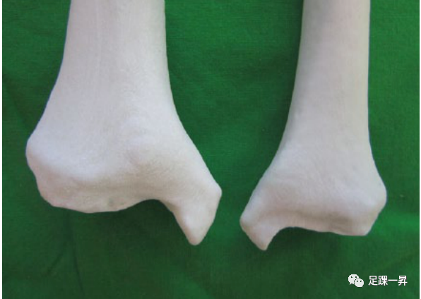 Figure 1 The shape and size differences between two tibias of a male 178 cm tall (left) and a female 146 cm tall (right) are apparent.Ankle avulsion fractures are typically fixed with tension bands or lag screws. However, elderly patients or those with osteoporosis due to diabetes may not be able to achieve stable fixation using Kirschner wires or screws, which may lead to internal fixation failure. Hook plates can be used to securely fix the avulsion fracture fragments. However, due to the relatively thin soft tissue covering this area, if the hook plate does not match the surface of the ankle joint, it may protrude and irritate the soft tissue. A real-sized 3D printed model of the patient’s ankle can serve as a template for customizing the hook plate. Preoperative molded hook plates allow for minimally invasive fixation without fully exposing the fracture ends.In this report, we describe the application of 3D printing technology in the management of complex tibial distal fractures and ankle avulsion fractures.Internal Fixation Surgery with Anatomical Plates for Complex Tibial Distal Fractures3D printing technology can be used for complex tibial distal fracture surgeries (Figure 2), in which one patient was less than 150 cm tall. Therefore, standard anatomical plates may have a mismatch between the plate and the bone. CT scans of both sides of the tibia were performed (Figure 3), generating 3D models of the affected and unaffected normal tibia (Figure 4), which were then used to obtain the fracture structure and select the most suitable anatomical plate (Figure 5). The plate placement simulation was conducted while considering the screw insertion trajectories. All cases successfully applied the plate without further bending or changing the plate (Figure 6), and there were no plate protrusions causing skin irritation. Additionally, the 3D models were very useful for training residents and medical students, as well as for communicating with patients.
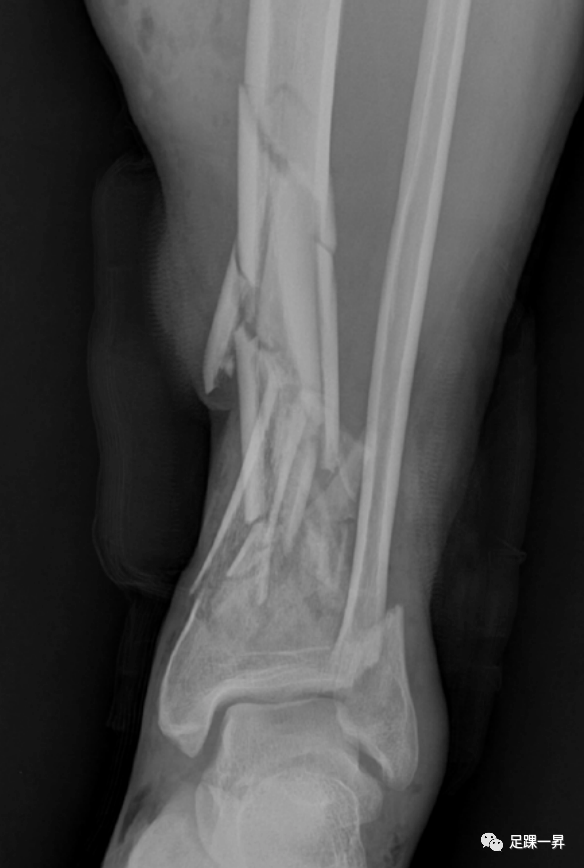 Figure 2 Preoperative X-ray shows a comminuted fracture of the distal tibia.
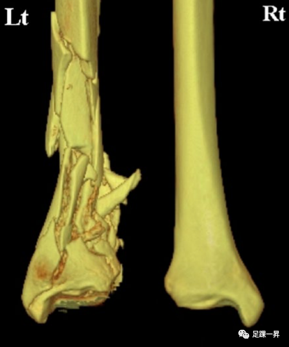 Figure 3 CT scan of both tibias. Left: fractured side; Right: healthy side.
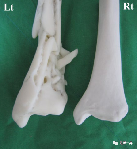 Figure 4 A real-sized 3D printed tibial fracture model (left). Using mirror imaging technology, the unaffected right tibia model was also printed as a reduction template (right).
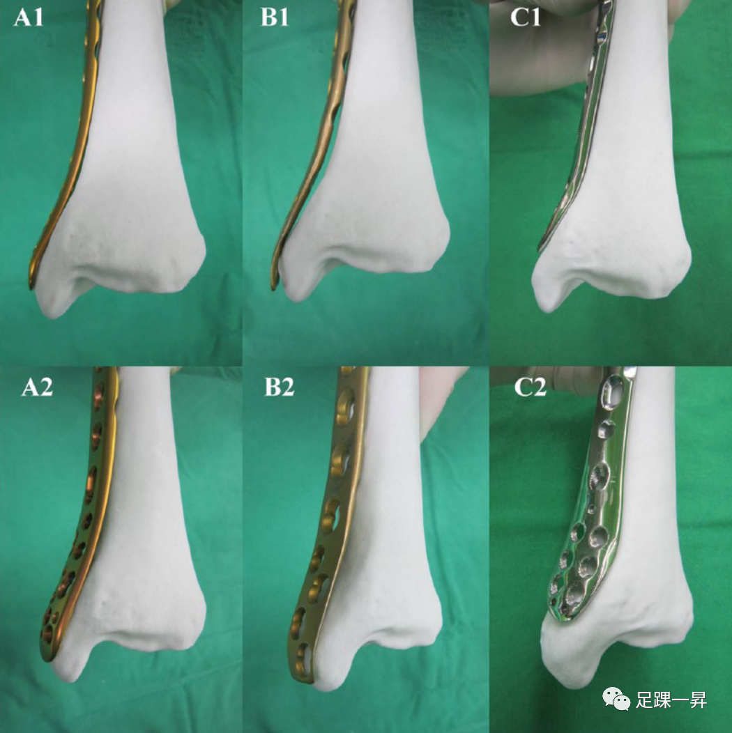 Figure 5 Using a real-sized 3D printed normal tibia model as a template, the best anatomical plate that fits the bone was selected from various manufacturers’ plates. The plate placement was then simulated while considering the screw movement trajectories on the plate.
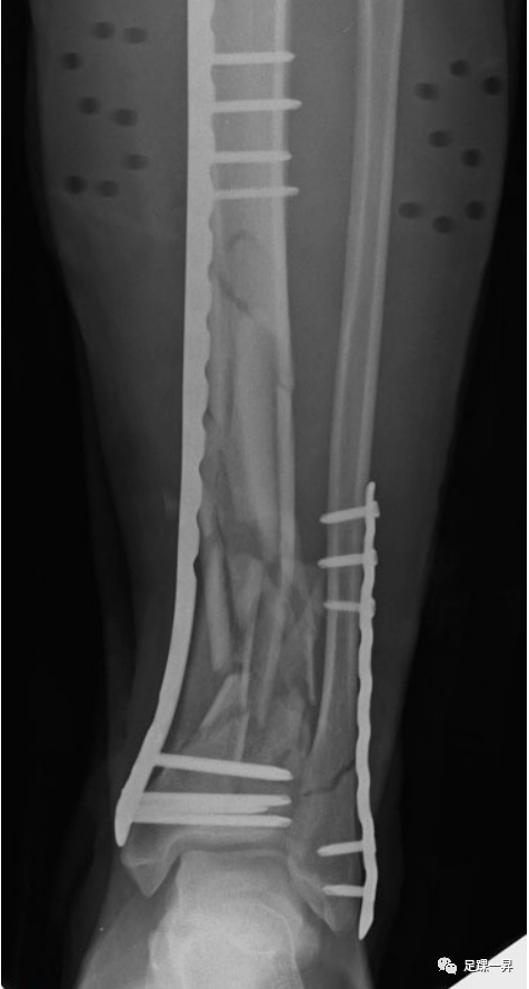 Figure 6 As per the preoperative plan, the best anatomical plate was chosen. No further bending or replacement of the plate was necessary, and no skin irritation was caused.Hook Plate Fixation for Ankle Avulsion FracturesFor patients with unilateral displaced ankle avulsion fractures, CT scans of both ankles were performed (Figure 7). Using mirror imaging technology, the opposite uninjured ankle was 3D printed into a real-sized fracture model. A locking compression hook plate is molded on the 3D printed model (Figure 8). For medial malleolus fractures, a small incision will be made at the distal end of the medial malleolus towards the fracture site. After reduction, a Kirschner wire is temporarily fixed, and the hook plate is inserted subcutaneously through the incision, separating the soft tissue, and the hook is inserted into the deltoid ligament to securely grasp the avulsed bone fragment. The plate is attached to the bone surface using cortical screws through the sliding holes of the hook plate (Figure 9). By applying pressure with cortical screws and locking sleeves at the proximal end of the plate, the hook plate is better attached to the medial malleolus, reducing skin irritation (Figure 10). One end of the reduction clamp fixes the cortical screw while the other end is fixed to the locking sleeve (Figure 11). After pressure is applied, locking screws are tightened, and the reduction clamp is released to place other screws (Figure 12).
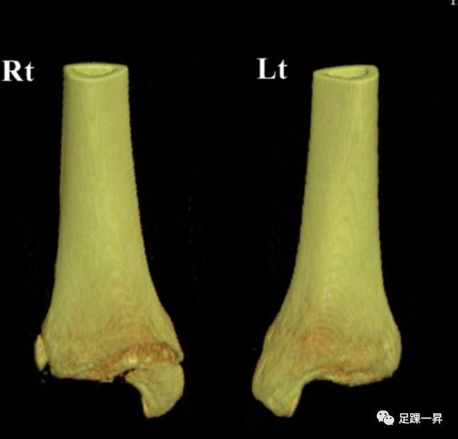 Figure 7 CT scan of both tibias of a patient with unilateral avulsion fracture of the medial malleolus. Left: fractured side; Right: healthy side.
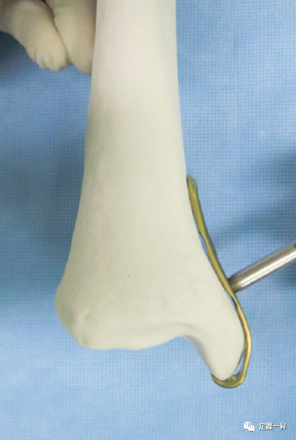 Figure 8 Using mirror imaging technology, the hook plate is pre-molded on the 3D printed model.
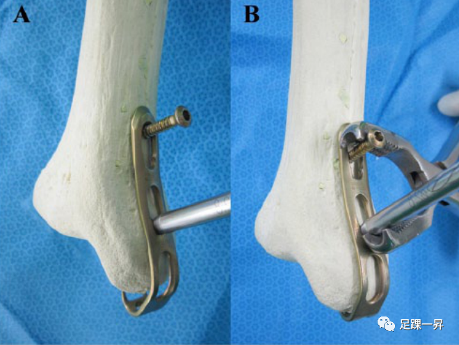 Figure 9 Axial compression model demonstration. (A) The hook plate is placed on the medial malleolus, and a cortical screw is screwed into the proximal end of the sliding hole, but not fully tightened. The locking sleeve is installed in the second locking hole. (B) The reduction clamp is used to gradually apply pressure to the cortical screw and sleeve.
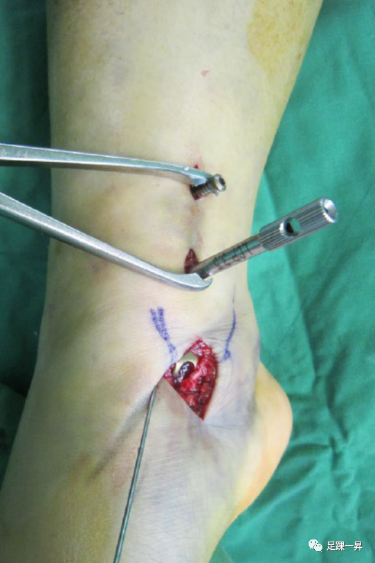 Figure 10 Intraoperative demonstration of the hook plate insertion into the subcutaneous layer. Cortical screws and locking sleeves are inserted through a small incision and fixed to the hook plate. The reduction clamp is used to gradually apply axial pressure.
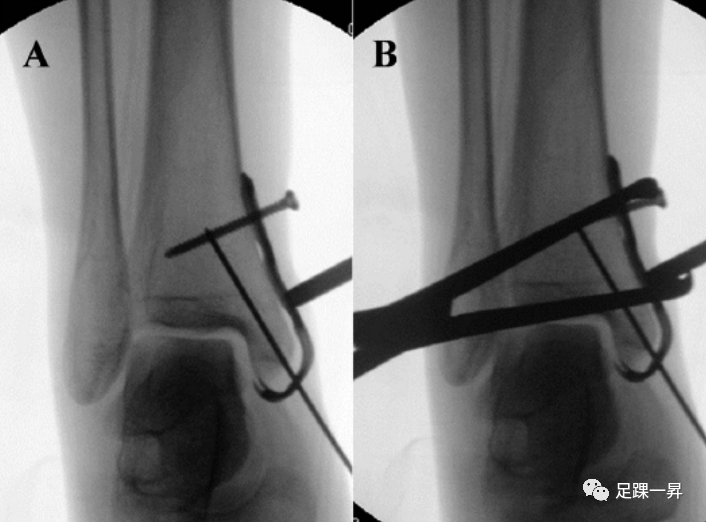 Figure 11 (A) Intraoperative fluoroscopic image showing the placement of the pre-molded hook plate, cortical screws, and locking sleeves. (B) Pressure is applied using the reduction clamp.
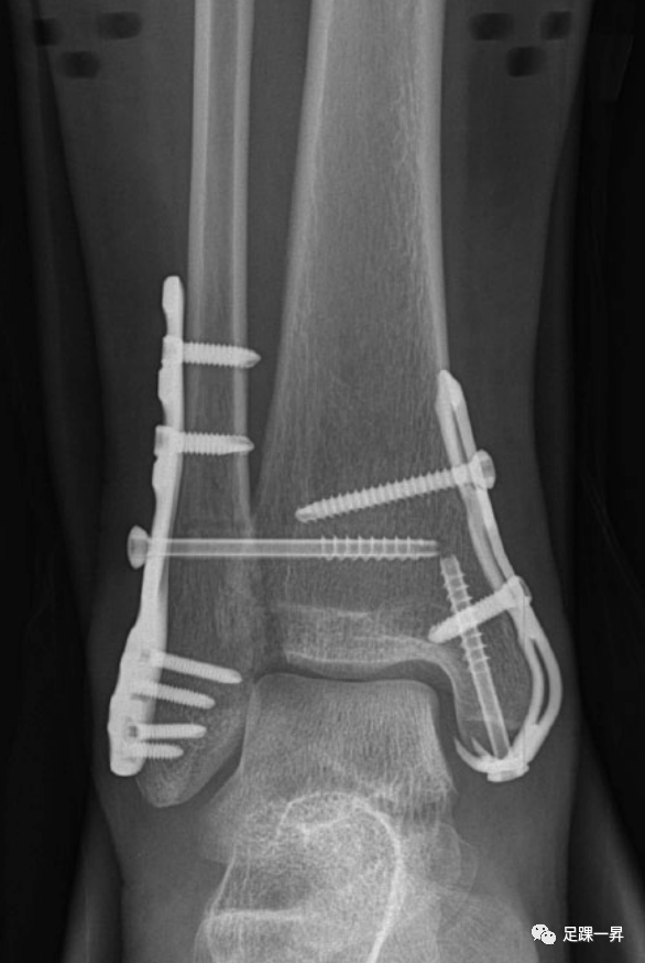 Figure 12 Postoperative X-ray shows that the pre-molded hook plate is closely attached to the medial malleolus, with an additional screw inserted into the distal hook to increase stability.ConclusionThe new techniques described in this article using 3D printing technology to manage complex tibial distal fractures and ankle avulsion fractures can serve as useful tools and techniques. However, the adoption of this new technology requires further evaluation of its cost-effectiveness.References: DOI: 10.1177/1071100715595695
Statement:This article is reproduced from <Foot and Ankle Yisheng>, and the copyright belongs to the original author. It is used for learning and communication purposes only, and reproduction without authorization is prohibited!
 Good Medical Skills Welcome Your Contributions
If you would like to share your clinical diagnosis and treatment experiences with more peers and let more people see you and benefit from you, we welcome your contributions!
Submission Requirements: Original academic content of more than 1500 words
Submission Method: haoyishu-zy03 (WeChat)
Medicine knows no bounds; every effort we make will be remembered in another way by this world. We look forward to your excellence!
Recommended Reading
Morning Read | The Fourth Generation of Minimally Invasive Bunion Surgery Has ArrivedMorning Read | [Surgical Techniques] A New Method to Identify Anatomical Relationships Under Single-Channel Spinal Endoscopy – “Manipulative Principles”Morning Read | How Does “Arm Wrestling” Lead to Humeral Shaft Fractures? – Analysis of Injury Mechanisms and Clinical Features
Click the card below to quickly follow us
👇 Click on 【Read the Original】 below to download the Good Medical Skills APP and start your learning journey!