
【Citation Format】GUO Can, YANG Chunxia, SUN Ning. Advances in near-infrared brain functional imaging of emotional tasks in affective disorder[J]. CHINESE JOURNAL OF NERVOUS AND MENTAL DISEASES, 2024, 50(1): 54-59.
【Cite this article】GUO Can, YANG Chunxia, SUN Ning. Advances in near-infrared brain functional imaging of emotional tasks in affective disorder[J]. CHINESE JOURNAL OF NERVOUS AND MENTAL DISEASES, 2024, 50(1): 54-59.
DOI:10.3969/j.issn.1002-0152.2024.01.010
Advances in near-infrared brain functional imaging of emotional tasks in affective disorder
GUO Can YANG Chunxia SUN Ning
Department of Mental Health, the First Hospital of Shanxi Medical University
Abstract:Emotional tasks (emotional task) are one of the main methods to study the attention bias and emotional function in affective disorders (affective disorder). Research based on functional near-infrared spectroscopy (functional near-infrared spectroscopy, fNIRS) under emotional tasks indicates that facial emotion recognition tasks, emotional stroop tasks, and emotion induction tasks combined with fNIRS technology have clinical value in the diagnosis and treatment of affective disorders. The deficits in attention function and emotional processing in patients with affective disorders are associated with abnormal activation of the left prefrontal cortex, particularly the differences in brain activation patterns are related to depressive symptoms in patients with major depressive disorder. The future direction of using fNIRS to study emotional tasks focuses on combining various neuroimaging techniques to conduct large-sample longitudinal cohort studies to obtain more objective diagnostic and therapeutic evidence, as well as comparing the differences in activation areas of different emotional stimuli.Keywords:
Depression; Bipolar disorder; Affective symptoms; Near-infrared brain functional imaging; Frontotemporal; Oxyhemoglobin; Emotional task
Affective disorders (affective disorder) are common mental illnesses, primarily including major depressive disorder and bipolar disorder (bipolar disorder, BD). Current studies show that the lifetime prevalence of major depressive disorder in adults in China is as high as 6.8%, with a lifetime prevalence of 0.6% for BD. Moreover, affective disorders are characterized by high recurrence rates, high suicide rates, and a high disease burden.[1-4] The etiology of these disorders remains unclear.[5-6] Currently, clinical diagnosis and treatment of affective disorders still lack objective and accurate biomarkers. Functional near-infrared spectroscopy (fNIRS) is an emerging clinical detection technology that provides quantitative measurement tools for exploring brain function differences in mental disorders, showing good application prospects.[7-8] Previous emotional-related studies based on fNIRS have mostly focused on exploring cortical activation during verbal fluency tasks, with limited research on emotional tasks. Emotional tasks present emotional information or stimuli that evoke subjective feelings in subjects, using different evaluation metrics to reflect attention bias and cognitive function, making them a key task paradigm for studying affective disorders. Therefore, this article reviews the research on major depressive disorder and BD based on emotional tasks and fNIRS technology, investigating the activation of the cortical areas during emotional tasks and analyzing the hemodynamic characteristics in the context of diagnosis, treatment, and assessment of affective disorders.
1 Overview of fNIRS
fNIRS is a light-based technology that emits near-infrared light in the wavelength range of 700-900 nm from the surface of the subject’s head to detect blood flow responses in the cortex. According to the Beer-Lambert law, it can measure changes in the relative concentration of hemoglobin[9-10], providing information on the activity of various brain regions. Due to its non-invasive nature, repeatability, suitability for natural settings, and high temporal resolution, fNIRS can be used for auxiliary diagnosis, cognitive function assessment, and treatment prediction in major depressive disorder and BD, making it an indispensable quantitative assessment platform in the field of affective disorders.
2 fNIRS Research in Affective Disorders Based on Emotional Tasks
The modulation of cognitive function by emotional information is one of the key issues in studying affective disorders. Currently, emotional task paradigms primarily include facial emotion recognition tasks (facial emotion recognition task, EFRT), emotional stroop tasks (emotional stroop effect, ESE), facial emotion gender judgment tasks, emotional autobiographical tasks, image recall tasks, emotional image tasks, negative emotional image description tasks, and emotional GO/NOGO tasks. This article summarizes three representative emotional tasks: facial emotion recognition tasks, emotional stroop tasks, and emotion induction tasks. These three emotional tasks measure subjects’ ability to recognize human facial emotions, their emotional attention bias towards objects, and their psychological behavior in various emotional states, with fNIRS studying emotional function and brain activation patterns based on these tasks.Table 1 summarizes the content of the three task paradigms,Table 2 organizes fNIRS studies on emotional tasks applied to affective disorders.Table 1 Content of the Three Task Paradigms for Emotional TasksTab.1 Specific content of three task paradigms for emotional tasks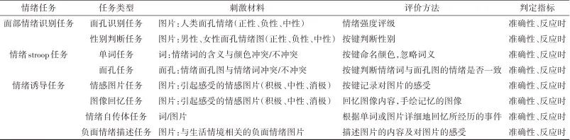 Table 2 fNIRS Studies on Emotional Disorders Based on Emotional TasksTab.2 fNIRS studies on emotional disorders based on emotional tasks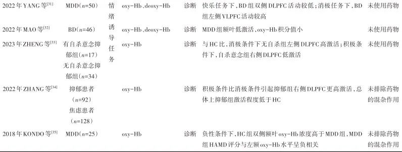
Note: MDD, Major Depressive Disorder; BD, Bipolar Disorder; HC, Healthy Control; HR, Healthy Siblings of BD Patients; BIP, At-Risk for Bipolar Disorder; oxy-Hb, Oxyhemoglobin; deoxy-Hb, Deoxyhemoglobin; FTC, Frontotemporal Cortex; PFC, Prefrontal Cortex; DLPFC, Dorsolateral Prefrontal Cortex; VLPFC, Ventrolateral Prefrontal Cortex.
2.1 Facial Emotion Recognition Task The facial emotion recognition task involves recognizing facial expressions to understand others’ emotional expressions, reflecting subjects’ perception, attention, and response capabilities to emotional faces. Behavioral and neuropsychological studies indicate that patients with affective disorders have recognition impairments and labeling errors in processing emotional faces[11-14]. Current consensus suggests that compared to healthy subjects, patients with major depressive disorder exhibit longer response times and generally lower accuracy in recognizing facial emotions. They tend to have higher accuracy in recognizing negative emotional faces compared to neutral ones, reflecting their attention bias towards negative stimuli and impaired ability to recognize neutral expressions[15-18].During the facial emotion recognition task, fNIRS detection of cortical blood flow can yield corresponding changes in hemoglobin levels. In terms of diagnosis, ZHANG et al.[15] included 35 patients with major depressive disorder (MDD) and 39 healthy controls, while MANELIS et al.[16] selected 51 depressed patients and 36 healthy controls, both using neutral face stimuli as a baseline to detect changes in oxyhemoglobin (oxy-Hb) concentration and activation of the prefrontal cortex (PFC) under happy and fearful face stimulus conditions. Results indicated that the difficulties in distinguishing neutral from other facial expressions in depressed patients were associated with reduced activation of the dorsolateral prefrontal cortex (DLPFC) during emotional processing. In identifying the severity of depression, one study selected 27 depressed patients and 24 normal controls to investigate hemodynamic changes in the PFC during the facial emotion recognition task, indicating that MDD patients showed significantly lower activity in the left PFC and lower average cross-wavelet coefficients and average wavelet coherence coefficients between bilateral oxy-Hb compared to the HC group[17]. These results suggest that MDD patients exhibit abnormal activation in the left PFC, and there are functional connectivity impairments between the bilateral PFC, indicating that hemodynamic changes in the PFC may be a further direction for research on diagnostic biomarkers for major depressive disorder.Additionally, in the areas of treatment mechanism research and effect evaluation, SEGAR et al.[19] used drug intervention and included 14 patients in the remission phase of their first episode mania (BD group), 14 healthy siblings of BD patients (HR group), and 13 healthy subjects (HC group), discovering that compared to the HC and BD groups, the HR group exhibited a higher degree of DLPFC activation while observing emotional faces, whereas the BD group did not show excessive activation. The former’s excessive activation may indicate that the observed prefrontal activation pattern during the facial emotion recognition task could serve as a risk marker for BD, but further research and evaluation in larger samples are needed. The latter BD group result may be attributed to the composition of patients in the BD group being in the pre-disease stage, remission phase, and receiving medication, leading to normalization of activation compared to the HR group. In summary, patients exhibit abnormal brain functional impairment in the PFC during tasks, and the facial emotion recognition task can be used to explore the emotional processing of affective disorders. Furthermore, emotional tasks combined with drug treatment may better reflect the drug’s impact on brain activation patterns[20-22]. Using fNIRS to detect blood oxygen signal changes during drug treatment may optimize clinical treatment plans for patients.2.2 Emotional Stroop Task The principle of the stroop effect primarily involves the brain’s integration and processing of various pieces of information. When the brain receives two conflicting pieces of information, the processing is interfered with, leading to increased response times and error rates. The emotional stroop task is a variant of the classic stroop effect, reflecting the influence of emotional information in stimuli on non-emotional information[23], and is widely applied in studying cognitive function and attention bias in patients with emotional disorders, making it an important area of stroop task research.In the diagnosis of affective disorders, MATSUBARA et al.[24] pointed out that patients with affective disorders had slower response times and lower accuracy in the emotional stroop task compared to healthy controls, but there was no significant difference in accuracy between MDD and BD patients. NISHIZAWA et al.[25] included 14 MDD patients and 20 normal subjects in a study on emotional words in the stroop task, finding that under negative word conditions, MDD patients exhibited a significant increase in oxy-Hb in the left frontal lobe compared to the HC group, while there was no significant difference in oxy-Hb concentrations between the two groups under positive stimuli, indicating that the induced wave related to the left superior frontal lobe was negatively correlated with the severity of depression. This result suggests that the left superior frontal lobe may be a potential site reflecting the severity of depressive symptoms. Additionally, another color word test compared 16 individuals at risk for bipolar disorder (BIP) with 46 healthy subjects, revealing lower activity in the left DLPFC of the BIP group and significantly reduced oxy-Hb levels in the frontotemporal lobe (FTL)[26]. Some results collectively indicate that patients with affective disorders exhibit reduced activation in the prefrontal cortex during emotional tasks[16-17,26-28]. However, this contradicts some neuroimaging studies which found increased activation in the left frontal middle and lower regions in BD and MDD patients under negative tasks[24]. The reason for this inconsistency may be differences in task paradigms (such as using watching tasks, face matching, or gender judgment tasks). Furthermore, recent meta-analyses indicate that different stimuli may affect top-down emotional regulation, with negative words possibly activating the frontal lobe more actively, while negative images may more easily activate the amygdala[29].In studies differentiating MDD patients from healthy controls, MATSUBARA et al.[30] selected 18 BD patients and 27 MDD patients to assess changes in brain activity during cognitive and emotional tasks. They found that patients exhibited less brain activity during cognitive tasks, while during emotional tasks, affective disorder patients showed more pronounced frontal-temporal activation compared to HC. ROC analysis revealed that frontal activation during cognitive tasks and happy word tasks was significantly associated with MDD. These findings suggest that comparing differences in brain activation during cognitive tasks and happy word tasks may help distinguish patients with major depressive disorder from healthy controls. In some results derived from emotional color word stroop tests, whether different stimuli affect activation in different regions and whether dual tasks can serve as effective means to differentiate major depressive disorder from healthy controls still require further large-sample studies for validation.2.3 Emotion Induction Task The emotion induction task involves presenting emotional materials to subjects to evoke a prolonged emotional state that influences subsequent cognitive activities/task responses (such as image recall tasks, emotional image tasks, image description tasks, and emotional autobiographical memory tasks), characterized by strong self-referentiality.In identifying features of affective disorders, YANG et al.[31] assessed the hemodynamic characteristics of 50 MDD patients across four combinatory tasks, including emotional picture tasks (emotional picture tasks, EPT) and negative emotional picture description tasks (negative emotional picture description task, NEPDT). Results showed that MDD patients exhibited completely opposite left-right frontal asymmetry characteristics compared to the control group, with differences between patients and controls being more significant in the combinatory tasks than in individual tasks. In single tasks, during NEPDT, MDD patients showed greater hemodynamic changes and higher sensitivity. However, during EPT, there was no significant difference between MDD and HC. The study also compared the box plots of bilateral frontal oxy-Hb concentrations under different tasks, revealing that the frontal asymmetry difference was greatest during NEPDT. This may suggest that this task is more suitable for inducing different degrees of activation in bilateral frontal lobes. Additionally, research involving 46 BD patients and 45 HC indicated that compared to HC, BD patients exhibited less hemodynamic changes in bilateral DLPFC when viewing pleasant images, while showing stronger hemodynamic changes in the left ventrolateral prefrontal cortex (VLPFC) when viewing unpleasant images[32]. This result aligns with findings from MATSUBARA et al.[24] using the emotional stroop paradigm, indicating that the neural response to emotional stimuli may be a characteristic marker of altered emotional processing in BD patients, independent of specific emotional tasks.Furthermore, in studies comparing differences in brain activation related to affective disorder symptoms, there are also some findings. ZHENG et al.[33] used emotional autobiographical memory tasks to investigate frontal activation differences between 17 depressed patients with suicidal ideation and 34 without. Under negative emotions, the activation rate of the left DLPFC in the non-suicidal ideation depression group was higher than that of the HC group. Under positive emotions, the activation rate of the right DLPFC in the suicidal ideation depression group was lower than that of the HC group. This indicates that patients with suicidal ideation may have certain deficits in emotional processing in the right DLPFC, while non-suicidal ideation depression patients may exhibit a resource compensation mechanism in the left DLPFC, suggesting that DLPFC abnormalities may be limited to the left hemisphere. Moreover, this study aligns with findings from ZHANG et al.[34] exploring bilateral DLPFC abnormal activation in 128 anxious and 92 depressed college students. Although these two studies contradict some findings of reduced left DLPFC activation in MDD patients, and are also contrary to some fNIRS studies using verbal fluency tasks or arithmetic tasks, the results consistently demonstrate cognitive function deficits in the areas of selective attention, executive function, and emotional processing in patients with major depressive disorder. The emotion induction task can not only explore patients’ neural activity but also has practical effects on improving patients’ symptoms and cognitive functions. KONDO et al.[35] studied 25 MDD patients and 25 matched healthy controls, finding that in unpleasant image recall tasks, the total score on the Hamilton scale was significantly negatively correlated with fluctuations in oxy-Hb concentration in the left frontal region. This result suggests that frontal functional impairment in patients with major depressive disorder may be related to state-dependent markers, and the image recall task measured by fNIRS may be an effective method for understanding the decline in frontal function in MDD patients. In summary, detecting frontal functional impairments in patients through fNIRS may provide potential biomarker clues for clinical assessment of cognition, emotion, and suicidal ideation. In task design and combined use, attention should be paid to the conditions under which different tasks are used, conducting detailed analysis and comparison of tasks, utilizing oxyhemoglobin integral values to illustrate hemodynamic changes in MDD during different tasks. Future research could incorporate brain fatigue patterns over time and their impact on hemodynamic analysis, as well as how the symptomatology of patients with major depressive disorder may affect DLPFC processing, which has reference value for future diagnosis and classification of major depressive disorder.
3 Summary and Outlook
In conclusion, fNIRS, as an emerging neuroimaging tool, provides objective quantitative indicators and technical support for exploring cognitive functions and emotional processing in affective disorders. Various emotional tasks collectively indicate that the hemodynamic changes observed in the left prefrontal cortex and bilateral prefrontal cortex can serve as effective tools for assessing cognitive and emotional functions in patients with affective disorders. The combination of emotional tasks and fNIRS signal detection may further reveal different pathophysiological mechanisms in patients with affective disorders.However, research on fNIRS in the field of affective disorders still has some limitations, and many issues remain to be addressed. First, in related studies, fNIRS technology has limited penetration into the brain, making it unable to detect deep regions involved in emotional processing, such as the amygdala and anterior cingulate gyrus. Future studies should combine fNIRS with other neuroimaging tools like fMRI to compensate for fNIRS’s shortcomings and explore changes in brain function from different dimensions. Second, research on fNIRS in the field of affective disorders is still immature, with some studies having small sample sizes and potential confounding effects of medications, which may lead to contradictions in results and reduce their generalizability. Future studies should conduct in-depth analyses in areas such as expanding sample sizes, improving variable data, and individual homogeneity to further support related research findings. Lastly, in emotional issues of patients with affective disorders, it is challenging to determine whether the abnormal activation patterns observed in the brain during studies are related to their emotional states or characteristics, and there is a lack of objective evaluation criteria for patients’ feelings towards stimuli in emotional tasks. Future research could incorporate the emotional valence of stimulus materials into assessments, employ longitudinal cohort studies with clinical interventions, and investigate how changes in patients’ emotional states affect facial expression recognition abilities and how these changes influence PFC activation patterns to further elucidate the neural mechanisms involved in emotional processing.
1. LU J, XU X, HUANG Y, et al. Prevalence of depressive disorders and treatment in China: A cross-sectional epidemiological study[J]. Lancet Psychiatry, 2021, 8(11): 981-990.
2. HUANG Y, WANG Y, WANG H, et al. Prevalence of mental disorders in China: A cross-sectional epidemiological study[J]. Lancet Psychiatry, 2019, 6(3): 211-224.
3. YANJUN T, RUIHAO W, LIN L, et al. The regulatory effects of the apelin/APJ system on depression: A prospective therapeutic target[J]. Neuropeptides, 2023, 102: 102382.
4. STANCA S, ROSSETTI M, BONGIOANNI P. The cerebellum’s role in affective disorders: The onset of its social dimension[J]. Metabolites, 2023, 13(11): 1113.
5. MATSUO K, HARADA K, FUJITA Y, et al. Distinctive neuroanatomical substrates for depression in bipolar disorder versus major depressive disorder[J]. Cereb Cortex, 2019, 29(1): 202-214.
6. CHEN L, WANG Y, NIU C, et al. Common and distinct abnormal frontal-limbic system structural and functional patterns in patients with major depression and bipolar disorder[J]. Neuroimage Clin, 2018, 20: 42-50.
7. KASSUBEK J. The application of neuroimaging to healthy and diseased brains: Present and future[J]. Front Neurol, 2017, 8: 61.
8. Wang Shensheng, Tan Zhonglin, Wu Yue, et al. Advances in the application of brain-computer interfaces in the diagnosis and treatment of depression[J]. Chinese Journal of Nervous and Mental Diseases, 2023, 49(4): 251-256.
9. LIU Hong, LI Yanyan, LUO Haijun, et al. Research progress on functional near-infrared spectroscopy blood oxygen measurement systems[J]. Journal of Chongqing Normal University (Natural Science Edition), 2023, 40(3): 155-163.
10. ZHANG Fuxu, WEI Yanyan, CHEN Qi, et al. Study on hemodynamic characteristics of brain blood flow in major depressive disorder patients during verbal fluency tasks[J]. Chinese Journal of Nervous and Mental Diseases, 2020, 46(10): 587-591.
11. CHIN FATT C R, JHA M K, COOPER C M, et al. Effect of intrinsic patterns of functional brain connectivity in moderating antidepressant treatment response in major depression[J]. Am J Psychiatry, 2020, 177(2): 143-154.
12. ROLLS E T. The orbitofrontal cortex and emotion in health and disease, including depression[J]. Neuropsychologia, 2019, 128: 14-43.
13. BYLSMA L M. Emotion context insensitivity in depression: Toward an integrated and contextualized approach[J]. Psychophysiology, 2021, 58(2): e13715.
14. REIS J A S, ROSSI G N, L OSÓRIO F, et al. Interventions for deficits in recognition of emotions in facial expressions in major depressive disorder: An updated systematic review of clinical trials[J]. Neurosci Biobehav Rev, 2023, 153: 105367.
15. ZHANG F, LIU W, ZHENG Y, et al. Decreased hemodynamic response to fearful faces relative to neutral faces in the medial frontal cortex of first-episode drug-naïve major depressive disorder[J]. J Affect Disord, 2023, 326: 57-65.
16. MANELIS A, HUPPERT T J, RODGERS E, et al. The role of the right prefrontal cortex in recognition of facial emotional expressions in depressed individuals: fNIRS study[J]. J Affect Disord, 2019, 258: 151-158.
17. GAO L, CAI Y, WANG H, et al. Probing prefrontal cortex hemodynamic alterations during facial emotion recognition for major depression disorder through functional near-infrared spectroscopy[J]. J Neural Eng, 2019, 16(2): 026026.
18. WANG L J, CHENG T H, GENG J S, et al. Comparisons of facial emotion recognition in different social contexts among patients with schizophrenia, major depressive disorder and bipolar disorder[J]. Asian J Psychiatr, 2023, 83: 103566.
19. SEGAR R, CHHABRA H, SREERAJ V S, et al. fNIRS study of prefrontal activation during emotion recognition-A Potential endophenotype for bipolar I disorder?[J]. J Affect Disord, 2021, 282: 869-875.
20. CATTARUNUSSI G, DELVECCHIO G, PRUNAS C, et al. Effects of pharmacological treatments on neuroimaging findings in first episode affective psychosis: A review of longitudinal studies[J]. J Affect Disord, 2020, 276: 1046-1051.
21. PRICE R B, DUMAN R. Neuroplasticity in cognitive and psychological mechanisms of depression: An integrative model[J]. Mol Psychiatry, 2020, 25(3): 530-543.
22. LI L, JI E, TANG F, et al. Abnormal brain activation during emotion processing of euthymic bipolar patients taking different mood stabilizers[J]. Brain Imaging Behav, 2019, 13(4): 905-913.
23. ARIOLI M, BASSO G, POGGI P, et al. Fronto-temporal brain activity and connectivity track implicit attention to positive and negative social words in a novel socio-emotional Stroop task[J]. Neuroimage, 2021, 226: 117580.
24. MATSUBARA T, MATSUO K, NAKASHIMA M, et al. Prefrontal activation in response to emotional words in patients with bipolar disorder and major depressive disorder[J]. Neuroimage, 2014, 85(Pt 1): 489-497.
25. NISHIZAWA Y, KANAZAWA T, KAWABATA Y, et al. fNIRS assessment during an emotional stroop task among patients with depression: Replication and extension[J]. Psychiatry Investig, 2019, 16(1): 80-86.
26. ALEKSANDROWICZ A, HAGENMULLER F, HAKER H, et al. Frontal brain activity in individuals at risk for schizophrenic psychosis and bipolar disorder during the emotional Stroop task – an fNIRS study[J]. Neuroimage Clin, 2020, 26: 102232.
27. HASSEL S, ALMEIDA J R, KERR N, et al. Elevated striatal and decreased dorsolateral prefrontal cortical activity in response to emotional stimuli in euthymic bipolar disorder: No associations with psychotropic medication load[J]. Bipolar Disord, 2008, 10(8): 916-927.
28. KERESTES R, BHAGWAGAR Z, NATHAN P J, et al. Prefrontal cortical response to emotional faces in individuals with major depressive disorder in remission[J]. Psychiatry Res, 2012, 202(1): 30-37.
29. FENG C, GU R, LI T, et al. Separate neural networks of implicit emotional processing between pictures and words: a coordinate-based meta-analysis of brain imaging studies[J]. Neurosci Biobehav Rev, 2021, 131: 331-344.
30. MATSUBARA T, CHEN C, HIROTSU M, et al. Prefrontal cortex activities during verbal fluency and emotional words tasks in major depressive, adjustment, and bipolar disorders with depressive states[J]. J Affect Disord, 2022, 316: 109-117.
31. YANG J, FAN J, CHENG A, et al. Hemodynamic analysis of the frontal cortex using multi-task fNIRS in major depressive disorder[J]. J Affect Disord, 2022, 315: 206-212.
32. MAO M, CHEN G, FENG K, et al. Characteristics of prefrontal activity during emotional and cognitive processing in patients with bipolar disorder: A multi-channel functional near-infrared spectroscopy study[J]. Front Neurosci, 2022, 16: 946543.
33. ZHENG M, DA H, PAN X, et al. Dorsolateral prefrontal activation in depressed young adults with and without suicidal ideation during an emotional autobiographical memory task: A fNIRS study[J]. J Affect Disord, 2023, 326: 216-224.
34. ZHANG Y, LI X, GUO Y, et al. Dorsolateral prefrontal activation in emotional autobiographical task in depressed and anxious college students: An fNIRS study[J]. Int J Environ Res Public Health, 2022, 19(21): 143.
35. KONDO A, SHOJI Y, MORITA K, et al. Characteristics of oxygenated hemoglobin concentration change during pleasant and unpleasant image-recall tasks in patients with depression: Comparison with healthy subjects[J]. Psychiatry Clin Neurosci, 2018, 72(8): 611-622.