
Orthopedic 3D Printing Technology: Turning Virtual into Reality

In daily life, many people have heard the term 3D.
The most familiar might be the three-dimensional 3D movies.
In fact, 3D printing technology has been applied in orthopedics.
It uses CT scan data for “three-dimensional reconstruction”.
Not only can it assist in surgical design,
but it can also directly print models or navigation templates.
Our orthopedic department has successfully applied 3D printing technology
for hundreds of patients with fractures and skeletal deformities
to implement personalized and customized surgeries.

Luo Yuanchao
Orthopedic Attending Physician
Consultation Time: Saturday Morning
Engaged in orthopedic work for over 10 years, specializing in spinal trauma, degenerative spinal diseases, spinal tumors, and orthopedic trauma diagnosis and treatment. He was the first in Zigong to carry out minimally invasive treatment of patellar fractures and Achilles tendon ruptures using percutaneous tension screws combined with tension bands, possessing rich clinical experience. Currently serves as a youth committee member of the Sichuan Provincial Spinal Cord Injury Specialized Committee and has received the “Excellent Medical Record Hospital in Zigong” award and the hospital’s “Advanced Individual” award. He has studied spine surgery at West China Hospital of Sichuan University, Southwest Hospital of Third Military Medical University, and the Third Hospital of Beijing.

What is 3D Printing Technology?
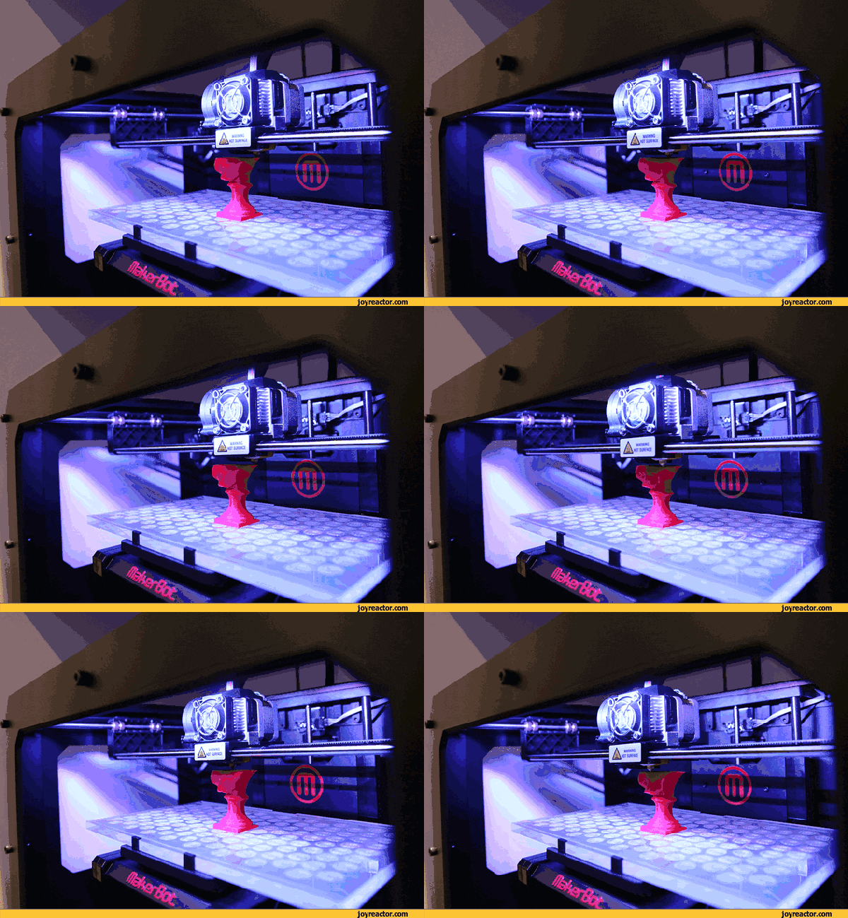
 3D printing is the process of manufacturing and producing three-dimensional objects from digital models in reality, as if bringing virtual items from computers and televisions into the real world.
NEWS
“
Advantages of 3D Printing Technology

1
Improves Surgical Precision
By simulating reconstruction through 3D printing, a bone model that is 1:1 in size to the injury site is created. Surgical simulations can clearly show the degree, type, and displacement of fractures, allowing for the formulation of scientifically reasonable personalized surgical plans, improving the precision of actual surgeries and enabling orthopedic surgeries to be “custom-made”. This makes difficult fracture surgeries both ‘precise’ and ‘minimally invasive’.
2
Shortens Recovery Time
Using 3D printing technology to assist patients in treatment, it presents and replicates in a realistic three-dimensional manner. This can shorten surgical time, reduce blood loss, and minimize surgical procedures. More direct surgical methods can be chosen, significantly shortening the patient’s recovery period.
Rehabilitation Case

1
Medical History Review
68-year-old Grandma Zhang
has suffered from neck pain and limited movement for over 10 years
and has had difficulty walking for 1 year.
She began to experience muscle atrophy in her limbs a year ago,
making her unable to care for herself.
Grandma Zhang has an old dislocation of the atlantoaxial joint
caused by rheumatoid arthritis,
which has compressed her spinal cord,
leading to paralysis of her limbs.
She has multiple and severe underlying diseases.
The surgery requires the placement of screws for reduction
and fixation,
which carries a high risk.
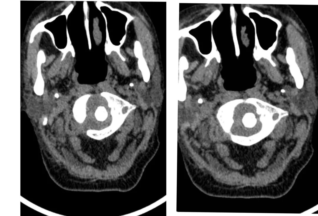
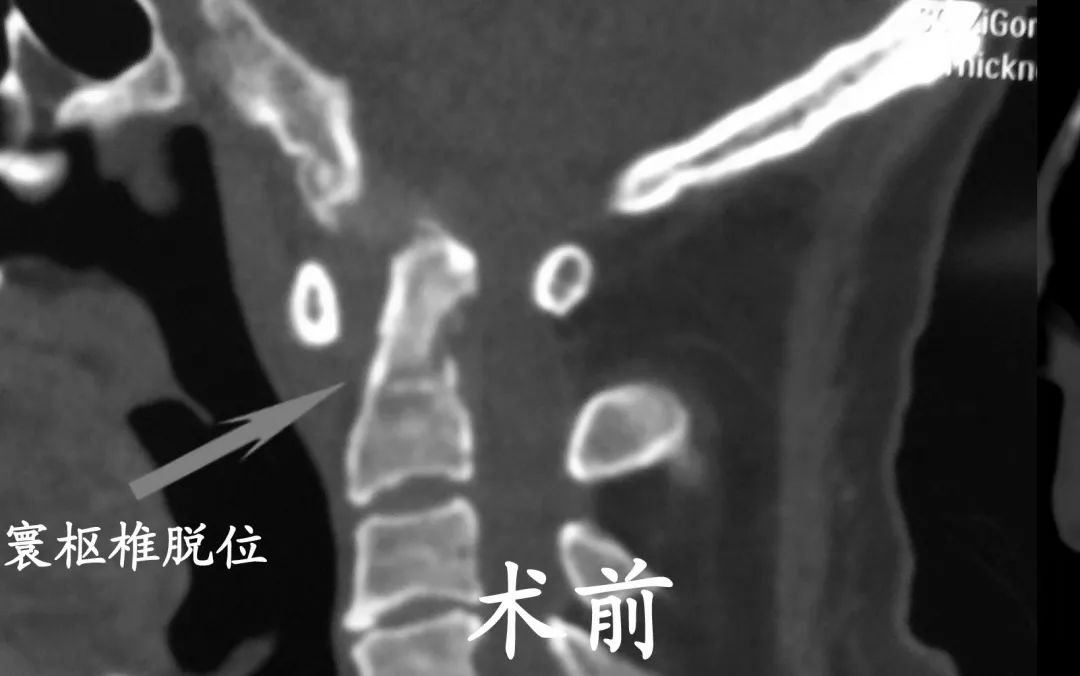
Pre-operative CT
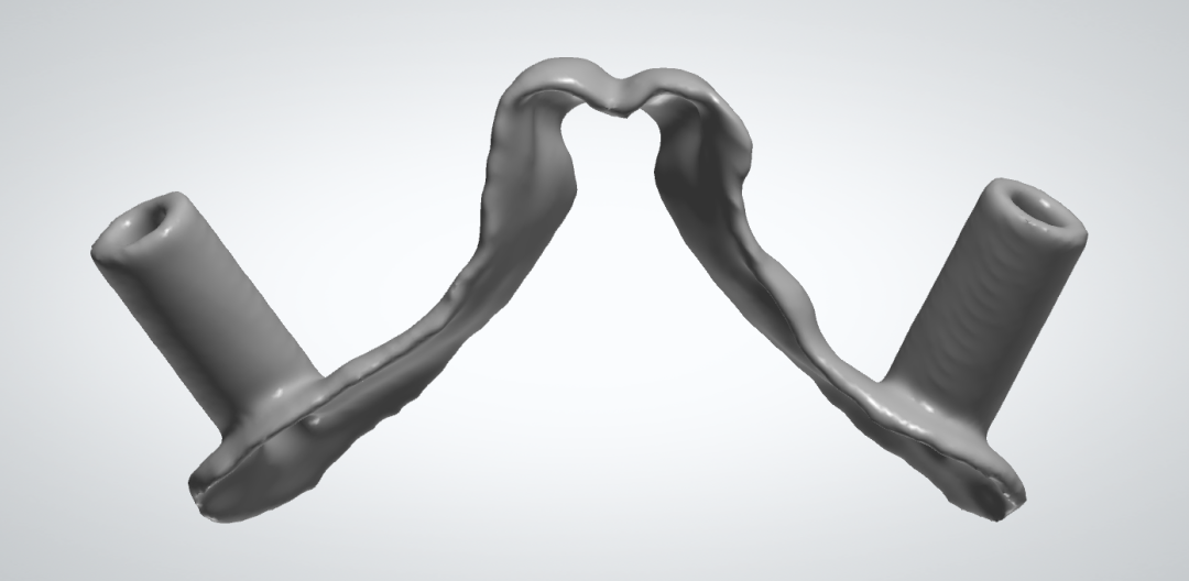
Pre-operative designed atlantoaxial guide plate
1
Formulating the Best Plan Using 3D Printing Technology
However, with 3D printing technology,
we designed a customized atlantoaxial screw guide plate for Grandma Zhang before surgery,
which will reduce the time for screw placement and lower the risk.
We can choose a more ideal screw placement path.
Our orthopedic team performed a posterior approach atlantoaxial dislocation decompression
and a screw-rod system internal fixation surgery for Grandma Zhang.
Post-operative rehabilitation exercises were supported
and Grandma Zhang’s paralysis symptoms gradually improved,
allowing her to walk normally and care for herself.
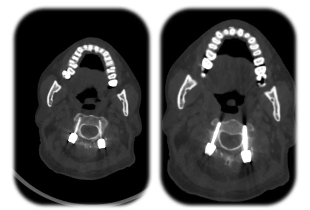
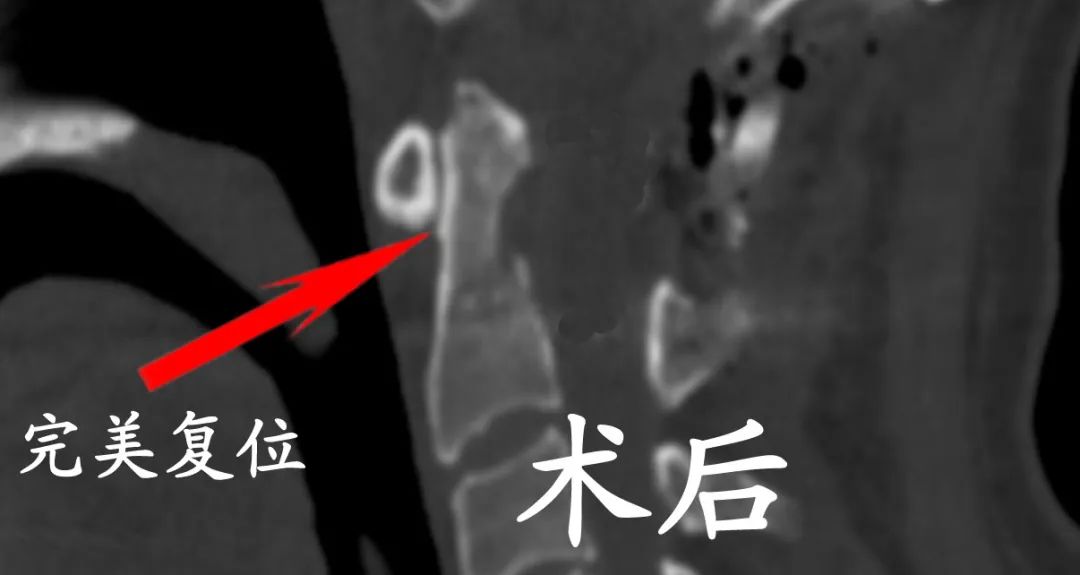
Post-operative CT (after screw placement)
3D Printing Technology Imaging
Let’s See It to Believe It
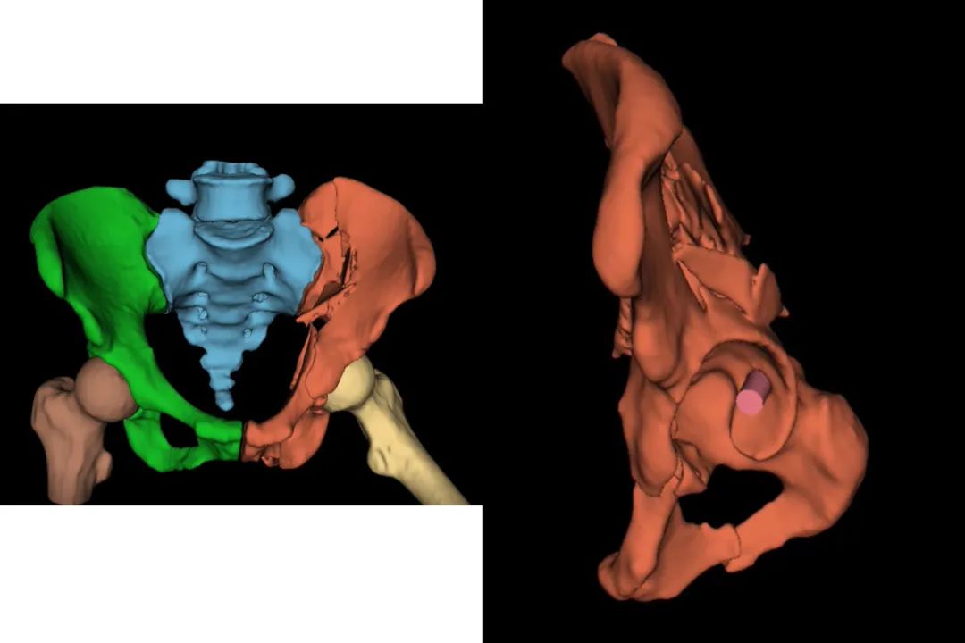
Pelvic fracture pre-operative 3D modeling
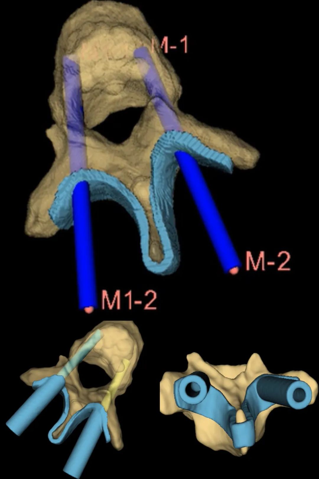
Pre-operative pedicle screw preparation
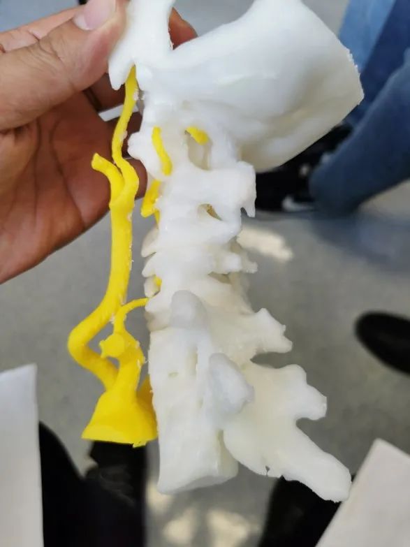
3D Printing Model for Cervical Deformity Patients, Including Carotid and Vertebral Arteries


Contributed by: Orthopedic Luo Yuanchao
Edited by: Zhao Yurong
Reviewed by: He Renjian, Gong Yong
Guided by: Yin Minggang
Statement: Some images and texts are sourced from the internet; please contact for removal if there is any infringement.