ClickBlueWords to Follow Us
Hello everyone, the article we are sharing today is titled “Chemotype- and Target-Driven Genome Mining for a New Natural Product Inhibitor of Bacterial Peptide Deformylase,” published in June 2025 in the Journal of the American Chemical Society.
The use of antibiotics drives the evolution of antimicrobial resistance (AMR), which limits the long-term efficacy of any drug. Therefore, it is crucial to develop antibiotics with entirely new structures and action targets. Actinonin (actinonin), an inhibitor of bacterial peptide deformylase (PDF), relies on a hydroxamate ester group that is toxic in vivo. The sequencing of numerous bacterial genomes provides an opportunity for “target-based natural product discovery,” where mining biosynthetic pathways may lead to the identification of molecules with the desired activity but lacking known pharmacophores. This study employs bioinformatics to conduct a chemotype-sensitive, target-based screening for natural product inhibitors of bacterial peptide deformylase (PDF). The research details the discovery process, heterologous expression, biosynthesis, total synthesis, and activity of “γ– gammanonins” (gammanonins), which are actinonin homologs derived from the phylum γ– Proteobacteria.
1.Analysis and Identification of Candidate Biosynthetic Gene Clusters (BGCs) for Peptide Deformylase (PDF) Inhibitors
UsingClusterScout tool, we searched over 135,000 completed sequenced and annotated bacterial genomes in the Integrated Microbial Genomes (IMG) database. The study used non-ribosomal peptide synthetase (NRPS) structural domains as the “screening anchor” to search for biosynthetic gene clusters (BGCs) adjacent to the PDF gene location. A preliminary search yielded 158 candidate gene clusters, which were further filtered to obtain 73 potential target gene clusters by excluding isolated domains, siderophore-related gene clusters, and pathways adjacent to the unique PDF gene in the genome. Using the CORASON tool, these potential target gene clusters were classified into 21 different NRPS biosynthetic gene cluster families, all of which carried PDF genes located nearby, suggesting that the PDF gene may be a potential self-protective gene (Figure 1b). Among these families, 18 gene clusters originated from high-yielding antibiotic-producing Gram-positive actinomycetes (Actinomycetes), while one candidate gene cluster was found in Gram-negative cyanobacteria (Cyanobacteria), α-Proteobacteria (Alphaproteobacteria), and γ-Proteobacteria (Gammaproteobacteria). The most promising potential target gene cluster originated from γ-Proteobacteria, encompassing multiple species of Vibrio (Vibrio) and Photorhabdus (Photorhabdus). This “minimal pathway” includes the following core components: 1 polyketide synthase (PKS) module, 3 non-ribosomal peptide synthetase (NRPS) modules, 1 initiator phosphopantetheinyl transferase (PPTase), and 1 pathway-specific methyltransferase (MT), with a key feature being the incorporation of a PDF gene within this biosynthetic operon (Figure 1c). The predicted chemical product of this biosynthetic gene cluster (BGC) is a “polyketide-extended tripeptide”: its structure is comparable to actinonin (actinonin), but lacks hydroxamate (hydroxamate) or other metal chelating groups. The pathway-specific methyltransferase (MT) was used for NCBI BLAST searches, revealing more identical pathways in the genera Xenorhabdus (Xenorhabdus) and Brenneria (Brenneria). Previous studies have indicated that this gene cluster is associated with interspecies antibacterial activity within the genus Vibrio (Vibrio), suggesting it may encode molecules with antibacterial properties. Based on these characteristics, we selected this biosynthetic gene cluster (BGC) for further investigation to discover a novel, hydroxamate-free bacterial peptide deformylase (PDF) inhibitor.
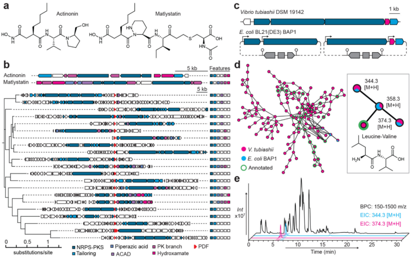
Figure 1
2. Study of Candidate Strains from the γ-Proteobacteria Phylum BGC
We obtained the strain Vibrio tubiashii (Vibrio tubiashii) DSM 19142 from the German Collection of Microorganisms and Cell Cultures (DSMZ) and employed previously reported culture and extraction conditions that were used to enrich bioactive components from the strain Vibrio ordalii (Vibrio ordalii) 12B09. No UV signal (300 nm) was detected for the product that was supposed to be reported, and the characteristic diagnostic mass of this product has never been reported in related studies. Due to the difficulty of gene knockout experiments in Vibrio tubiashii (V. tubiashii), we chose to perform heterologous expression of this biosynthetic pathway in the E. coli (E. coli) BL21 (DE3) derivative strain BAP1 to search for products associated with this gene cluster. The biosynthetic gene cluster (BGC) was cloned into two pET Duet plasmids, using an inducible T7 promoter to drive the expression of each “synthetic assembly line” gene while also driving the co-transcription of the peptide deformylase (PDF) gene and the methyltransferase (MT) gene (Figure 1c). The E. coli strain BAP1 carrying this reconstructed pathway was cultured in M9 minimal medium, and after induction, incubated overnight at 37°C, 30°C, or 16°C. The cultures were then centrifuged, and the components from the cell pellets and supernatants were extracted and analyzed using high-resolution liquid chromatography-mass spectrometry (HR-LCMS). To identify the metabolites associated with the expression of this biosynthetic gene cluster (BGC), we compared the extracts from Vibrio tubiashii (V. tubiashii) with those from engineered E. coli using the Global Natural Products Social Molecular Networking (GNPS) platform. The results showed that when induced at 16°C, a series of peptide metabolites related to “leucine-valine dipeptide” annotated in the GNPS database were present in both Vibrio tubiashii and BAP1 strains (Figure 1d, e). These three molecules are modified tripeptides, with very low content in both the cell supernatant and cell pellet. Using the matching biosynthetic gene cluster from Photorhabdus asymbiotica (Photorhabdus asymbiotica) DSM 15149, we constructed expression plasmids, and the same products were also detected in this strain, with similarly low content. Since the yield of the Vibrio tubiashii biosynthetic gene cluster (BGC) in the heterologous expression system was higher than in its natural host, and E. coli utilized a chemically defined medium (facilitating product separation and purification), E. coli was chosen as the production strain for this type of product in all subsequent experiments. To determine which metabolite is the final product of the target biosynthetic gene cluster (BGC), stable isotope-labeled methionine and acetate were added, which could reveal the involvement of the methyltransferase (MT) and terminal polyketide synthase (PKS) modules, respectively. The results showed that labeled methyl groups were detected in all metabolites, but only in the ion with a mass-to-charge ratio (m/z 374.3 [M+H]⁺ (corresponding to metabolite 1) was detected with labeled acetate associated with PKS. By predicting the substrates of the adenylation domain, it was speculated that the composition of this type of metabolite consists of 1 L-leucine (L-leucine) residue and 2 L-valine (L-valine) residues, which was confirmed by the secondary mass spectrometry (MS/MS) analysis result of the tripeptide with a mass-to-charge ratio of 344.3 [M+H]⁺ (corresponding to metabolite 2). To further verify this composition, a series of tripeptide variants were synthesized using solid-phase peptide synthesis (SPPS), which contained leucine, valine, isoleucine, and N-methylvaline at different positions. Among them, the synthesized tripeptide N-methyl-L-valine-L-valine-L-leucine was found to be identical to the natural tripeptide (metabolite 2). Additionally, a series of minor ions (mass-to-charge ratio 358.3 [M+H]⁺) were detected, and secondary mass spectrometry analysis indicated that these ions corresponded to tripeptide variants where valine was replaced by leucine/isoleucine. To elucidate the complete structure of the natural product (metabolite 1), we cultured, extracted, and processed 100 L of the engineered E. coli production strain. The mixed extract of cell pellets and supernatants was separated using size-exclusion chromatography and semi-preparative liquid chromatography-mass spectrometry (semipreparative LCMS), ultimately obtaining 7.8 mg of metabolite 2 and 1.8 mg of metabolite 1 (Figure 2a), which was sufficient for structural elucidation via nuclear magnetic resonance (NMR). The structure of metabolite 2 matched perfectly with the synthesized tripeptide; while metabolite 1 exhibited a “polyketide-extended leucine diol” structure, reminiscent of the statine (statine) residue analog — statinol.
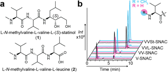
Figure 2
3. Synthesis of Compound 1
First, N-methylated dipeptides were prepared via solid-phase peptide synthesis (SPPS), which were then coupled with specific isomers of statinol (statinol). The synthesis of statinol followed the known synthetic route of statine (statine), yielding the R/S non-racemic isomer of Fmoc-L-statine ethyl ester (Fmoc-L-statine ethyl ester), which is a key intermediate in the synthesis process. NMR analysis of Mosher esters confirmed that the S-type isomer was the major product. After isolating each non-racemic isomer, secondary reduction and deprotection reactions were performed to obtain pure statinol. The major isomer (L-S-statinol) was coupled with the prepared dipeptide, and the retention time and secondary mass spectrometry (MS/MS) fragmentation pattern of the resulting product were found to be completely consistent with compound 1 (Figure 2a).
4. Biosynthetic Analysis of Compound 1
The biosynthetic gene cluster (BGC) possesses several special features that are hoped to be elucidated. The most notable is that its α-N-methylation appears to be achieved through trans-acting, which sharply contrasts with the synthesis mechanisms of almost all bacterial non-ribosomal peptides, where the N-methyltransferase structural domain is typically embedded in a cis-acting form within non-ribosomal peptide synthetases (NRPS). To study the substrate preference of the methyltransferase (MT), N-acetylcysteamine (SNAC) derivatives of each intermediate were prepared, and the methyltransferase was expressed and purified in vitro, followed by methylation reaction experiments. Liquid chromatography-mass spectrometry (LCMS) analysis showed that this methyltransferase could not catalyze the methylation of a single aminoacyl-SNAC substrate, but preferentially acted on enzyme-bound polyketide-extended tripeptides (Figure 2b). When the methyltransferase gene was removed from the expression plasmid, no compound 1, compound 2, or unmethylated compound 2 was detected, only the unmethylated compound 1 was observed. The absence of the signal for unmethylated compound 2 suggests that this tripeptide may be degraded by the cell, or the overexpression of the methyltransferase led to the premature release of compound 2 from the synthetic assembly line. Next, the function of the tandem acyl carrier protein (ACP) structural domains in the polyketide synthase (PKS) was investigated. One of the ACP structural domains may supply the cis-acting PKS structural domain, while the other may participate in the trans-methylation process. After mutating the key serine residues in each ACP structural domain to alanine, only a decrease in the yield of compound 1 was observed, with no new intermediates detected — indicating that these two ACP structural domains are functionally largely redundant.
5. Activity Evaluation of Compound 1
Attempts were made to detect the activity of compound 1 and the role of the biosynthetic gene cluster (BGC) related peptide deformylase (PDF). First, the PDF gene was removed from the plasmid, and the culture extracts were analyzed to verify whether this gene was involved in biosynthesis. The results showed that the removal of the PDF gene did not lead to the production of new intermediates, only a slight effect on the yield of compound 1. The removal of this presumed “self-protective gene” (PDF gene) did not affect the growth of E. coli after induction. To verify whether E. coli has inherent resistance to compound 1, the PDF enzyme from E. coli was overexpressed and purified. The experiment employed an in vitro endpoint method to detect the ability of the PDF enzyme to release primary amines from N-formylated tripeptide substrates, which was monitored using the fluorescent reporter molecule fluorescamine. The results showed that, unlike actinonin (actinonin, positive control), compound 1 did not exhibit inhibitory activity against E. coli PDF enzyme. Given that previous studies reported that this gene cluster has anti-Vibrio activity, and that there is a clear self-protective gene in its operon, further tests were conducted to see if compound 1 could inhibit Vibrio PDF enzymes. The PDF enzymes related to the gene cluster were expressed and purified, along with the “housekeeping” PDF enzymes from Vibrio tubiashii (V. tubiashii) and Vibrio crassostreae (V. crassostreae), where Vibrio crassostreae is known to be sensitive to the products of this gene cluster. However, consistent with the results from the E. coli experiments, compound 1 did not show inhibitory activity against these Vibrio PDF enzymes. To explore whether the “lack of metal-binding groups” is the reason for the inactivity of compound 1, N-carboxymethyl-L-valine-L-valine-L-(S)-statinol (compound 3) was synthesized, and antibacterial activity and enzyme inhibition experiments were repeated. The results showed that this modified product exhibited neither significant PDF enzyme inhibitory activity nor antibacterial activity. Theoretically, if carboxy-S-adenosylmethionine (Cx-SAM) were used instead of S-adenosylmethionine (SAM) as a co-substrate, the biosynthetic process might yield compound 3; however, this compound was not detected in the culture extracts of either Vibrio or engineered E. coli. Furthermore, overexpressing the biosynthetic genes related to Cx-SAM during the gene cluster induction did not yield the expected products. In summary, these studies indicate that neither the natural host nor the heterologous expression host produced bioactive products from this gene cluster.
6. Metabolomic Analysis of Bioactive Samples
To find the corresponding molecules associated with the reported antibacterial activity of this gene cluster, we obtained the original producing strain (Vibrio ordaliiV. ordalii 12B09) and its corresponding gene cluster knockout strain. A “spot-on-lawn assay” was conducted using Vibrio crassostreae as the indicator strain. A clear inhibition zone appeared around the wild-type 12B09 strain, while no inhibition zone was observed around the gene cluster knockout strain or Vibrio tubiashii (Figure 3a). The culture extract of the 12B09 strain was subjected to extraction, high-resolution liquid chromatography-mass spectrometry (HR-LCMS analysis, and Global Natural Products Social Molecular Networking (GNPS) analysis, revealing a series of abundant molecules related to compound 1 that had not been previously detected (Figure 3b, c). These metabolites were also present in the cultures of Photorhabdus asymbiotica (P. asymbiotica), but were not detected when the same genes were expressed in E. coli. Based on the mass of these molecules, it was speculated that their difference from compound 1 lies in the terminal group being an aldehyde (instead of the hydroxyl in compound 1) (corresponding to compounds 4-5). Further analysis indicated that all detected molecular masses could be explained by the following processes: the active β-hydroxyaldehyde (compound 6) spontaneously dehydrating, and intracellular aldehyde reductases converting the aldehyde group into a hydroxyl group (compound 7).
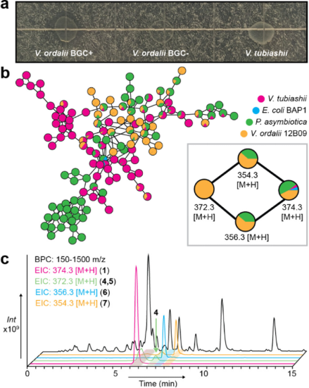
Figure 3
7. Discovery and Analysis of Gammanonin To search for products associated with antibacterial activity, extracts from the strain Vibrio ordalii (V. ordalii) 12B09 were repeatedly fractionated, and bioactivity assays were conducted using Vibrio crassostreae (V. crassostreae) as the indicator strain. Antibacterial activity was only observed in correlation with a single peak detected in high-resolution liquid chromatography-mass spectrometry (HR-LCMS analysis (mass-to-charge ratio 372.3 [M + H]⁺, corresponding to compound 4; Figure 3c). During the separation process, this molecule slowly converted to an ion with a mass-to-charge ratio of 354.3 [M + H]⁺, as well as another ion eluting shortly after compound 1 with a mass-to-charge ratio of 372.3 [M + H]⁺. Through two-dimensional nuclear magnetic resonance (2D NMR analysis, this active molecule was confirmed to be a cyclic hemiaminal (cyclic hemiaminal) — this structural feature explains its inherent reactivity and the unique in-source fragmentation pattern observed in LCMS analysis. Given its structural similarity to actinonin (actinonin) and its origin from the phylum γ-Proteobacteria (Gammaproteobacteria), this new molecule was named Gammanonin (compound 4, Figure 4).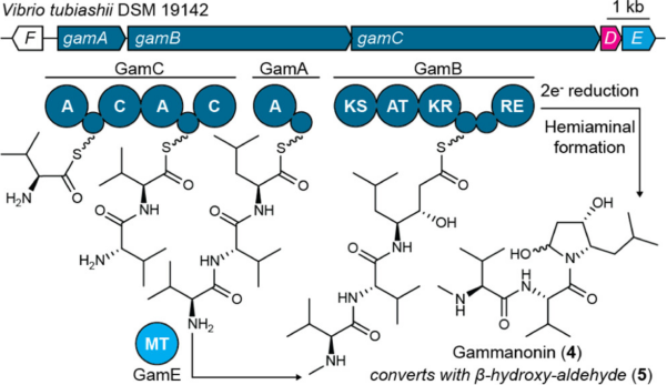 Figure 4
The cyclic hemiaminal structure of Gammanonin resembles tetrahydroisoquinoline-type alkaloids, where this structural feature can serve as a covalent electrophilic site (electrophilic warhead). To verify whether the formation of the hemiaminal is a spontaneous process, a linear aldehyde (compound 5) was synthesized. Solid-phase peptide synthesis (SPPS) was initiated using Weinreb amide resin loaded with Fmoc-L-(S)-statine (Fmoc-L-(S)-statine) as the starting material, extending the peptide chain to a tripeptide structure, and then cleaving it from the resin using lithium aluminum hydride (lithium aluminum hydride) to obtain the aldehyde form of the product. High-resolution liquid chromatography-mass spectrometry (HR-LCMS) analysis showed that the reaction produced a linear aldehyde (corresponding to the earlier eluting ion with a mass-to-charge ratio of 372.3 [M + H]⁺, i.e., compound 5), as well as a presumed α,β-unsaturated aldehyde (compound 6, mass-to-charge ratio 354.3 [M + H]⁺), and dimers produced from intermolecular hydroaldol condensation side reactions. The cyclic hemiaminal (compound 4) was only a minor product, indicating that its biosynthetic process may at least partially occur spontaneously.
8. Biological Activity of Gammanonin After isolating Gammanonin (compound 4) through “bioactivity-guided fractionation,” further tests were conducted to determine whether it could inhibit bacterial peptide deformylase (PDF). Using the previously established in vitro endpoint method, it was found that Gammanonin could effectively inhibit the activity of PDF enzymes from E. coli (E. coli), Vibrio crassostreae (V. crassostreae), and Vibrio tubiashii (V. tubiashii) “housekeeping” PDF enzymes (Figure 5a–c). Dose-response comparisons showed that Gammanonin (compound 4) exhibited weaker inhibitory activity than actinonin (actinonin); however, if the PDF enzyme was pre-incubated with Gammanonin before adding the substrate, the inhibition effect was significantly enhanced — this phenomenon aligns with the “covalent mechanism” (i.e., Gammanonin inhibits by forming a covalent bond with the enzyme). The PDF enzyme associated with the biosynthetic gene cluster (BGC) (named GamD) exhibited some resistance to Gammanonin (Figure 5d). Phylogenetic analysis of the PDF sequences from γ-Proteobacteria revealed that different bacterial sources of GamD genes formed an independent branch, consistent with the hypothesis that “GamD is a protective protein evolved to resist Gammanonin.”
Comparative analysis of PDF enzyme sequences also revealed that some conserved residues in GamD had undergone mutations, including amino acids located near the actinonin binding site. In all non-GamD type PDF sequences from γ-Proteobacteria, a conserved cysteine is present, suggesting that this site may be the target of Gammanonin (i.e., where Gammanonin reacts). However, experiments showed that mutating this cysteine to valine, which is unique to GamD, did not confer resistance to Gammanonin on the PDF enzyme.
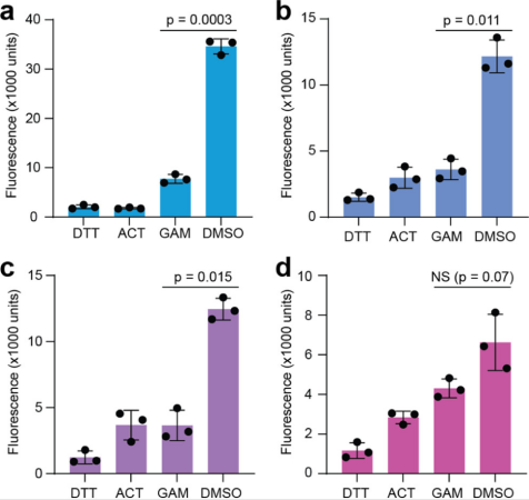 Figure 5